DOI: http://dx.doi.org/10.1590/1516-1439.288514
Materials Research. 2014; 17(6): 1434-1441 © 2014
*e-mail: [email protected]
1. Introduction
Paleobiology studies the history and evolution of life on Earth by the means of fossil record. However, this is not an easy task. During the fossilization process, taphonomy (everything that occurs after the death of an organism, until its burial and discovery by the paleontologist)1 can alter the morphology of the organism, hide important structures and build artifacts that can lead paleontologists to misinterpretations2-4.
Worldwide, in just a few years, the use of a series of advanced and/or high resolution techniques, mostly non-destructive, has proved to be important for the study of very old and rare well preserved fossils, in order to assist the work of paleontologists. The application of these techniques (e.g. Raman and FT-IR spectroscopies5,6, X ray microCT7-9, NanoSIMs10) to the study of fossils has expanded research in paleobiology and led it to a higher level of sophistication, and it is called paleometry11.
Inspired in the well established Brazilian archaeometry which studies archaeological, etnographical and the so called patrimonial materials12-16, the application of paleometrical techniques to the study of many Brazilian fossils is still growing17-21 and opening new perspectives to deepen our
knowledge of the biological afinities and paleoecological
aspects. Some exceptionally well-preserved fossils (e.g. the Ediacaran Corumbá Group and the Cretaceous Araripe
Basin) have become, in fact, scientiic challenges to the development of twenty-irst century Brazilian paleobiology.
Now, our new paleontology requires, not only the basic description of the oldest forms of life on Earth, but also an understanding and foundation of most modern concepts and methodological assumptions to bring extinct contexts to life. For Brazilian paleontologists, paleometrical techniques have proved to be important, for example, both for the elucidation of the chemical composition of paleoinvertebrate skeletons22,23, and to understand the processes of fossilization and paleoenvironment in contexts of climate and geochemical changes in the past20.
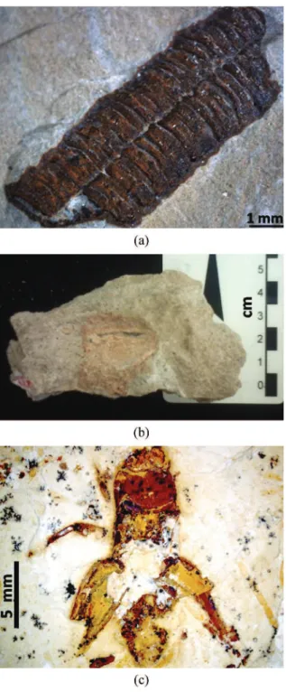
In this work, the potential of some of these techniques to the study of invertebrate fossils collected in different and important paleontological sites in Brazil, as seen in Figure 1: Corumbella werneri from the Corumbá group (Ediacaran); Unionoida freshwater mollusk from the Bauru Group (Cretaceous); and insects of the Crato Formation from the Araripe Basin (Cretaceous) is presented. The data here compiled is important both to the paleobiological insights
Paleometry: A Brand New Area in Brazilian Science
Adriana de Oliveira Delgadoa*, Pedro Victor Buckb, Gabriel Ladeira Osésc, Renato Pirani Ghilardid, Elidiane Cipriano Rangele, Mirian Liza Alves Forancelli Pachecob
aCentro de Ciências e Tecnologias para Sustentabilidade, Universidade Federal de
São Carlos – UFSCar, Rod. João Leme dos Santos, Km 110, CEP 18052-780, Sorocaba, SP, Brazil
bCentro de Ciências Humanas e Biológicas, Universidade Federal de São Carlos – UFSCar,
Rod. João Leme dos Santos, Km 110, CEP 18052-780, Sorocaba, SP, Brazil
cInstituto de Geociências, Universidade de São Paulo – USP, Rua do Lago, 562,
Cidade Universitária, CEP 05508-080, São Paulo, SP, Brazil
dFaculdade de Ciências, Universidade Estadual Paulista “Júlio de Mesquita Filho” – UNESP,
Av. Eng. Luiz Edmundo Carrijo Coube, 14-01, CEP 17033-360, Bauru, SP, Brazil
eUniversidade Estadual Paulista “Júlio de Mesquita Filho” – UNESP, Campus Experimental de
Sorocaba, Avenida Três de Março, 511, CEP 18087-180, Sorocaba, SP, Brazil
Received: April 8, 2014; Revised: December 18, 2014
Paleometry is a promising research ield that brings together different areas, such as physics and
chemistry, applied to paleobiological issues. In spite of being recognized abroad, it is a new research
ield in Brazil. The most important characteristic is the application of mostly non-destructive techniques
to the study of fossils. This work compiles some paleometrical applications to different geological contexts, such as the synthesis of hard skeleton in Corumbella werneri, geochemical aspects about fresh water bivalves from the Bauru Group and the exceptional preservation of arthropods from the
Crato Member. Diffuse Relectance Infrared (DRIFT) and Energy Dispersive X-ray Spectroscopy
(EDX) were complementary to elucidate the types of skeletogenesys in Corumbella. In the case of the bivalves, DRIFT revealed to be important to elucidate aspects about death and fossilization. Among arthropods, morphological analysis with Scanning Electron Microscopy (SEM) associated with EDX
was more proitable to understand fossilization process and paleoenvironmental implications.
and in the ield of materials characterization in order to boost
paleometry in Brazil.
2. Paleontological Aspects and Motivation
Corumbella werneri (Ediacaran, Corumbá Group) is a fossil preserved in marls and shales. It was considered as
an elongated polyhedral tube: a kind of ixed life form of
cnidarian medusas that lived ca. 543 million years ago22. Their fossils were firstly documented in Ladário and Corumbá, Brazil24. Since it was one of the irst animals on Earth capable of building a real skeleton, studies with
Corumbella are important to understand the origin and evolution of skeletonized animals on Earth.
Unionoida mollusks (Cretaceous, Bauru Group) are typically preserved in freshwater sandstones, indicative of energetic processes in paleoenvironmental reconstructions25. Our samples were collected in the municipality of Monte Alto, SP, Brazil. Paleometrical analysis has proved to be important to elucidate how these bivalves died and why the vast majority of specimens have articulated valves with sedimentary matrix inside.
The fossil insects from the Crato Formation (Cretaceous, Araripe Basin) are worldwide known for their high level of preservation in carbonates, including three-dimensional specimens with soft tissues preserved26-28. These fossils have important paleobiological and paleoenvironmental information and it is, therefore, crucial to understand the taphonomic processes that led to their exceptional
preservation. Despite having been briely discussed in
previous studies26,29, the taphonomy of the insects of the Crato Formation is still an unresolved question.
3. Experimental
The fossils have been characterized at the Laboratory of Characterization of Materials (LMCMat) at UNESP (Sorocaba, Brazil), in collaboration with the Group of Plasmas and Materials and at the Brazilian Nanotechnology National Laboratory (LNNano) at CNPEM (Campinas, Brazil).
The chemical composition of Corumbella werneri, (Figure 2a) and the Unionoida freshwater mollusk (Figure 2b) were investigated by using Diffuse Relectance Infrared Fourier Transform Spectroscopy (DRIFT) at LMCMat. DRIFT analysis revealed to be the most appropriate for IR spectroscopy due to the small amount of available samples and to the configuration of the spectrometer sample holder. For the analysis, grated powder of the fossils and the respective rock matrices were collected and dried at 50 oC. The dried powder was placed in a
cylindrical sample cup that was partially illed with KBr
powder, forming an upper layer with the material of interest. According to J. Ji et al.30, the use of unmixed layers saves
a signiicant amount of time and does not interfere with
the sensitivity to carbonates and sandstones, which are the main components in the analyzed fossils. The ratio of the
amount of sample and KBr powder was kept approximately
to 1:9. The layers were carefully pressed into the cup for the measurement of the sample spectra and another sample cup
illed with only KBr powder was used for the measurement
of the background spectrum. The analyses were carried out with a Jasco FT-IR 410 spectrometer in the range of 600-4000 cm–1, with a resolution of 4 cm–1 and average of 100 scans.
The micromorphology characterization of Corumbella werneri was performed at LNNano, with an electron
Delgado et al.
1436 Materials Research
microscope FEI Quanta 650 FE in the mode of secondary electrons detection, with acceleration voltages of 10 kV. Energy Dispersive X-ray Spectroscopy (EDX) was also carried out using an X-Max detector in semi-quantitative and mapping mode, in order to identify the distribution of chemical elements and compare it to FTIR results.
The fossil cricket of the Crato Formation (Figure 2c) was kindly lent to the researchers by IGc/USP collection
(specimen n. GP/1E-7105). Its ultrastructure was characterized at LMCMat, analyzed by Scanning Electron Microscopy (SEM), with a Jeol JSM6010 microscope, with acceleration voltage of 10 kV20. The fossil samples were coated with Au/Pd thin layer in order to improve the quality of the micrographs. EDX measurements with a Jeol Dry SD Hyper Detector were also applied to the sample, in order to identify the chemical composition of the observed structures on the fossil surface and inside the specimen20.
4. Results and Discussion
4.1. Corumbella werneri
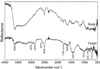
Diffuse reflectance analyses, in Figure 3, give us information about the ultrastructure of the carapace of
Corumbella werneri and the respective rock matrix. The main IR bands are indicated in Figure 3 and correspond to calcite30-32. It is worth pointing out that in the relectance spectrum, the weak IR bands appear stronger than expected for the absorption and transmission spectra. This stems
from the fact that in relectance spectra there is no linear
relationship between band intensity and concentration (as it occurs in transmission), and quantitative analyses by the DRIFT method are therefore rather complicated. The band in 2509 cm–1, for example, is described as very weak and weak in the transmission spectra presented in works of Miller and Wilkins33 and Huang and Kerr31, respectively, while it can be considered strong in our work in consistence
with the relectance data presented by Ji et al.30. Since the quantitative approach is not necessary at this point of our study, our research will focus on qualitative analysis that can provide paleobiologists with new important information.
Table 1 presents the comparison between the IR
relectance bands of the fossil of C. werneri and its rock matrix (Figure 3). The bands in the IR region from 600 to 1500 cm–1 occur due to the four fundamental modes of vibration of the carbonate ion (CO3–2): ν
1 is the symmetrical
stretching of CO (νs); ν2 is the out-of-plane bending of CO3
(γ); ν3 is the asymmetrical stretching of CO (νas); and ν4 is
the in-plane bending of OCO (δ)31,34 . The ν
1 mode should be inactive in FTIR spectra due to the symmetry of the molecule31,35, but since the studied sample (rock + fossil) presents several other minor constituents and impurities (common in sedimentary rocks), it is possible that bands at 1010 cm–1 for the fossil and 1160 cm–1 for the rock matrix
are due to the ν1 mode, which theoretically would occur at 1087 cm–1. The vibrations at 875 and 711 cm–1 correspond to out-of-plane and in-plane bending, respectively. Both
are observed in the carapace relectance spectrum, but not
in the rock matrix spectrum, despite being a marl (kind of shale carbonate rich rock). From the difference in spectra of the fossil and the rock, we can wonder if the synthesis of this hard exoskeleton leads to higher concentrations of the calcite in the carapace than in the surrounding rock medium. Finally, the asymmetrical stretching mode occurs at 1452 cm–1 as a broad band in the fossil relectance spectrum with no obvious correspondent band in the rock spectrum. This band is actually a double degenerated band that is observed for pure materials as a doublet. The mixture of minor components in the matrix can be responsible for the
enlargement of each line of the doublet, in a manner that they cannot be interpreted. The other observed bands of IR
Relectance are all regarded as vibrations of the carbonate
ion in overtone modes and combinations of the fundamental modes30,36. Most of them appear both in the relectance spectra of the Corumbella and in the rock with different relative intensities.
On the other hand, the large shoulders observed in 1000-1200 cm–1 and 1600-1800 cm–1 can be putative chitin,
when compared with the relectance data of analyzed chitin
from black corals37. These shoulders are clearly not present in the spectrum of rock matrix, hence the assignment and interpretation of these will be further investigated in future work.
The EDX mapping (Figure 4) supports the assumption that there is higher concentration of calcium (attributed to calcite) in the carapace of Corumbella in comparison with the rock matrix. It is also possible to see in Figure 4c, that Fe atoms are more concentrated (density in gray scale) in the
rock than in the fossil. Si, Al and O atoms were also detected by EDX and presented the same surface distribution as Fe atoms. The existence of O is related to the oxides present in the matrix. On the other hand, in Figure 4d, it is seen that Ca ions present a higher concentration in the carapace, with C atoms accompanying this tendency. The presence of Ca and C in the carapace denotes the presence of calcite in an
organic carapace and, together with IR relectance analysis,
gives us evidence of a biomineralized exoskeleton. As a technique for elemental characterization, EDX is important to complement IR Reflectance or FTIR Absorption spectroscopy. Moreover, the EDX mapping analysis allows the investigation of the distribution of element concentration with higher resolution than it could be achieved with IR spectroscopy.
The collected results from DRIFT and EDX spectroscopies reinforce, if not an entirely organic tegument23, at least, a weakly mineralized Corumbella carapace22, among one of the irst skeletonized animals. The evolution of animal skeletogenesis could be linked to environmental changes, such as the oceanic chemistry, as well as selective pressures correlated with the appearance of new ecological relations, such as predator/prey ones38.
4.2. Unionoida bivalve
The same approach previously described was applied to
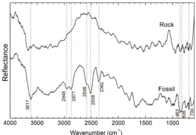
the bivalve. The identiied IR relectance bands are indicated
in the spectrum in Figure 5 and listed in Table 1.
It is noticeable that the IR spectrum of rock does not contain any information about its chemical composition, since the continuous noise is quite high when compared to
candidates to relection bands. This occurs due to the fact
that sedimentary rocks, such as sandstones, can be composed of different kinds of minerals and organic matter.
The IR relectance spectrum of the fossil, on the other
hand, presents bands that are attributed to calcite, as seen in Table 1. Since the reddish internal part of the mollusk should correspond to soft tissue fossilized in the interior
Figure 3. Diffuse relectance infrared spectra of Corumbella werneri carapace and rock matrix. The positions of the absorption bands are indicated in the igure.
Table 1. Comparison of IR relectance bands for analyzed fossils
and their rock matrices.
Wavenumber (cm–1)
C. werneri matrix molusk matrix
3626 3622 3617 3668
2978 2983 2948 *
2878 2878 2877 *
2575 2594 2588 *
2509 2511 2508 *
--- 2360 2362 *
1795 1796 * *
1645 1641 * *
1452 * * *
1010 1160 * *
875 * 863-836 *
711 --- 700 *
Delgado et al.
1438 Materials Research
Figure 4. SEM/EDX analysis of Corumbella werneri: (a) Image of the analyzed portion of carapace and rock matrix. The scale in photograph refers to the all four images. (b) SEM micrograph of fossil and rock; (c) EDX map of the distribution of Fe on the sample; (d) EDX map of the distribution of Ca on the sample. For the igures (c) and (d), the lighter spots indicate higher concentration of elements, while the dark regions indicate the absence of the element.
Figure 5. Diffuse relectance infrared spectra of inner part of Unionoida bivalve and surround rock matrix. The positions of absorption
of the shell, the presence of calcite suggests models of fossilization via incorporation of calcite available in the internal part of the shell39,40. This can occur when the specimen is fossilized with closed and articulated valves, creating an internal system isolated from the environment. After death, in decomposition, closed valves may have solubilized, and calcite might have been trapped in the sediment within the valves.
4.3. Insect of the Crato Formation
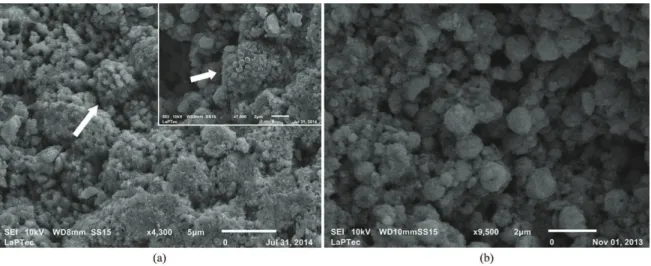
SEM analysis of the fossil cricket from the Crato Formation was performed in order to investigate the characteristics of the minerals that replaced the original organic matter. It was found that the texture of the exoskeleton or carapace (Figure 6a) is different from the inner part of the specimen (Figure 6b). While the exoskeleton consists of pseudomorphs of framboidal pyrite
>5µm in diameter, the inner portion of the fossil is illed with
framboidal pyrite pseudomorphs with ca. 1µm of diameter. Pyrite is a metallic mineral with chemical formula FeS2
(iron sulide).The observed grains are interpreted as being
pseudomorphs since their EDX analysis (Table 2) does not show the presence of sulfur in chemical composition. The pseudomorphs are covered with pliable structures here interpreted as mineralized extracellular polymeric substance (EPS) (Figure 7).
Data presented in Table 2, should be interpreted as an estimation of the chemical composition of the exoskeleton and the inner part of the fossil. Despite not being a quantitative analysis, the data gives valuable information about the presence or absence of key elements that can be associated with taphonomic and environmental processes.
EDX data supports that both external and internal portions of the fossils have iron and oxygen, suggesting that pyrite has been replaced by iron oxides/hydroxides due to weathering20. Furthermore, the inner part of the insects also presents small amounts of phosphorous and magnesium, that are not observed in the exoskeleton. These elements must be
further investigated since there are is no deinite evidence
Figure 6. Photomicrographs of the minerals which constitute the fossils from the Crato Formation. (a) Exoskeleton replaced by pseudomorphs of framboidal pyrite (indicated by arrow; detail of a well preserved pseudomorph). (b) Interior of the fossil, with preserved framboidal pyrite pseudomorps.
Figure 7. Pseudomorphs of framboidal pyrite covered by mineralized EPS.
Table 2. Estimated chemical composition of the exoskeleton and inner part of the insect of Crato Formation. Data were obtained with EDX in semi-quantitative mode. The errors were automatic calculated by the analysis software.
Atomic composition (%)
element exoskeleton inner part
C 23.735 ± 0.010 24.697 ± 0.010 O 56.60 ± 0.04 60.63 ± 0.05 Na 0.575 ± 0.021 0.523 ± 0.021
Mg --- 0.369 ± 0.021
Al 0.914 ± 0.021 0.503 ± 0.021 Si 2.14 ± 0.03 1.067 ± 0.021
P --- 1.78 ± 0.03
Ca 5.51 ± 0.06 8.29 ± 0.07 Fe 10.533 ± 0.020 2.13 ± 0.13
that could relate them with intrinsic characteristics of the organisms neither extrinsic conditions of the environment or with taphonomic processes41.
The textural differences veriied between the carapace
Delgado et al.
1440 Materials Research
mechanisms of fossilization. The presence of framboidal
pyrite pseudomorphs, replacing and inilling the fossil
, which possibly indicates the oxidation of previously precipitated pyrite framboids, as well as their coating by EPS, strongly suggest the activity of sulfate reducing bacteria during diagenesis, as suggested elsewhere29. The very small size of the minerals may also account for the high
degree of idelity of preservation of the Crato Formation
fossil insects20.
5. Conclusion and Perspective
IR Reflectance and SEM/EDX analysis were successfully applied to the study of Brazilian fossils from different geological contexts contributing to the progress and
broadening of paleometry in Brazil. The use of IR Relectance
instead of Transmittance allowed the observation of the bands that would appear weaker in transmission/absorption modes, which can be a great advantage for complex materials such as rocks and fossils. The EDX mapping showed to be an important complementary technique to:
(a) Relectance spectroscopy, bringing information about
the distribution of elements on fossil surface enabling even higher resolution with IR analysis; (b) SEM morphological
characterization, allowing elemental analysis of speciic
biological structures. Interpretative results contributed to
clarify outstanding questions still open in paleobiology and in taphonomy, such as: (1) the understanding of evolutionary and geochemical trends that led to the origin
and diversiication of skeletonized animals such as the
Corumbella; (2) the establishment of speciic conditions
that culminated in different types of fossilization in high energy environments, such as the Bauru Group; (3) the interpretation and comprehension of exceptional and rare well preserved soft parts, just like the case of insects from the Crato Formation. In this sense, paleometry has shed new light on material sciences studies and has opened a window to a brand new and exciting past for paleobiologists.
Acknowledgements
We would like to acknowledge Rafael Parra from LMCMat for SEM micrographs of insects and Professor Setembrino Petri from the University of São Paulo for having supported part of this work by supervising Gabriel
L. Osés’ inal undergraduate monograph.
We also acknowledge LNNano/CNPEM for the use of SEM (Quanta) microscope; Fapesp (proc. 2009/02312-4; 2013/16545-6, 2013/06719-7 and 2013/06718-0) and CNPq
for the inancial support and M. Amália S. Ladeira and Izabel
M. S. Ladeira from English for You (São Paulo, Brazil) for the language revision.
References
1. Behrensmeyer AK and Hook RW. Paleoenvironmental contexts and taphonomic modes in the terrestrial fossil record. In: Behrensmeyer AK, Damuth JD, DiMichele WA, Potts R, Hans-Dieter S and Wing SL. Terrestrial ecosystems through time: evolutionary paleoecology of terrrrestrial plants and animals. Chicago: University of Chicago Press; 1992. p. 15-136.
2. Waggoner BM. Interpreting the earliest metazoan fossils: What can we learn? American Zoologist. 1998; 38(6):975-982.
3. Lucas SG. Taphotaxon. Lethaia. 2001; 34(1):30. http://dx.doi. org/10.1080/002411601300068198.
4. Simões M, Rodrigues S, Leme JM and Van Iten H. Some Middle Paleozoic Conulariids (Cnidaria) as possible examples of taphonomic artifats. Journal of Taphonomy. 2003; 1(3):165-186.
5. Schopf JW and Kudryavtsev AB. Confocal laser scanning microscopy and Raman imagery of ancient microscopic fossils.
Precambrian Research. 2009; 173(1-4):39-49. http://dx.doi. org/10.1016/j.precamres.2009.02.007.
6. Igisu M, Ueno Y, Shimojima M, Nakashima SM, Awramik SM, Ohta H, et al. Micro-FTIR spectroscopic signatures of bacterial lipids in proterozoic microfossils. Precambrian Research. 2009; 173(1-4):19-26. http://dx.doi.org/10.1016/j. precamres.2009.03.006.
7. Chen JY, Bottjer DJ, Davidson EH, Li G, Gao F, Cameron RA, et al. Phase contrast synchrotron X-ray microtomography of Ediacaran (Doushantuo) metazoan microfossils: phylogenetic diversity and evolutionary implications. Precambrian Research. 2009; 173(1-4):191-200. http://dx.doi.org/10.1016/j. precamres.2009.04.004.
8. Pidassa B. High-resolution X-ray imaging of fossil samples. [Thesis]. Munich: Technische Universität München; 2013.
9. Pacheco MLAF, Galante D, Leme J, Rodrigues F, Bidola P, Hagadorn W, et al. Insights into the skeletonization, lifestyle, and affinity of the unusual Ediacaran fossil Corumbella. PLoS ONE. 2014; (in press)
10. Oehler DZ, Robert F, Walter MR, Sugitani K, Allwood A, Meibom A, et al. NanoSIMS: Insights to biogenicity and syngeneity of Archean carbonaceous structures. Precambrian Research. 2009; 173(1-4):70-78. http://dx.doi.org/10.1016/j. precamres.2009.01.001.
11. Riquelme F, Ruvalcaba-Sil JL and Alvarado-Ortega J. Palaeometry: Non-destructive analysis of fossil materials.
Boletín de la Sociedad Geológica Mexicana. 2009; 61(2):177-183.
12. Rosalie David A, Edwards HGM, Farwell DW and De Faria DLA. Raman Spectroscopic Analysis of Ancient Egyptian Pigments. Archaeometry. 2001; 43(4):461-473. http://dx.doi. org/10.1111/1475-4754.00029.
13. Silva FA, Appoloni CR, Quiñones FRE, Santos AO, Da Silva LM, Barbieri PF, et al. A arqueometria e a análise de artefatos cerâmicos: um estudo de fragmentos cerâmicos etnográficos e arqueológicos por fluorescência de Raios X (EDXRF) e transmissão Gama. Revista de Arqueologia. 2004; 17(1):41-61.
14. Rizzuto MA, Tabacniks MH, Added N, Barbosa MDL, Curado JF, Pascholati PR, et al. Pixe externo para análises de objetos de arte e arqueologia. Revista Brasileira de Arqueometria, Restauração e Conservação. 2007; 1:309 -312.
15. Lima SC, Rizzutto MA, Added N, Barbosa MDL, Trindade GF and Fleming MIDA. Pre-Hispanic ceramics analyzed using PIXE and radiographic techniques. Nuclear Instruments & Methods in Physics Research. Section B, Beam Interactions with Materials and Atoms. 2011; 269(24):3025-3031. http:// dx.doi.org/10.1016/j.nimb.2011.04.064.
central highland Ecuador. Archaeometry. 2013; 55(2):198-213. http://dx.doi.org/10.1111/j.1475-4754.2012.00683.x.
17. Lima RJC, Saraiva AAS, Lanfredi S, Nobre MAL, Freire PTC and Sasaki JM. Caracterização espectroscópica de peixe do período cretáceo (Bacia do Araripe). Quimica Nova. 2007; 30(1):22-24. http://dx.doi.org/10.1590/S0100-40422007000100005.
18. Fairchild TR, Sanchez EAM, Pacheco MLAF and Leme JM. Evolution of Precambrian life in the Brazilian geological record. International Journal of Astrobiology. 2012; 11(4):309-323. http://dx.doi.org/10.1017/S1473550412000183.
19. Freire PTC, Abagaro BTO, Sousa Filho FE, Silva JH, Saraiva AAF, Brito DDS, et al. Pyritization of fossils from the Lagerstätte Araripe Basin, Northeast Brazil, from the Cretaceous Period. In: Whitley N and Vinsen PT, editors.
Pyrite: synthesis, characterization and uses. New York: Nova Science Publishers Inc; 2013. p. 123-140.
20. Osés GL. Artrópodes fósseis do Membro Crato (Formação Santana, Bacia do Araripe, Eocretáceo, NE do Brasil): levantamento taxonômico, tafonômico e paleoecológico utilizando técnicas não-destrutivas. [Monografia]. São Paulo: Universidade de São Paulo; 2013.
21. Buck PV. Paleometria aplicada ao estudo de fósseis brasileiros: implicações evolutivas e tafonômicas. [Monografia]. Sorocaba: Universidade Federal de São Carlos; 2013.
22. Pacheco MLAF, Leme J and Machado A. Taphonomic analysis and geometric modelling for the reconstitution of the Ediacaran metazoan Corumbella werneri Hahn et al. 1982 (Tamengo Formation, Corumbá Basin, Brazil). Journal of Taphonomy. 2011; 9(4):269-283.
23. Warren LV, Pacheco MLAF, Fairchild TR, Simões MG, Riccomini C, Boggiani PC, et al. The Dawn of animal skeletogenesis: ultrastructural analysis of Ediacaran metazoan
Corumbella werneri.Geology. 2012; 40(8):691-694. http:// dx.doi.org/10.1130/G33005.1.
24. Hahn G, Hahn R, Leonardos OH, Pflug HD and Walde DHG. Kfrperlich erhaltene Scyphozoen Reste aus dem Jungprekambrium Brasiliens. Geologica et Paleontologica. 1982; 16:1-18.
25. Ghilardi RP, D’Ágosta FCP, Alves K and Campos ACA. Tafonomia de moluscos fósseis do Grupo Bauru (Cretáceo Superior, bacia Bauru), na região do Município de Monte Alto, São Paulo, Brasil. Boletim do Museu Paraense Emílio Goeldi.
Ciências Naturais. 2011; 6(2):197-206.
26. Grimaldi D. Insects from the Santana Formation, lower cretaceous, of Brazil. New York: Bulletin of the AMNH; 1990. 27. Grimaldi D. The Santana Formation insects. In: Maisey JG.
Santana fossils: an illustrated atlas. Neptune City: T.F.H. Publications; 1991. p. 379-406.
28. Martill DM. Fossils of the Santana and Crato Formations, Brazil. London: Palaeontological Association; 1993.
29. Menon F and Martill DM. Taphonomy and preservation of Crato Formation arthropods. In: The crato fossil beds of brazil: window to an ancient world. Cambridge: Cambridge University Press; 2007, p. 79-96.. http://dx.doi.org/10.1017/ CBO9780511535512.008.
30. Ji J, Ge Y, Balsam W, Damuth JE and Chen J. Rapid identification of dolomite using a Fourier Transform Infrared Spectrophotometer (FTIR): A fast method for identifying Heinrich events in IODP Site U1308. Marine Geology. 2009; 258(1-4):60-68. http://dx.doi.org/10.1016/j. margeo.2008.11.007.
31. Huang CK and Kerr PF. Infrared study of the carbonate minerals. The American Mineralogist. 1960; 45(3-4):311-324.
32. National Institute of Standards and Technology. Calcium carbonate (calcite). Material measurement laboratory; 2011. Available from: <http://www.nist.gov/mml/>. Access in: 24/03/2014.
33. Miller FA and Wilkins CF. Infrared spectra and characteristic frequencies of inorganic Ions. Analytical Chemistry. 1952; 24(8):1253-1294. http://dx.doi.org/10.1021/ac60068a007. 34. Bessler KE and Rodrigues LC. Os Polimorfos de carbonato
de cálcio: Uma síntese fácil de aragonita. Quimica Nova. 2008; 31(1):178-180. http://dx.doi.org/10.1590/S0100-40422008000100032.
35. Reichenbächer M and Popp J. Challenges in molecular structure determination. Springer; 2012.. http://dx.doi. org/10.1007/978-3-642-24390-5.
36. Clark RN. Spectroscopy of rocks and minerals, and principles of spectroscopy. In: Rencz AN, editors. Remote sensing for the earth sciences – Manual of remote sensing. New York: John Wiley & Sons; 1999. p. 3-58. v. 3.
37. Bo M, Bavestrello G, Kurek D, Paasch S, Brunner E, Born R, et al. Isolation and identification of chitin in the black coral Parantipathes larix (Anthozoa: Cnidaria). International Journal of Biological Macromolecules. 2012; 51(1-2):129-137. http:// dx.doi.org/10.1016/j.ijbiomac.2012.04.016. PMid:22546360
38. Wood RA. Paleoecology of the earliest skeletal metazoan communities: Implications for early biomineralization.
Earth-Science Reviews. 2011; 106(1-2):184-190. http://dx.doi. org/10.1016/j.earscirev.2011.01.011.
39. Jacob DE, Wirth R, Soldati AL, Wehrmeister U and Schreiber A. Amorphous calcium carbonate in the shells of adult Unionoida.
Journal of Structural Biology. 2011; 173(2):241-249. http:// dx.doi.org/10.1016/j.jsb.2010.09.011. PMid:20850546 40. Yang W, Kashani N, Li XW, Zhang GP and Meyers MA.
Structural characterization and mechanical behavior of a bivalve shell (Saxidomus purpuratus). Materials Science and Engineering C. 2011; 31(4):724-729. http://dx.doi. org/10.1016/j.msec.2010.10.003.