*e-mail: [email protected]
Article presented at the II Simpósio Mineiro de Ciências dos Materiais November 12-14, 2007, Ouro Preto - MG.
Development and Characterization of an Intraocular Biodegradable Polymer
System Containing Cyclosporine-A for the Treatment of Posterior Uveitis
Juliana Barbosa Salibaa, André Augusto Gomes Faracoa, Maria Irene Yoshidab,
Wander Luiz de Vasconcelosc, Armando da Silva-Cunhaa, Herman Sander Mansurc*
a
Departamento de Produtos Farmacêuticos, Universidade Federal de Minas Gerais – UFMG,
Av. Antônio Carlos, 6627, 31270-010 Belo Horizonte - MG, Brazil
b
Departamento de Química, Universidade Federal de Minas Gerais – UFMG
Av. Antônio Carlos, 6627, 31270-010 Belo Horizonte - MG, Brazil
c
Departamento de Engenharia Metalúrgica e de Materiais,
Universidade Federal de Minas Gerias – UFMG,
Rua Espírito Santo, 35/316, Centro, 30160-030 Belo Horizonte - MG, Brazil
Received: December 4, 2007; Revised: February 29, 2008
The aim of this study was to synthesize and characterize the biodegradable intraocular implants based on poly (D,L–lactide–co–glycolide) (PLGA 75:25) with Cyclosporine-A (CyA) and to evaluate their in vitro drug delivery profile. Thermal analysis was conducted by using Thermogravimetry (TG) and Differential Scanning Calorimetry (DSC). Phase analysis and crystallinity of the polymer-CyA samples were assessed through X ray diffraction (XRD) and Fourier transform infrared spectroscopy (FTIR). Finally, microstructure and morphology of the systems were investigated by Scanning Electron Microscopy (SEM). The results showed that CyA was successfully incorporated into PLGA network with drug loading of approximately 31.6%. Also, based on FTIR and thermal analyses (TGA/DSC) no significant physical-chemical interaction was detected at the micro-nanoscale level between polymer/drug. SEM micrographs have indicated a uniform drug distribution in PLGA matrix. XRD patterns have showed that the incorporated semi-crystalline structure of CyA has not significantly altered the polymeric mainly amorphous network. In addition, the results have confirmed the chemical and biological drug stability, the drug distribution into the polymeric matrix and the possibility of cyclosporine prolonged delivery system profile.
Keywords: nanostructured drug delivery system, biodegradable intraocular implants, biodegradable polymer
1. Introduction
Biodegradable polymers have been largely used in pharma-ceuticals and biomedical field, respectively as delivery systems or as biomaterials1. The poly (D,L-lactide-co-glycolide) is a classic
example amongst the synthetic polymers and has been well applied as drug delivery system due to its satisfactory biocompatibility and absence of significant toxicity in vivo studies2,3. Due to biodegradable
intraocular implants nanoparticle constitution, the nanoscience can be an important tool to study their properties based on the polymer/ drug interaction analysis.
The drug/polymers interface has been previously reported and characterized by conformational, spectroscopic, thermal and mor-phological techniques in order to detect possible polymer and drug interaction and to justify the preliminary in vitro release profile. The PLGA 75:25, presents linear chain containing 75% glucolide and 25% lactide. The lactide differs from glycolide by the presence of methyl group, characterizing its higher hydrophobicity. The polymer propor-tion and the chain size will define the in vitro and in vivo copolymer degradation. Besides that, this copolymer presents an amorphous state and its glass transition temperature occurs at, approximately 50 °C. This material also presents thermoplastic property where
copolymer chains are held together by relatively weak van der Waals and dipole-dipole forces1.
The CyA is a cyclic endecaptide commonly used as a selective immunosuppressive drug, with anti-inflammatory properties effec-tive in the treatment of the posterior uveitis isolated from fungal. The lyophilized drug exhibits characteristics consisted with a glass thermotropic liquid in a semi-crystalline state and has solid-liquid phase transition in range of 118-125 °C11. The presence of β-sheets
intermolecular interaction indicates the molecular stability and bio-logical activity was maintained4. Cyclosporine A has been widely
and effectively used for the treatment of various forms of chronic uveitis. However, poor penetration of topical CsA into the eye and serious systemic toxicity produced by oral administration require a new approach to overcome the hurdles of blood–ocular block and low bioavailability of CyA12.
The lyophilized mixture was obtained by dissolving PLGA 75:25 and cyclosporine A (CyA) in a adequate quantity of acetonitrile as organic solvent (CH3CN). The formed solution was then lyophilized during 24/48 hours.
2.2. X ray diffraction analysis (XRD)
The lyophilized samples PLGA 75:25, CyA and lyophilized mixture was analyzed by (PHILIPS, PW1710) using CuKα radiation with λ= 1.54056 Å. XRD analyses were conducted in the 2θ range from 3.03 to 89.91° with steps of 0.06°. Narrow peaks identified within the scan range were confirmed using previously published literature9.
2.3. Thermogravimetry characterization (TG)
The thermal stability was evaluated by thermogravimetry analysis (TG, Shimatzu TGA 50H). The lyophilized samples PLGA 75:75 and CyA were heated in sealed platinum pans at a heating rate of 10 ° C. min–1 from room temperature to 750 °C under nitrogen
atmos-phere at rate of 50 mL.min-1.
2.4. Differential scanning calorimetry characterization (DSC)
To evaluate the possible interaction between the compounds, the thermal analysis was performed by differential scanning calorimetry (DSC, Shimatzu DSC-50). The samples CyA, PLGA 75:25 and lyophilized mixture was heated in sealed aluminum pans, and the first scan was measured at a heating rate of 10 °C.min–1 from 100 to
180 °C under helium atmosphere. Calibration of the system was performed using indium standard.
2.5. Fourier transform infrared spectroscopy
characterization (FTIR)
Fourier transform infrared spectroscopy (FTIR) characteriza-tion of pure PLGA 75:25 and CyA mixture in ratio of 100:10 and the lyophilized mixture was conducted in order to investigate the presence of specific chemical groups and interactions between the components. FTIR spectra were obtained within the range between 4000 and 400 cm−1 (Perkin-Elmer, Paragon 1000), using the diffuse
reflectance spectroscopy method (DRIFTS-FTIR). Samples were mixed with dried KBr powder (1.0 wt %), then placed in a sampling cup and 64 scans were acquired at 2 cm−1 resolution with the
subtrac-tion of KBr background.
2.6. The preliminary in vitro study
The preliminary in vitro study was conducted by placing a tube containing 2.0 mL of BSS (balanced salt solution, Bausch & Lomb) and the implant was placed in an incubator at 37 °C at 30 rpm. At pre-determined intervals, the entire medium was sampled and other 2.0 mL of fresh medium was immediately added to each tube. The release profile was evaluated as a cumulative percentage of cyclosporine-A released in the medium. The amount of the drug released was meas-ured by high-performance liquid chromatography (HPLC) using a Waters® apparatus.
3.1. Synthesis and preparation of the intraocular implants
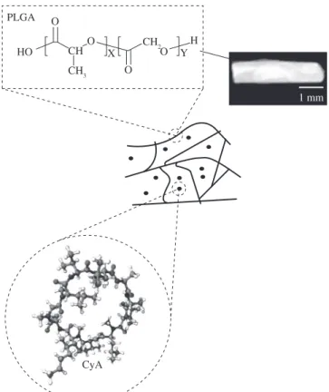
Figure 1 shows the mixture lyophilized implants characteristics which presented cylindrical shape with approximately 4.00 mm of length and 1.00 mm of diameter. The average weight of implants was 5.9 ± 0.1 mg (n = 10) and the average incorporation of CyA into the polymeric matrix was of 31.6% (n = 3).
In addition, SEM micrographs of PLGA-CyA lyophilized samples showed a morphological aspect of uniform and homogeneous surface and the absence of significant superficial irregularities (Figure 2a,b). Hence, the micrographs have proven to be an important tool for an adequate understanding the implant under in vitro delivery profile.
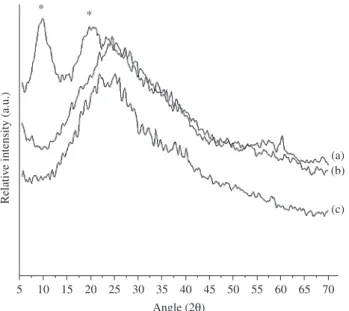
3.2. The X ray diffraction
Figure 3 shows the XRD results of lyophilized mixture and lyophilized samples of CyA and PLA 75:25.The peaks between 7° and 4° (2è) correspond to the CyA semi-crystalline state ( Figure 3a)8 and no peak has been observed for the PLGA 25:25
(Figure 3b). Therefore, the results confirmed its amorphous state9.
In addition, the crystallinity of lyophilized CyA (Figure 3a) was not observed in the lyophilized mixture sample (Figure 3c). This fact can be explained by the possibility of the CyA distribution into the poly-meric matrix. It is believed that the drug presents a small molecular
PLGA
HO O
O
O O CH
CH3
CH2
X
H Y
CyA
1 mm
mobility capacity among the polymeric chains, characterizing the observed amorphous state of lyophilized mixture.
3.3. Thermogravimetry characterization
The TG curves of PLGA 75:25 e CyA. The onset of mass loss for PLGA occurs at 247 °C and for CyA occurs at 254 °C. In addition, the 100% mass loss occurs at 378 °C and 600 °C for PLGA 75:25 e CyA, respectively (Figure 4).
A thermal stability at 100-120 °C, temperature used in implants preparation, shows that both samples present thermal stability dur-ing the implant preparation, indicatdur-ing the viability of the developed implant preparation method.
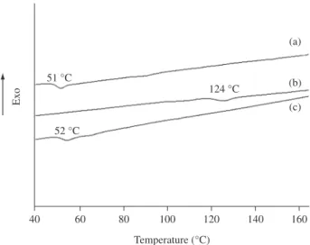
3.4. Differential scanning calorimetry characterization
The glass transition temperature (Tg) of the polymer can be used as a measurement for the mobility of the macromolecules9 and
the evaluation of the solid-to-liquid transition of the CyA character-istic consistent with a glass thermotropic liquid crystal10 also give
information about the CyA molecular mobility and changes at its semi-crystalline state. These characteristics can be used to explain the presence or absence of physical interaction.
The evaluations of the glass transition temperature of the lyophi-lized drug and polymer precursors and in the systems were obtained from the DSC curves (Figure 5).
15 kV x200 100 m DEMET/UFMG 15 kV x1,000 100 m DEMET/UFMG
(a) (b)
Figure 2. SEM pictures from the mixture lyophilized implant before in vitro incubation a) magnification 200x; b) magnification 1,000x, with just minor porosity at the surface.
Relati
ve intensity (a.u.)
5 10 15 20 25 30 35 40 45 50 55 60 65 70
* *
(a) (b)
(c)
Angle (2θ)
Figure 3. XRD of samples: a) lyophilized CyA, b) lyophilized PLGA 75:25 and c) lyophilized mixture.
Mass loss (%)
100
75
50
25
0
0 200 400 600 800
Temperature (°C)
(a) (b)
mobility from polymers chains. These samples, either in the pure form or associated in systems, have presented the same capacity of chain mobility, leading to an indication of the absence of physical interac-tion between polymers and drug. The inexistence of the CyA solid-to-liquid transition in the lyophilized mixture (Figure 5c) is probably related to the rearrangement of drug molecular conformation into the polymeric matrix or maybe to its impossibility to be detected under the experimental conditions, result that confirms the XRD hypothesis. During the association of polymer to drug, no detectable change on chain and molecular conformation could be verified.
3.5. The fourier transform infrared spectroscopy (FTIR)
characterization
The possibilities of chemical and physical interactions were also evaluated by FTIR (Figure 6). FTIR analysis was mostly based at the amide-I and ester region from the spectra once they are the specific chemical group present in each molecule under investigation, which would be a suitable indication for any chemical interaction that may occur among them. Also, the physical interaction between polymer network and drug was evaluated at the methyl asymmetric stretch-ing region.
The FTIR spectra of the powders PLGA 75:25 and CyA with ratio 100:10 (Figure 6a) have given evidence of both PLGA (polymer) and CyA (drug) structure information, showing all major character-istic bands at 1750 cm–1 and 1628 cm–1 correlated to the presence
of amides and ester groups in CyA and PLGA 75:25, respectively. These results are endorsed by FTIR spectra obtained from both pure CyA and PLGA 5:25 (data not showed) and with the previously reported literature4, 11.
The lyophilized mixture (Figure 6b) presented similar vibrational band at 1740 cm–1 (C = O ester stretching) and band at approximately
1630 cm–1 (C = O primary amide stretching). On the other hand,
im-of β-sheets intermolecular interaction present in the drug and, con-sequently, CyA stability and biological activity was maintained11.
Although the presence of a minor change in the methyl stretching band in polymer/drug samples, we have not considered this value as associated with some physical interaction. Such results are attributed to some probable alteration on CyA conformation, endorsed by the presence of N-H amide-I stretching. The observed high intensity band associated with N-H amide-I stretching in all investigated samples are likely to be due to a CyA molecular rearrangement through hy-drophilic interaction aiming to reach the most stable thermodynamic conformation at polymer/drug interface, supporting the results from XRD and DSC techniques.
TG curves have proved the thermal stability of samples at the implant preparation temperature. Moreover, the XRD patterns as-sociated with DSC and FTIR results showed the drug distribution into the polymeric matrix.
In FTIR spectra no extra bindings or chemical shifts were observed, indicating that there is no strong chemical interaction between polymer and drug into the polymer/drug network. In addi-tion no detectable changes in polymer thermal behavior from DSC data are attributed to the absence of physical interactions between polymer and drug. These results confirm the drug chemical stability, the permanence of their biological activity and the possibility of drug sustained delivery system profile.
3.6. The preliminary in vitro studies
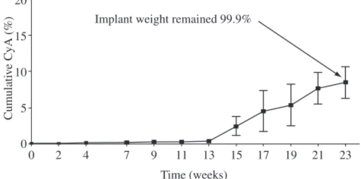
The preliminary in vitro release study of the intraocular implant developed was performed to determinate the initial CyA delivery profile. A slow and prolonged release was observed during the 23 weeks (Figure 7).
The cumulative CyA released was 8.4% to mixture lyophilized implants (Figure 7). The ratio of CyA released is most probably
Exo
40 60 80 100 120 140 160
Temperature (°C) 52 °C
51 °C
124 °C
(a)
(b)
(c)
Figure 5. DSC curves of the samples: a) PLGA 75:25 lyophilized; b) lyophi-lized CyA and c) lyophilyophi-lized mixture PLGA 75:25.
Wavenumber (cm1)
500 1000 1500 2000 2500 3000 3500 4000
1628 1628
1749 1750
2944 2995
2959
3319 (a)
(b)
regulated by the diffusion of the drug in the swelled polymer network and not by the polymer matrix disintegration. Such affirmation can be made once it was not observed any significant implant mass loss dur-ing the in vitro release assay (Figure 7). Besides that, the drug release profile can be also attributed to the homogeneous implant surface obtained as showed in SEM micrograph (Figure 2). Consequently, such results indicate the possibility of the biodegradable implants to deliver CyA in a prolonged way when incubated in similar posterior segment of the eye conditions.
4. Conclusions
In the present work biodegradable polymeric intraocular implants were developed and obtained from lyophilized mixture sample. Based on the nanostructure evaluation of the polymer/drug network, we have successfully characterized the system with several different techniques. We have showed by XRD the CyA distribution into the implant polymeric matrix. These findings were confirmed by DSC and FTIR results. Besides that, DSC and FTIR showed the absence of some detectable physical and chemical interaction between drug and polymer and the presence of the CyA in structural biological activity. The morphological characterization showed the homoge-neous implant and the preliminary in vitro release studies proved the implant capacity to release the drug in a prolonged profile when incubated into a medium condition similar to the eye constituents. In summary, the delivery system developed and fully characterized in the present work can be applied, in the future, for the treatment of posterior uveitis.
Acknowledgements
The authors wish to thank Prof André Faraco by assisting in sample preparation and laboratory protocols. This work was financially sup-ported by CNPQ/FAPEMIG/CNPq.
References
1. Merkli A, Tabatabay C, Gurny R, Heller J. Biodegradable polymers
for the controlled release of ocular drugs. Prog. Polym. Sci. 1998;
23(3):563-580.
2. Yasukawa MDT, Kimura H, Tabata Y, Ogura Y. Biodegradable scleral
plugs for vitreoretinal drug delivery. Adv. Drug. Del. Ver. 2001;
52(1):25-36.
3. Avitable T, Marano F, Castiglione F, Bucolo C, Cro M, Ambrosio L, et al. Biocompatibility and biodegradation of intravitreal hyaluronan implants
in rabbits. Biomaterials 2001; 22(3):195-200.
4. Stevenson C, Tan MM, Ballesteros DL. Pharmaceutical liquid crystals:
The relevance of partially ordered systems. J. Pharm. Sci. 2003;
94(9):1861-1880.
5. Fialho SL, Cunha-Júnior AS. Manufacturing techniques of biodegradable
implants intended for intraocular application. Drug. Deliv. 2005;
12(2):109-116.
6. Kunou N, Ogura Y, Yasukawa T, Kimura H, Miyamoto H, Honda Y, Ikada Y. Controlled intraocular delivery of ganciclovir with use of biodegradable
scleral implant in rabbits. J. Cont. Rel. 1995; 37:143-150.
7. Yasukawa T, Kimura H, Tabata Y, Ogura Y. Biodegradable scleral plugs
for vitreoretinal drug delivery. Advanced Drug Delivery Reviews 2001;
52(1):25-36.
8. Ballesteros DL, Fattah AA, Stevenson CL, Bennet DB. Properties and stability of a liquid crystal form of cyclosporine-the first reported
naturally occurring peptide that exists as a thermotropic liquid. Journal
of pharmaceutical Science 2003; 92(9):1821-1831.
9. Elkharraz K, Faisant N, Guse C, Siepmann F, Arica-Yegin B, Oger J.-M, Gust R, Goepferich A, Benoit JP, Siepmann J. Paclitaxel-loaded microparticles and implants for the treatment of brain cancer: preparation
and physicochemical characterization. International Journal of
Pharmaceutics 2006; 314(2):127-136.
10. Chan HK, Clark AR, Feeley JC, Kuo MC, Lehrman SR, Pikal-Cleland K, Miller DP, Vehring R, Lechuga-Ballesteros D. Physical stability of salmon calcitonin spray-dried powders for inhalation. J. Phar. Sci. 2005; 93(3):792-804.
11. Jayasuriya AC, Assad M, Jayatissa AH, Ebraheim NA. Dissolution
behavior of biomimetic minerals on 3D PLGA scaffold. Surface and
Coating Technology 2006; 200(22-23):6336-6339.
12. Dong X, Shi W, Yuan G, Xie L, Wang S, Lin P. Intravitreal implantation of the biodegradable cyclosporin A drug delivery system for experimental
chronic uveitis. Graefe’s Arch. Clin. Exp. Ophthalmol. 2006;
244(4):492-497.
Cumulati
ve CyA (%)
0 2 4 7 9 11 13 15 17 19 21 23
Time (weeks) Implant weight remained 99.9%
5
0 10 15 20
Figure 7. Cumulative release of CyA from: mixture lyophilized implants in