Submitted20 February 2016
Accepted 2 May 2016
Published26 May 2016
Corresponding author
Ricardo Coentre, [email protected]
Academic editor
Jafri Abdullah
Additional Information and Declarations can be found on page 7
DOI10.7717/peerj.2069
Copyright
2016 Coentre et al.
Distributed under
Creative Commons CC-BY 4.0
OPEN ACCESS
Retrospective study on structural
neuroimaging in first-episode psychosis
Ricardo Coentre1,2, Amilcar Silva-dos-Santos3and Miguel Cotrim Talina3,4
1First-Episode Psychosis Program, Department of Psychiatry, Hospital Vila Franca de Xira, Vila Franca de Xira, Portugal
2Faculty of Medicine, University of Lisbon, Lisbon, Portugal
3Department of Psychiatry, Hospital Vila Franca de Xira, Vila Franca de Xira, Portugal 4CEDOC, Chronic Diseases Research Centre, Nova Medical School, Lisbon, Portugal
ABSTRACT
Background.No consensus between guidelines exists regarding neuroimaging in first-episode psychosis. The purpose of this study is to assess anomalies found in structural neuroimaging exams (brain computed tomography (CT) and magnetic resonance imaging (MRI)) in the initial medical work-up of patients presenting first-episode psychosis.
Methods.The study subjects were 32 patients aged 18–48 years (mean age: 29.6 years), consecutively admitted with first-episode psychosis diagnosis. Socio-demographic and clinical data and neuroimaging exams (CT and MRI) were retrospectively studied. Diagnostic assessments were made using the Operational Criteria Checklist +. Neu-roimaging images (CT and MRI) and respective reports were analysed by an experienced consultant psychiatrist.
Results.None of the patients had abnormalities in neuroimaging exams responsible for psychotic symptoms. Thirty-seven percent of patients had incidental brain findings not causally related to the psychosis (brain atrophy, arachnoid cyst, asymmetric lateral ventricles, dilated lateral ventricles, plagiocephaly andfalx cerebri calcification). No further medical referral was needed for any of these patients. No significant differences regarding gender, age, diagnosis, duration of untreated psychosis, in-stay andcannabis
use were found between patients who had neuroimaging abnormalities versusthose without.
Discussion. This study suggests that structural neuroimaging exams reveal scarce abnormalities in young patients with first-episode psychosis. Structural neuroimaging is especially useful in first-episode psychosis patients with neurological symptoms, atypical clinical picture and old age.
SubjectsNeurology, Psychiatry and Psychology, Radiology and Medical Imaging
Keywords First-episode psychosis, Neuroimaging, Magnetic resonance imaging, Brain computed
tomography
INTRODUCTION
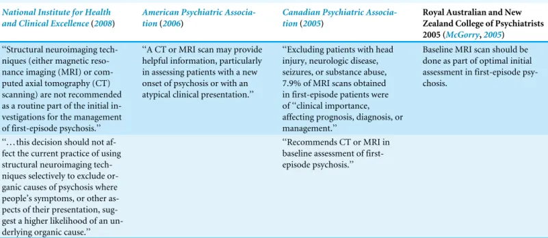
Table 1 Some current guidelines regarding neuroimaging in first-episode psychosis.
National Institute for Health and Clinical Excellence(2008)
American Psychiatric Associa-tion(2006)
Canadian Psychiatric Associa-tion(2005)
Royal Australian and New Zealand College of Psychiatrists 2005 (McGorry,2005)
‘‘Structural neuroimaging tech-niques (either magnetic reso-nance imaging (MRI) or com-puted axial tomography (CT) scanning) are not recommended as a routine part of the initial in-vestigations for the management of first-episode psychosis.’’
‘‘A CT or MRI scan may provide helpful information, particularly in assessing patients with a new onset of psychosis or with an atypical clinical presentation.’’
‘‘Excluding patients with head injury, neurologic disease, seizures, or substance abuse, 7.9% of MRI scans obtained in first-episode patients were of ‘‘clinical importance, affecting prognosis, diagnosis, or management.’’
Baseline MRI scan should be done as part of optimal initial assessment in first-episode psy-chosis.
‘‘. . . this decision should not af-fect the current practice of using structural neuroimaging tech-niques selectively to exclude or-ganic causes of psychosis where people’s symptoms, or other as-pects of their presentation, sug-gest a higher likelihood of an un-derlying organic cause.’’
‘‘Recommends CT or MRI in baseline assessment of first-episode psychosis.’’
management and treatment (Woolley,2005). These include brain injury, demyelinating disease, tumours, multiple sclerosis or stroke. In recent years, much research has been made in both structural and functional neuroimaging in early phases of psychosis (Dazzan et al.,2015;Hager & Keshavan,2015;Jardri,2013;Jung et al.,2010;Strakowski et al.,2008; Tognin et al.,2014). However, the impact of these studies in clinical practice has been disappointing.
Guidelines are divergent regarding neuroimaging in first-episode psychosis. Table 1
summarises some of the current major guidelines (American Psychiatric Association,2006; Canadian Psychiatric Association,2005;McGorry,2005;National Institute for Health and Clinical Excellence,2008). When admitting young patients with first-episode psychosis with no positive neurological findings, physicians are faced with the decision if brain imaging should be performed. Early identification of ‘‘organic’’ lesions responsible for psychotic symptoms could lead to a change in the treatment because the lesions may be surgically or medically treatable. Initial reports seemed promising, with abnormality rates of 30%, but with time most of these findings in structural neuroimaging were incidental and not causal to the psychotic picture (Goodstein,1985;Weinberger,1984).
three studies included CT and MRI neuroimaging (Goulet et al.,2009;Khandanpour, Hoggard & Connolly,2013;Robert Williams, Yukio Koyanagi & Shigemi Hishinuma,2014). Therefore, divergent results in structural neuroimaging abnormalities rates could be mainly explained by different patient age included. As expected, studies where older patients with first-episode psychosis were included more neuroimaging abnormalities were found. For exampleGewirtz et al. (1994) studied first-episode psychosis patients with a mean age of 35, ranging between 18 and 66 years. CT neuroimaging was studied and 42.3% revealed benign and nonspecific abnormalities (diffuse cortical atrophy, arachnoid cysts, ventricular enlargement and venous angioma) and 2.4% of abnormalities link to psychosis (arachnoid cyst in right temporal area, bilateral parietal and subinsular infarcts, bilateral parietal ischemic changes and colloidal cyst in the third ventricule with obstrutction of the foramen of Munro) (Gewirtz et al.,1994). By the contrary in one published study that included only young first-episode psychosis patients (age between 12 and 30 years) (Robert Williams, Yukio Koyanagi & Shigemi Hishinuma,2014) revealed 5.2% of incidental neuroimaging findings with none considered to be causal to psychosis. When included only the few studies published with young first-episode psychosis patients, overall non causal structural neuroimaging abnormalities results revealed rates from 2.2% to 13.2%, and abnormalities link to psychosis from 0% to 1.3% (Bain,1998;Goulet et al.,2009;Lubman et al.,2002; Robert Williams, Yukio Koyanagi & Shigemi Hishinuma,2014).
CT and MRI are the most used structural neuroimaging techniques in daily clinical prac-tice. CT scans are assessable and take little time, but deliver radiation. MRI carries no risk of radiation exposure, provides better spatial resolution and grey-white matter differentiation than CT. MRI is not widely available mainly because of the cost compared with CT. MRI is particularly useful in epilepsy, multiple sclerosis, small brain tumours and vasculitis.
The aim of this study is to evaluate abnormalities found in neuroimaging exams in a sample of young patients with first-episode psychosis. Complementing the few studies published, we try to contribute to more robust evidence-based decisions about structural neuroimaging in clinical practice in young first-episode psychosis patients. We also tried to overcome existing limitations of previous published studies, including only patients with first-episode psychosis diagnosis, young patients (age < 50 years), consecutive patient series and referred all details of study design. We included as well CT and MRI neuroimaging and in- and outpatients approaching to daily clinical routine.
MATERIALS AND METHODS
Sample
Vila Franca de Xira Ethics Committee. In addition to the written informed consent to the brain imaging that all patients give during the daily routine of neuroimaging, each participant gave a specific written informed consent to the present study.
Data collection
The medical electronic files of the patients were reviewed retrospectively. Socio-demographic and clinical data, neuroimaging images (CT and MRI) and respective reports were analysed by an experienced consultant psychiatrist. CT scans were performed in Siemens SOMATON Emotion 16 CT machine with 2.4 mm section thickness (Siemens Healthcare). All CT studies were considered for contrast administration after review of non-contrasted study. In 12 patients contrast for contrast-enhanced CT scan was administered. MRI were performed in Toshiba Titan 1.5 T machine (Toshiba Medical Systems Europe). All participants evaluated using MRI had sagittal T1-weighted, axial T2-weighted, axial T2-weighted Flair, axial gradient-echo, axial diffusion-weighted and coronal T2-weighted imaging. Gadolinium-enhanced MRI was used in all MRI studies performed (axial, sagittal and T1-weighted coronal sequences). CT and MRI images were analyzed using Impax 6.5. Software (Agfa Healthcare Inc.). All CT and MRI reports had been written by a consultant neuroradiologist. When a doubt persisted after examination of the neuroimaging images and reports, collaboration of a consultant neuroradiologist was used to reanalyse the exams. Brain images (CT and/or MRI) are used in daily practice in the clinical evaluation of all first-episode psychosis presenting to the Department of Psychiatry as part of the initial medical work-up. Patients with only non-brain abnormalities in CT scan or MRI (sinus disease, sebaceous cyst or dermoid cyst) were included in patients without neuroimaging abnormalities. We used the Operational Criteria Checklist + (OPCRIT+) instrument to achieve DSM-IV diagnosis (McGuffin, Farmer & Harvey, 1991;Rucker et al.,2011). OPCRIT+ is a checklist including items of psychiatric history and psychopathology. Checklist ratings are entered into the OPCRIT+ software, which generates a diagnosis for the main categories of affective and psychotic disorders defined according to major classification systems, including DSM-IV. OPCRIT+ has been shown to have good reliability when used by different raters. The rater was an experienced consultant psychiatrist, trained in the use of OPCRIT+.
Statistical analysis
Statistical analysis was made using SPSS statistics 21 for Windows (SPSS, Chicago, IL, USA). Descriptive analyses are presented as proportions for count data and as means with standard deviations (SD) for continuous data. We separated the samples in two groups, according to the existence of neuroimaging abnormalities (i.e., with vs. without neuroimaging abnormalities), and compared some socio-demographic and clinical characteristics using chi-squared test for categorical variables (or Fisher exact tests as appropriate) and student’s
t-test for continuous variables. The level of statistical significance wasp<0.05.
RESULTS
18.8% married and 15.6% divorced. Twenty-two percent of the participants were students, 53.13% unemployed and 25% employed. Of the sample, 6.3% participants lived alone. There was cannabis use in 53.13% of the participants, and 65.63% had an in-stay during acute phase of the illness. Mean duration of untreated psychosis (DUP) was 76.5 weeks (SD = 107.4). According to DSM-IV diagnosis, 31.25% had the diagnosis of schizophrenia, 40.63% psychotic disorder not otherwise specified, 21.88% cannabis-induced psychotic disorder and 6.25% delusional disorder. All patients were on first days of antipsychotic medication when CT scan or MRI was performed. Regarding the type of medication, 31.25% patients were treated with risperidone oral, 21.88% with olanzapine oral, 15.63% paliperidone palmitate long-acting injectable, 9.38% aripiprazole oral, 9.38% paliperidone oral, 6.25% haloperidol oral, 3.13% risperidone long-acting injectable and 3.13% quetiapine fumarate extended-release oral.
Twenty-nine (90.63%) patients received a CT, 1 (3.13%) an MRI and 2 (6.25%) both CT and MRI. None of the neuroimaging findings was considered to be a potential or significant contributory cause to the psychotic episode. Twelve patients (37.5%) had incidental brain lesions not causally related to the psychosis: brain atrophy (n=4), arachnoid cyst (n=3),
asymmetric lateral ventricles (n=2), dilated lateral ventricles (n=1), plagiocephaly (n=1)
andfalx cerebricalcification (n=1).
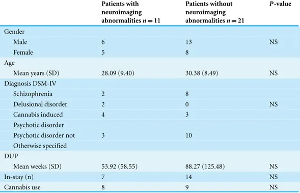
There were no statistically significant differences between participants groups (with vs. without neuroimaging abnormalities) regarding gender (p=0.687), age (p=0.490), DUP (p=0.399), psychiatric diagnosis (p=0.069), in-stay (p=0.582) and cannabis use (p=0.108) (Table 2).
DISCUSSION
In our sample, no neuroimaging lesions responsible for psychotic symptoms were found. Minor incidental abnormalities were present in 37.5% of the patients. Despite the relevant methodological differences compared to previous published studies, our results are in line with other studies that included young patients in first-episode psychosis. In 1998, Battaglia & Spector studied 45 patients with first-episode psychosis who received a CT scan, and three scans showed positive findings that correlated with neuropsychiatric symptomatology (Battaglia & Spector,1988).Goulet et al.(2009) studied 46 patients with first-episode psychosis, in which 44 had CT scans, two had MRI and only one patient showed a lipoma above the pineal gland with no relation with clinical picture. Like our research, previous published studies show that findings in structural brain imaging in young patients with first-episode psychosis are limited regarding clinical utility.
Table 2 Socio-demographic and clinical characteristics of patients with and without neuroimaging abnormalities.
Patients with neuroimaging abnormalitiesn=11
Patients without neuroimaging abnormalitiesn=21
P-value
;Gender
; Male 6 13 NS
; Female 5 8
;Age
; Mean years (SD) 28.09 (9.40) 30.38 (8.49) NS
;Diagnosis DSM-IV
; Schizophrenia 2 8
; Delusional disorder 2 0 NS
; Cannabis induced 4 3
; Psychotic disorder
; Psychotic disorder not 3 10
; Otherwise specified ;DUP
; Mean weeks (SD) 53.92 (58.55) 88.27 (125.48) NS
;In-stay (n) 7 14 NS
;Cannabis use 8 9 NS
Notes.
NS, not significant; SD, standard deviation; DSM-IV, Diagnostic and Statistical of Mental Disorders, fourth edition; DUP, duration of untreated psychosis.
had incidental brain lesions not responsible to the psychosis. These results seem to indicate that in older (>50 years) first-episode psychotic patients, structural neuroimaging exams identify a higher number of organic lesions responsible for the psychosis, even so in low rate.
As expected, we did not find any significant differences between the group of patients with incidental brain findings and those without, reflecting that these neuroimaging findings do not reflect a subgroup of patients with particular socio-demographic or clinical characteristics (e.g., longer DUP or more in-stay) (Table 2).
None of the studies chose patients randomly, including our own. But contrary to most of published studies, we include patients consecutively, reducing selection bias.
Only one study that specifically investigated economical factors of structural neuroimag-ing in psychosis was found (Albon et al.,2008). The authors concluded that if screening with structural neuroimaging was implemented in all patients presenting with psychotic symptoms before 65 years, little would be found to affect clinical management. The authors also emphasised that there is a paucity in good-quality evidence on the clinical benefits of structural neuroimaging on which to base economic research; thus, the outcome from an economic perspective is not clear.
prevalence and type of the radiological findings. Third, few patients included had an MRI, but taking into account previous studies, this would likely not significantly alter our findings.
CONCLUSIONS
In conclusion, our results are concordant with the few previous studies in which clinically relevant abnormalities in structural neuroimaging in young patients with first-episode psychosis are scarce. We think that neurological examination is a valuable adjunct instrument once it can help to determine if a patient has signs of a brain lesion, so structural neuroimaging should be performed. However, sometimes the psychiatric status did not permit the elaboration of a good-quality neurological examination, so it would be desirable to improve clinical status first and after doing a neurological examination. Neuroimaging exams should specially be performed in some clinical pictures, such as in patients with neurological symptoms or signs, atypical clinical picture, delirium/organic suggestive symptoms (visual hallucinations, disorientation, memory loss and blurred conscience) and old age (age > 50 years). When a neuroimaging exam is indicated, an MRI should probably be performed because of previously mentioned advantages. MRI should be especially preferred if epilepsy, multiple sclerosis, small tumours and vasculitis are major diagnostic hypotheses.
ADDITIONAL INFORMATION AND DECLARATIONS
Funding
The authors received no funding for this work.
Competing Interests
The authors declare there are no competing interests.
Author Contributions
• Ricardo Coentre conceived and designed the experiments, performed the experiments,
analyzed the data, contributed reagents/materials/analysis tools, wrote the paper, prepared figures and/or tables, reviewed drafts of the paper.
• Amilcar Silva-dos-Santos conceived and designed the experiments, analyzed the data,
wrote the paper, reviewed drafts of the paper.
• Miguel Cotrim Talina conceived and designed the experiments, analyzed the data, wrote
the paper.
Human Ethics
The following information was supplied relating to ethical approvals (i.e., approving body and any reference numbers):
Hospital Vila Franca de Xira Ethics Committee.
Data Availability
Supplemental Information
Supplemental information for this article can be found online athttp://dx.doi.org/10.7717/ peerj.2069#supplemental-information.
REFERENCES
Albon E, Tsourapas A, Frew E, Davenport C, Oyebode F, Bayliss S, Arvanitis T, Meads C. 2008.Structural neuroimaging in psychosis: a systematic review and economic evaluation.Health Technology Assessment 12: iii–iv, ix–163.
American Psychiatric Association. 1994.Diagnostic and statistical manual of mental disorders. Fourth edition. Washington, D.C.: American Psychiatric Association.
American Psychiatric Association. 2006.American Psychiatric Association practice guidelines for the treatment of psychiatric disorders: Compendium 2006. Washington, D.C.: American Psychiatric Association.
Bain BK. 1998.CT scans of first-break psychotic patients in good general health.
Psychiatry Services49:234–235DOI 10.1176/ps.49.2.234.
Battaglia J, Spector IC. 1988.Utility of the CAT scan in a first psychotic episode.General Hospital Psychiatry10:398–401DOI 10.1016/0163-8343(88)90062-X.
Canadian Psychiatric Association. 2005.Clinical practice guidelines: treatment of schizophrenia.Canadian Journal of Psychiatry 50(S1):7S–57S.
Dazzan P, Arango C, Fleischacker W, Galderisi S, Glenthøj B, Leucht S,
Meyer-Lindenberg A, Kahn R, Rujescu D, Sommer I, Winter I, McGuire P. 2015.Magnetic resonance imaging and the prediction of outcome infirst-episode schizophrenia: a review of current evidence and directions for future research.Schizophrenia Bulletin 41:574–583DOI 10.1093/schbul/sbv024.
Freudenreich O, Schulz SC, Goff DC. 2009.Initial medical work-up of first-episode psychosis: aconceptual review.Early Intervention in Psychiatry3:10–18
DOI 10.1111/j.1751-7893.2008.00105.x.
Gewirtz G, Squires-Wheeler E, Sharif Z, Honer WG. 1994.Results of computerised tomography during first admission for psychosis.British Journal of Psychiatry 164:789–795DOI 10.1192/bjp.164.6.789.
Goodstein RK. 1985.Guide to cat scanning in hospital psychiatry. Overview ofclinical practice and criteria for use.General Hospital Psychiatry7:367–376
DOI 10.1016/0163-8343(85)90054-4.
Goulet K, Deschamps B, Evoy F, Trudel J-F. 2009.Use of brain imaging (computed tomography and magnetic resonance imaging) in first-episode psychosis: review and retrospective study.Canadian Journal of Psychiatry54:493–501.
Hager BM, Keshavan MS. 2015.Neuroimaging biomarkers for psychosis.Current Behavioral Neuroscience Reports2:102–111DOI 10.1007/s40473-015-0035-4.
Jardri R. 2013.Brain imaging of first-episode psychosis.Encephale39(Suppl 2):S93–S98
Jung WH, Jang JH, Byun MS, An SK, Kwon JS. 2010.Structural brain alterations in individuals at ultra-high risk for psychosis: a review of magnetic resonance imaging studies and future directions.Journal of Korean Medical Science25:1700–1709
DOI 10.3346/jkms.2010.25.12.1700.
Khandanpour N, Hoggard N, Connolly DJA. 2013.The role of MRI and CT of the brain in first episodes ofpsychosis.Clinical Radiology68:245–250
DOI 10.1016/j.crad.2012.07.010.
Lubman DI, Velakoulis D, McGorry PD, Smith DJ, Brewer W, Stuart G, Desmond P, Tress B, Pantelis C. 2002.Incidental radiological findings on brain magnetic resonance imaging in first-episode psychosis and chronic schizophrenia.Acta Psychiatrica Scandinavica106:331–336DOI 10.1034/j.1600-0447.2002.02217.x.
McGorry PD. 2005.Royal Australian and New Zealand college of psychiatristsclinical practice guidelines for the treatment of schizophrenia and related disorders.
Australian & New Zealand Journal of Psychiatry39:1–30
DOI 10.1111/j.1440-1614.2005.01516.x.
McGuffin P, Farmer A, Harvey I. 1991.A polydiagnostic application of operational criteria in studiesof psychotic illness. development and reliability of the OPCRIT system.Archives of General Psychiatry48:764–770
DOI 10.1001/archpsyc.1991.01810320088015.
National Institute for Health and Clinical Excellence. 2008.Structural neuroimaging in first-episode psychosis.Available athttps:// www.nice.org.uk/ guidance/ ta136
(accessed 19 January 2016).
Robert Williams S, Yukio Koyanagi C, Shigemi Hishinuma E. 2014.On the usefulness of structural brain imaging for young first episode inpatients with psychosis.
Psychiatry Research224:104–106DOI 10.1016/j.pscychresns.2014.08.001.
Rucker J, Newman S, Gray J, Gunasinghe C, Broadbent M, Brittain P, Baggaley M, Denis M, Turp J, Stewart R, Lovestone S, Schumann G, Farmer A, McGuffin P. 2011.OPCRIT+: an electronic system for psychiatric diagnosis and data collec-tion in clinical and research settings.British Journal of Psychiatry199:151–155
DOI 10.1192/bjp.bp.110.082925.
Strahl B, Cheung YK, Stuckey SL. 2010.Diagnostic yield of computed tomography of the brain in first episode psychosis.Journal of Medical Imaging and Radiation Oncology 54(5):431–434DOI 10.1111/j.1754-9485.2010.02196.x.
Strakowski SM, Adler CM, Cerullo MA, Eliassen JC, Lamy M, Fleck DE, Lee JH, DelBello MP. 2008.Magnetic resonance imaging brain activation in first-episode bipolar mania during a response inhibition task.Early Intervention in Psychiatry 2:225–233DOI 10.1111/j.1751-7893.2008.00082.x.
Weinberger DR. 1984.Brain disease and psychiatric illness: when should a psychiatrist order a CAT scan?American Journal of Psychiatry141:1521–1527
DOI 10.1176/ajp.141.12.1521.