858
https://doi.org/10.1590/0004-282X20170170 ARTICLE
Convexity subarachnoid hemorrhage: clinical
features and etiology of an Argentinian cohort
Hemorragia subaracnóidea da convexidade: características clínicas e etiologia de uma
coorte argentina
Aníbal Chertcoff1, Lucrecia Bandeo1, Fátima Pantiu1, Luciana León Cejas1, Sol Pacha1, Claudia Uribe Roca1,
Manuel Fernández Pardal1, Ricardo Reisin1, Pablo Bonardo1
1Hospital Británico de Buenos Aires, Department of Neurology, Perdriel, Buenos Aires, Argentina.
Correspondence: Aníbal Chertcoff; Department of Neurology, Hospital Británico de Buenos Aires; Perdriel 74, 1280, Buenos Aires, Argentina; E-mail: [email protected]
Conflict of interest: There is no conlict of interest to declare.
Received 07 May 2017; Received in inal form 17 August 2017; Accepted 25 September 2017.
ABSTRACT
Nontraumatic convexity subarachnoid hemorrhage is an increasingly recognized subtype of subarachnoid bleeding. Objective: Our aim was to describe the etiology and clinical features of a cohort of patients with convexity subarachnoid hemorrhage. Methods: We retrospectively analyzed all cases of convexity subarachnoid hemorrhage admitted to our hospital between January 2012 and April 2017. Demographic features, clinical characteristics, complementary investigations, etiology and mortality were assessed. Twenty patients (65% females) were identiied. Mean age: 53 years (range, 15-86 years). Results: Symptoms on admission: headache (65%), sensory and/or motor symptoms (50%) and seizures (35%). Commonest causes: cerebral vein thrombosis (20%), reversible cerebral vasoconstriction syndrome (20%) and cerebral amyloid angiopathy (20%). Two patients died. Conclusion: Convexity subarachnoid hemorrhage may be related to a wide spectrum of etiologies. In our patients, an increased prevalence of cerebral vein thrombosis was observed. Mortality was low and not related to the bleeding itself.
Keywords: cerebral amyloid angiopathy, etiology; sinus thrombosis, intracranial; subarachnoid hemorrhage.
RESUMO
A hemorragia subaracnóidea não traumática da convexidade é um subtipo cada vez mais reconhecido de sangramento subaracnóideo. Objetivo: Nosso objetivo foi descrever a etiologia e as características clínicas de uma coorte de pacientes com hemorragia subaracnóidea da convexidade. Métodos: Foram analisados retrospectivamente todos os casos de hemorragia subaracnóidea da convexidade admitidos em nosso hospital entre janeiro de 2012 e abril de 2017. Foram avaliados os aspectos demográicos, características clínicas, investigações complementares, etiologia e mortalidade. Vinte pacientes (65% mulheres) foram identiicados. Média de idade: 53 anos (intervalo, 15-86). Resultados: Sintomas na admissão: dor de cabeça (65%), sintomas sensitivos e/ou motores (50%) e convulsões (35%). Causas mais comuns: trombose venosa cerebral (20%), síndrome de vasoconstrição cerebral reversível (20%) e angiopatia amilóide cerebral (20%). Dois pacientes morreram. Conclusão: A hemorragia subaracnóidea da convexidade pode estar relacionada a um amplo espectro de etiologias. Em nossos pacientes, observou-se uma maior prevalência de trombose venosa cerebral. A mortalidade foi baixa e não relacionada à própria hemorragia.
Palavras-chave: angiopatia amiloide cerebral, etiologia; trombose dos seios intracranianos; hemorragia subaracnóidea.
Nontraumatic nonaneurysmal convexity subarachnoid hemorrhage (cSAH) is an increasingly recognized subtype of sub-arachnoid bleeding localized in one or more cortical sulci of the brain without involvement of the neighboring parenchyma or
extension to the interhemispheric issure, basal cisterns or ven
-tricles1. hese features help to distinguish cSAH from aneurys
-mal bleeding and nontraumatic perimesencephalic
subarach-noid hemorrhage. Recently, many case series have described the clinical features, etiology and outcome of this entity although
information concerning Latin American populations is scarce2.
We therefore consider it valuable to describe the etiology and clinical features of cSAH in a hospital-based Argentinian cohort.
METHODS
All consecutive patients admitted to our hospital with
nontraumatic nonaneurysmal subarachnoid hemorrhage,
evaluated by the Department of Neurology from January 2012
to April 2017, were identiied from hospital records. Patients with evidence of blood in the interhemispheric issures,
basal cisterns or adjacent brain parenchyma were excluded.
Demographic features, clinical characteristics, complemen
-tary investigations, etiology and mortality were retrospec -tively assessed. Diagnosis of cerebral amyloid angiopathy
859
Chertcoff A et al. Convexity SAH in an Argentinian cohort
criteria3,4. Reversible cerebral vasoconstriction syndrome
(RCVS) was diagnosed under current criteria5. All other
diag-noses were recorded in accordance to medical records. he
ethics committee of the institution approved the study.
We identiied 31 patients with cSAH. Six were excluded due to cisternal, interhemispheric or adjacent parenchy
-mal bleeding. Information on ive patients was missing. Accordingly, data from 20 patients was analyzed. he Figure shows representative images of cSAH. he demograph -ics and medical history of our population is summarized
in Table 1. hirteen (65%) were females. he mean age was 53 years (range: 15–86 years). he most common comorbid conditions were arterial hypertension and dyslipidemia (30% and 25% respectively). Only two patients had a history of previ -ous cerebrovascular disease: one had an ischemic stroke and an intracerebral hemorrhage and the second an intracerebral hemorrhage. Two patients had acute oncohematological
dis-eases at cSAH presentation. Four patients were taking anti
-platelets, one was on oral anticoagulation and one received
vasoconstrictors (ergotamine and pseudoephedrine).
RESULTS
Results are summarized in Table 2. A comparison with recent series is performed.
Clinical features
he most common presenting symptom was headache in 13 patients. Among these, thunderclap headache was reported in ive. Four had progressive and severe headache and four had mild headache. Other frequent symptoms
were sensory-motor symptoms in 10 patients and seizures in seven.
Complementary investigations
Nineteen patients had a head CT scan. Brain MRI with MR-angiography was performed in 19 patients. Seven
patients underwent conventional brain angiogram and two had a CT-angiography. Regarding the localization of the
sub-arachnoid hemorrhage, frontal lobe was the most frequently afected in 15 patients, parietal lobe in six, occipital in six and
temporal in three. Ten patients had bilateral bleeding.
Table 1. Demographics and medical history of 20 patients with convexity subarachnoid hemorrhage.
Variable No.
Age, years* 53 ± 20 (15–86)
Female 13
Previous medical history
Smoking 7
Arterial Hypertension 6
Dyslipidemia 5
Diabetes mellitus 2
Coronary artery disease 3
Previous ischemic stroke 1
Previous intracerebral hemorrhage 2
Current oncohematological disease 2
Current antiplatelet therapy 4
Current oral anticoagulation 1
Current vasoconstrictors use 1
*values indicate mean ± SD (range); SD: standard deviation
Figure. Representative images of cSAH. A, B: MRI (Axial FLAIR sequence) of a 35-year-old female with headache and seizures due to cerebral vein thrombosis with right frontal cSAH. MR-venography exhibited partial thrombosis of the superior sagittal sinus (arrow). C, D: MRI (Axial FLAIR sequence) of a 40-year-old female with acute myeloid leukemia and bilateral cSAH due to posterior reversible encephalopathy syndrome. This patient died due to underlying disease.
860 Arq Neuropsiquiatr 2017;75(12):858-861
Etiology
he most common causes of cSAH were cerebral vein thrombosis (CVT), RCVS and CAA each one of them in four patients. Other causes were posterior reversible encepha
-lopathy syndrome in two patients, central nervous system vasculitis in two, infectious endocarditis in two, anticoagula -tion-related in one and undetermined in one patient.
Mortality
Two patients died. In both cases, cSAH was due to poste -rior reversible encephalopathy syndrome and both had acute
oncohematological diseases. he cause of death was unre -lated to the cSAH.
DISCUSSION
Although several etiologies have been suggested as causes
for cSAH, RCVS and CAA seem to be the most frequent in nearly all series. Previous studies have shown that the etiol -ogy and the clinical presentation of cSAH is mainly related
to age at presentation: younger patients (less than 60 years
old) present with headache with a thunderclap pattern com-monly due to RCVS and older patients typically complain
about sensory-motor symptoms secondary to CAA. Our
study showed a higher prevalence of CVT in comparison with
other studies1,2,6,7. In our population, CVT was as frequent as
RCVS and CAA. Cerebral vein thrombosis is an increasingly recognized cause of cSAH and some authors suggest that this might be related to technological advances made in
noninva-sive diagnostic radiology during the last decades8,9. Another
possible explanation may be that our cohort was younger than other series and had a female predominance; CVT is
more frequent in such populations10. Likewise, we think that the higher frequency of seizures reported in our series was
also probably related to the increased prevalence of CVT.
Regarding clinical presentation in CAA and RCVS patients,
two out of four patients with CAA presented with transient
paresthesias commonly known as ‘amyloid spells’11. hree out
of four patients with RCVS experienced thunderclap
head-ache. Only one CAA patient presented with headache but it
was described as mild.
Concerning diagnostic investigations, brain MRI with noninvasive MR-angiography seems to be the imaging
modality of choice for cSAH. Some authors consider that cSAH pattern of bleeding is not related with aneurysmal rupture and that procedural risks do not justify performing
a catheter angiography in these patients12. In accordance,
conventional angiogram was carried out only in 35% of our
patients and solely for excluding aneurysmal bleeding. Like
previous reports, the frontal lobe was the primary site of bleeding. Bilateral bleeding was as frequent as unilateral1,2,6,7.
With regard to outcome, although some studies sug -gest that cSAH has a worse prognosis than other types of Table 2. Clinical characteristics, complementary investigations, etiology and mortality of cSAH.
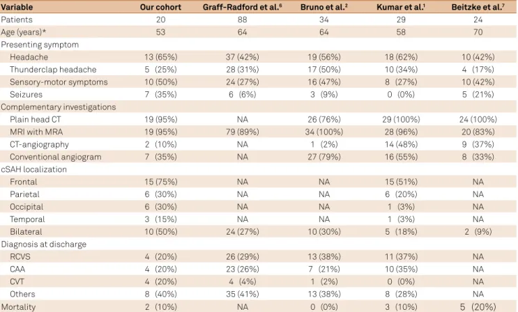
Variable Our cohort Graff-Radford et al.6 Bruno et al.2 Kumar et al.1 Beitzke et al.7
Patients 20 88 34 29 24
Age (years)* 53 64 64 58 70
Presenting symptom
Headache 13 (65%) 37 (42%) 19 (56%) 18 (62%) 10 (42%)
Thunderclap headache 5 (25%) 28 (31%) 17 (50%) 10 (34%) 4 (17%)
Sensory-motor symptoms 10 (50%) 24 (27%) 16 (47%) 8 (27%) 10 (42%)
Seizures 7 (35%) 6 (6%) 3 (9%) 0 (0%) 5 (21%)
Complementary investigations
Plain head CT 19 (95%) NA 26 (76%) 29 (100%) 24 (100%)
MRI with MRA 19 (95%) 79 (89%) 34 (100%) 28 (96%) 20 (83%)
CT-angiography 2 (10%) NA 1 (2%) 14 (48%) 9 (37%)
Conventional angiogram 7 (35%) NA 27 (79%) 16 (55%) 8 (33%)
cSAH localization
Frontal 15 (75%) NA NA 15 (51%) NA
Parietal 6 (30%) NA NA 6 (20%) NA
Occipital 6 (30%) NA NA 1 (3%) NA
Temporal 3 (15%) NA NA 1 (3%) NA
Bilateral 10 (50%) 24 (27%) 10 (30%) 5 (18%) 2 (9%)
Diagnosis at discharge
RCVS 4 (20%) 26 (29%) 13 (38%) 11 (37%) NA
CAA 4 (20%) 23 (26%) 7 (21%) 10 (35%) NA
CVT 4 (20%) 4 (4%) 1 (2%) 0 (0%) NA
Others 8 (40%) 35 (41%) 13 (38%) 8 (28%) NA
Mortality 2 (10%) NA 0 (0%) 3 (10%) 5 (20%)
861
Chertcoff A et al. Convexity SAH in an Argentinian cohort
nonaneurysmal SAH (e.g. perimesencephalic SAH), this did
not seem to be the case for our cohort and other series2,13.
One such study that exhibited a poorer prognosis of cSAH, had an older population (mean age: 70 years), with 83% of the patients being over 60 years old. In this cohort, neurological disease was identiied as the direct or indirect cause of death in 80% of the patients suggesting that older populations have
a worse outcome with cSAH7. he mean age of our cohort
was 53 years and only 45% of them were over 60 years old. Only two of our patients died and in both cases death was
unrelated to the cSAH but to progression of underlying onco-hematological disease. We believe that mortality in cSAH is low and probably linked to underlying disease and not the bleeding itself as opposed to aneurysmal or traumatic SAH.
Our study, however, presents certain drawbacks. First of all, this is a single center retrospective cohort. his design is likely to cause signiicant selection bias as patients are selected by admission to a speciic hospital and from a radio
-logical sign, which may be subtle, especially on CT. In addi
-tion to the younger age of this cohort, many patients with
CAA and cSAH often present with transient ischemic
symp-toms and, without MRI as the standard work up in transient ischemic stroke, it is unlikely that the cSAH would be identi
-ied in this population, as symptoms might not be alarming. To conclude, according to our indings, in young and female patients with cSAH, physicians should consider CVT as one of the main diferential diagnosis among others such
as CAA and RCVS.
References
1. Kumar S, Goddeau RP Jr, Selim MH, Thomas A, Schlaug G, Alhazzani A et al. Atraumatic convexal subarachnoid hemorrhage: clinical presentation, imaging patterns, and etiologies. Neurology. 2010;16;74(11):893-9. https://doi.org/10.1212/WNL.0b013e3181d55efa 2. Bruno VA, Lereis VP, Hawkes M, Ameriso SF. Nontraumatic
subarachnoid hemorrhage of the convexity. Curr Neurol Neurosci Rep. 2013;13(4):338. https://doi.org/10.1007/s11910-013-0338-3 3. Knudsen KA, Rosand J, Karluk D, Greenberg SM. Clinical diagnosis
of cerebral amyloid angiopathy: validation of the Boston criteria. Neurology 2001;56(4):537-9. https://doi.org/10.1212/WNL.56.4.537 4. Rooden S, Grond J, Boom R, Haan J, Linn J, Greenberg SM et al.
Descriptive analysis of the Boston criteria applied to a Dutch-type cerebral amyloid angiopathy population. Stroke 2009;40(9):3022-7. https://doi.org/10.1161/STROKEAHA.109.554378
5. Ducros A. Reversible cerebral vasoconstriction syndrome. Lancet Neurol 2012; 11(10): 906-17. https://doi.org/10.1016/S1474-4422(12)70135-7 6. Graff-Radford J, Fugate JE, Klaas J, Flemming KD, Brown RD,
Rabinstein AA. Distinguishing clinical and radiological features of non-traumatic convexal subarachnoid hemorrhage. Eur J Neurol. 2016;23(5):839-46. https://doi.org/10.1111/ene.12926
7. Beitzke M, Gattringer T, Enzinger C, Wagner G, Niederkorn K, Fazekas F. Clinical presentation, etiology, and long-term prognosis in patients with nontraumatic convexal subarachnoid hemorrhage. Stroke. 2011;42(11):3055-60. https://doi.org/10.1161/STROKEAHA.111.621847
8. Bittencourt LK, Palma-Filho F, Domingues RC, Gasparetto EL. Subarachnoid hemorrhage in isolated cortical
vein thrombosis: are presentation of an unusual condition. Arq Neuropsiquiatr. 2009;67(4):1106-8. https://doi.org/10.1590/S0004-282X2009000600029 9. Benabu Y, Mark L, Daniel S, Glikstein R. Cerebral venous
thrombosis presenting with subarachnoid hemorrhage: case report and review. Am J Emerg Med. 2009;27(1):96-106. https://doi.org/10.1016/j.ajem.2008.01.021
10. Saposnik G, Barinagarrementeria F, Brown RD Jr, Bushnell CD, Cucchiara B, Cushman M et al. Diagnosis and management of cerebral venous thrombosis: a statement for healthcare professionals from the American Heart Association/American Stroke Association. Stroke.
2011;42(4):1158-92. https://doi.org/10.1161/STR.0b013e31820a8364 11. Charidimou A, Linn J, Vernooij MW, Opherk C, Akoudad S,
Baron JC et al. Cortical supericial siderosis: detection and clinical signiicance in cerebral amyloid angiopathy and related conditions. Brain. 2015;138(Pt 8):2126-39. https://doi.org/10.1093/brain/awv162 12. Patel KC, Finelli PF. Nonaneurysmal convexity subarachnoid
hemorrhage. Neurocrit Care. 2006;4(3):229-33. https://doi.org/10.1385/NCC:4:3:229