Cop
yright
© ABE&M t
odos os dir
eit
os r
eser
vados
.
An intrasellar germinoma
with normal tumor marker
concentrations mimicking primary
lymphocytic hypophysitis
Germinoma intrasselar com marcadores tumorais normais mimetizando hipoisite linfocítica primária
Mariana F. Guzzo1, Cristina B. Formiga Bueno1, Thiago T.
Amancio2, Sergio Rosemberg2, Cleonice Bueno3, Edson L.
Arioli1,Andrea Glezer1, Marcello D. Bronstein1
SUMMARY
Intracranial germinomas (GE) are malignant neoplasms most commonly found in the suprasellar region, which may cause anterior and particularly posterior pituitary hormone deicits with central diabetes insipidus (DI). Differential diagnosis of pituitary stalk thickening includes granulomatous, inlammatory, infectious, and neoplastic lesions. Although careful analysis of clinical, laboratory, and imaging indings may facilitate the diagnosis, transsphenoidal biopsy is indicated to conirm the dise-ase, as the correct diagnosis directs the appropriate treatment. Arq Bras Endocrinol Metab. 2013;57(7):566-70
SUMÁRIO
Germinomas intracranianos (GE) são neoplasias malignas comumente na região suprasse-lar, podendo causar deiciência hormonal da hipóise anterior, em particular da hipóise poste-rior, com diabetes insípido central (DI). Entre os diagnósticos diferenciais do espessamento de haste hipoisária, incluem-se doenças granulomatosas, inlamatórias, infecciosas e neoplá-sicas. Embora as avaliações clínica, laboratorial e a ressonância magnética selar sugiram o diag-nóstico, a biópsia transesfenoidal está indicada para conirmação, visto que o diagnóstico correto direciona o tratamento. Arq Bras Endocrinol Metab. 2013;57(7):566-70
1 Neuroendocrine Unit, Division of
Endocrinology and Metabolism, Hospital das Clinicas, Faculdade de Medicina, Universidade de São Paulo (HC-FMUSP), São Paulo, SP, Brazil
2 Department of Pathology,
FMUSP, São Paulo, SP, Brazil
3 Laboratório de Investigação
Médica em Reumatologia (LIM-17), FMUSP, São Paulo, SP, Brazil
Correspondence to: Marcello D. Bronstein Unidade de Neuroendocrinologia, Divisão de Endocrinologia e Metabologia,
Hospital das Clínicas, Faculdade de Medicina, Universidade de São Paulo Av. Dr. Eneas de Carvalho, 255, 7º andar, sala 7037, Instituto Central 05403-000 – São Paulo, SP, Brazil [email protected]
Received on Oct/26/2012 Accepted on July/10/2013
INTRODUCTION
I
ntracranial germinomas (GE) are malignant neo-plasms that most likely arise from primitive germ cells that failed to migrate to the genital crest during embryonic development (1). They represent about 3.4% of all primary intracranial tumors, predominantly affect pre-pubertal children, and are more often local-ized in the pineal gland or suprasellar region, although bifocal lesions have also been described (1,2). Most commonly, they cause anterior (mainly GH deiciency) and particularly posterior pituitary hormone deicits with central diabetes insipidus (DI) (1).Other diseases of neoplastic, granulomatous, infec-tious and inlammatory origin could be dificult to
dif-ferentiate from GE, because of the similar clinical, im-aging and pathological features. In order to elucidate the etiopathogenesis in patients with dificult differen-tial diagnosis, a transsphenoidal biopsy is indicated (1).
Regarding the proper approach, corticosteroids for lymphocytic hypophysitis (LH) and radiotherapy (RaT) plus chemotherapy (ChT) for GE (2), a case of GE mimicking LH is presented.
CASE REPORT
History and clinical examination
Cop
yright
© ABE&M t
odos os dir
eit
os r
eser
vados
.
was stopped), polyuria, polydipsia, fatigue, galactor-rhea, dry skin, and hair loss. She described a pulsatile headache since adolescence. Weight and body mass in-dex (BMI) were 63.2 kg and 24.5 kg/m2,
respective-ly. Her medical and family history was unremarkable. Physical and neurological examination revealed no ab-normality. Biochemical evaluation was normal. Regard-ing basal hormonal evaluation, she presented hyperpro-lactinemia (prolactin: 50 ng/mL – normal range (NR): 2.0-15.0 ng/mL), hypogonadotropic hypogonadism, secondary hypothyroidism (fT4: 0.56 µU/mL – NR: 0.70 – 1.50 ng/mL; TSH: 2.3 µU/mL – NR: 0.40-4.5 µU/mL), and low basal serum cortisol (cortisol 8 am: 7.5 µg/dL – NR: 5-25 µg/dL). Central DI was diagnosed based on clinical presentation and response to desmopressin (DDAVP) on the water deprivation test, leading to oral DDAVP treatment. She was also replaced with L-T4 and hydrocortisone acetate. Serum and spinal cerebral luid (CSF) tumor markers (alpha-fetoprotein and β-HCG) were negative. Physical exam-ination, chest x-ray, blood angiotensin-converting en-zyme (ACE) was measured, and a PPD test (Tuberculin Puriied Protein Derivative Test) excluded sarcoidosis and tuberculosis, respectively. Other diagnostic work-up included a skeletal survey to rule out histiocytosis. Several serum autoimmune antibodies were positive: anti-thyroid, antinuclear and anti-pituitary (APA)
an-tibodies were positive. The detection of anti-pituitary antibodies was performed by indirect immunoluores-cence in tissue sections of human cadaveric pituitary glands based on a research protocol of the University of Sao Paulo Medical School.
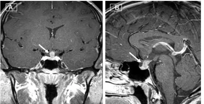
Sellar magnetic resonance imaging (sellar MRI) depicted a supraselar mass extending to the posterior pituitary with normal sellar space, leading to a diffuse thickening of the pituitary stalk (Figure 1A and 1B). Optic chiasm was normal. Invasion of the cavernous sinus was not evident.
Despite the clinical evidence pointing to lymphocy-tic hypophysitis, a pituitary biopsy through transsphe-noidal route was performed in order to rule-out other causes and, therefore, to choose the appropriate thera-py. Pathological examination showed a biphasic popula-tion of mature small lymphocytes and large neoplastic cells with abundant clear cytoplasm, round central nu-clei and prominent nucleoli (Figure 2A). Immunohis-tochemistry (IHC) was positive for placental alkaline phosphatase (PLAP) (Figure 2B) and c-kit protein (CD 117) in the neoplastic large cells (not shown) conirmed the diagnosis of an intrasellar GE. Immune markers also revealed a population of B-lymphocytes (CD 20 posi-tive – Figure 2C) and T-lymphocytes (CD 3 posiposi-tive – Figure 2D). The patient was referred to treatment with ChT and RaT with clinical improvement.
Cop
yright
© ABE&M t
odos os dir
eit
os r
eser
vados
.
Figure 2. (A) Large tumor cells with round nuclei and abundant clear cytoplasm. Presence of moderate lymphocytic iniltrate. (H & E, 200X); (B) Tumor cells showing PLAP immunoreactivity; (C) Presence of B-lymphocytes (CD 20 positive); (D) Presence of T-lymphocytes (CD3 positive).
DISCUSSION
This study deals with the dificulties in diagnosing pitu-itary stalk thickening lesions, reporting a patient with a typical clinical and laboratorial picture of LH in which the inal histopathological diagnosis was GE.
The differential diagnosis of masses affecting the pituitary stalk is broad and includes inlammatory and infectious diseases, germ cell tumors, gliomas, menin-gioma, metastatic tumors, and vascular lesions (3).
LH is a rare entity with estimated incidence of one case in nine million persons-year characterized by pitu-itary and/or stalk autoimmune inlammation. The ave-rage age at diagnosis is 34.5 years in females and 44.7 years in males. Lymphocytic adenohypophysitis (LAH) is strongly associated with pregnancy, 57% of cases oc-curring during gestation or in the postpartum period. This could be related to a pituitary antigens presenta-tion to the immune system, probably due to lactotroph hyperplasia and increase in pituitary blood low (4).
Clinical presentation of LAH is variable and includes symptoms related to mass compression of sellar
neigh-boring regions (optic chiasm, cavernous sinus), hypopi-tuitarism, and hyperprolactinemia. Its clinical suspicion should be raised if the degree of hypopituitarism con-licts with the appearance of pituitary gland in imaging exams, and rapidly installation of hormonal deiciencies, mainly in the corticotrophic axis, in women in the puer-peral pe riod. Central DI can occur if posterior pituitary or pituitary stalk are involved. Sellar MRI routinely shows homogeneous enhancement of the entire gland. The as-sociation with others autoimmune diseases happens in 20% of the cases, mostly with Hashimoto’s thyroiditis (5).
Cop
yright
© ABE&M t
odos os dir
eit
os r
eser
vados
.
Table 1. Ten cases of germinoma mimicking clinically lymphocytic hypophysitis described in the literature
Case Age Sex Clinical picture evaluationHormonal
Thickening of pituitary stalk
in sellar MRI APA
Mononuclear and lymphocytic iniltrate Serum Tumoral markers IHC Initial treatment
Ozbey and cols., 2006 (1)
24 Female Headache Panhyp Yes +
intrasellar mass
ND ND β-HCG PLAP GCE
Gutenberg and cols., 2011 (2)
11 Female Blurred vision, fatigue, polyuria, polydipsia and low stature
Panhyp + DI No and intra and suprasellar mass with posterior extension
Negative Yes Negative CD79 CD3 GCE
Saborowski and cols., 2007 (12)
12 Female Low stature Panhyp + DI yes ND Yes ND Nd GCE
Houdouin and cols., 2003 (13)
13 Male Visual ield defects
Panhyp + DI yes ND Yes ND PLAP
CD117
Surgery
Houdouin and cols., 2003 (13)
21 Male Visual ields defects, polyuria
Panhyp + DI yes ND Yes ND PLAP
CD117
Surgery
Fehn and cols., 1999 (14)
12 Female Polyuria Panhyp + DI Yes + intrasellar mass
ND Yes ND Nd GCE
Terasaka and cols., 2012 (9)
40 Female Headache, diplopia, amenorrhea
Panhyp + DI Yes + intra and suprasellar mass
ND Yes and marked ibrous tissue
PLAP CD43;
CD45RO; CD20
GCE
Mikami-Terao and cols., 2006 (10)
13 Female Headache and pubertal arrest
Panhyp + DI Yes + intra and suprasellar mass
Positive Yes PLAP CD20;
CD45RO; CD3; CD5; CD45RO
GCE
Torremocha and cols., 2002 (15)
45 Male headache and extraocular muscle palsy
FSH and LH deiciencies
Intrasellar mass extending in to rigth cavernous sinus
ND Yes β-HCG in
CSF
PLAP Vimentin
GCE
Endo and cols., 2002 (16)
12 Male Low stature, fadiga, bitemporal hemianopsia
Panhyp + DI Intra and suprasellar mass extension to right cavernous sinus
ND Yes with
multinucleated giant cells
Negative PLAP Surgery
CSF: cerebral spinal luid; DI: diabetes insipidus; Panhyp: panhypopituitarism; APA: antipituitary antibodies; ND: not done; Nd: not described; PLAP: placental alkaline phosphatase; IHC: immunohistochemistry; GCE: glucocorticoid.
The deinitive diagnosis of LH depends on histo-pathological evaluation. Nevertheless, a presumptive di-agnosis could be done in a typical case, and a therapeutic approach should be based on the grade of suspicious and clinical manifestations of LH (7). In the present case, we would like to emphasize the importance of histopatho-logical conirmation since pitfalls in diagnosis may occur. GE are rare lesions, affecting predominantly pre-pu-bertal children and are more often localized in the pi-neal gland and/or in suprasellar region. Clinically, they are present as a triad of central DI, hypopituitarism, and visual disturbances, which could mask other lesions
that affect sellar region. This form of brain neoplasm is a highly curable with RaT and ChT (8).
To date, about ten cases (Table 1) of LH clinically mimicking GE have been reported. In most of them, the initial diagnosis was LH, and treatment with cor-ticosteroids was prescribed. The unfavorable clinical follow-up followed by pituitary biopsy was critical for diagnosis. In most cases, the histological diagnosis of GE is not dificult due to its typical pathological ind-ing, the “two-cell pattern” (9).
Cop
yright
© ABE&M t
odos os dir
eit
os r
eser
vados
.
the inding of APA is rare. Besides, APA positivity in the reported patient harbored others autoimmune dis-orders, such as Hashimoto’s thyroiditis and positive an-tinuclear antibody (7,11).
In conclusion, diffuse lymphocytic iniltration in sellar masses and pituitary antibodies do not always in-dicate a diagnosis of LH, even with its typical clinical and radiological features. However, the precise diagno-sis can only be obtained with histological assessment in order to rule out others diseases, such as GE.
Disclosure: no potential conlict of interest relevant to this article was reported.
REFERENCES
1. Ozbey N, Sencer A, Tanyolac S, Kurt R, Sencer S, Bilgic B, et al. An intrasellar germinoma with normal cerebrospinal luid beta-HCG concentrations misdiagnosed as hypophysitis. Hormones (Athens). 2006;5(1):67-71.
2. Gutenberg A, Bell JJ, Lupi I, Tzou SC, Landek-Salgado MA, Kimura H, et al. Pituitary and systemic autoimmunity in a case of intrasellar germinoma. Pituitary. 2011;14(4):388-94.
3. Glezer A, Bronstein MD. Approach to the patient with persistent hyperprolactinemia and negative sellar imaging. J Clin Endocrinol Metab. 2012;97(7):2211-6.
4. Caturegli P, Newschaffer C, Olivi A, Pomper MG, Burger PC, Rose NR. Autoimmune hypophysitis. Endocr Rev. 2005;26(5):599-614.
5. Caturegli P, Lupi I, Landek-Salgado M, Kimura H, Rose NR. Pituitary autoimmunity: 30 years later. Autoimmun Rev. 2008;7(8):631-7.
6. Lupi I, Manetti L, Raffaelli V, Lombardi M, Cosottini M, Iannelli A, et al. Diagnosis and treatment of autoimmune hypophysitis: a short review. J Endocrinol Invest. 2011;34(8):e245-52.
7. Glezer A, Bronstein MD. Pituitary autoimmune disease: nuances in clinical presentation. Endocrine. 2012;42(1):74-9.
8. Jensen AW, Laack NN, Buckner JC, Schomberg PJ, Wetmore CJ, Brown PD. Long-term follow-up of dose-adapted and reduced-ield radiotherapy with or without chemotherapy for central nervous system germinoma. Int J Radiat Oncol Biol Phys. 2010;77(5):1449-56.
9. Terasaka S, Kawabori M, Kobayashi H, Murata J, Kanno H, Tanaka S, et al. Neurohypophyseal germinoma with abundant ibrous tissue. Brain Tumor Pathol. 2012;29(1):58-62.
10. Mikami-Terao Y, Akiyama M, Yanagisawa T, Takahashi-Fujigasaki J, Yokoi K, Fukuoka K, et al. Lymphocytic hypophysitis with central diabetes insipidus and subsequent hypopituitarism masking a suprasellar germinoma in a 13-year-old girl. Childs Nerv Syst. 2006;22(10):1338-43.
11. Glezer A, Paraiba DB, Bronstein MD. Rare sellar lesions. Endocrinol Metab Clin North Am. 2008;37(1):195-211.
12. Saborowski O, Radü E, Medelowitsch A, Tolnay M, Kirsch E. Suprasellar germinoma masked by lymphocytic hypophysitis: a case report. Clin Neuroradiol. 2007;17:259.
13. Houdouin L, Polivka M, Henegar C, Blanquet A, Delalande O, Mikol J. [Pituitary germinoma and lymphocytic hypophysitis: a pitfall. Report of two cases]. Ann Pathol. 2003;23(4):349-54. 14. Fehn M, Bettendorf M, Lüdecke DK, Sommer C, Saeger W.
Lymphocytic hypophysitis masking a suprasellar germinoma in a 12-year-old girl -- a case report. Pituitary. 1999;1(3-4):303-7. 15. Torremocha F, Hadjadj S, Menet E, Kas A, Bourgeois H, Levillain
P, et al. [Pituitary germinoma presenting as a pseudotumoral lymphocytic hypophysitis in a man]. Ann Endocrinol (Paris). 2002;63(1):13-7.