CASE REPORT
Surgical importance of variant hepatic blood vessels:
a case report
Importância cirúrgica de variações em vasos sangüíneos hepáticos:
relato de caso
Thejodhar Pulakunta,
1Bhagath Kumar Potu,
1Vasavi Rakesh Gorantla,
2Venkata Ramana Vollala,
3Jency Thomas
2Abstract
This report describes a variation in blood vessels of the liver and abnormal entry of hepatic arteries into the liver found during routine dissection in an approximately 43-year-old male cadaver. An accessory hepatic artery arose from the superior mesenteric artery and entered the liver at the porta hepatis, whereas the proper hepatic artery was seen entering the left liver lobe at the fissure for ligamentum venosum. Clinical implications of such variation are discussed in the article.
Keywords: Hepatic artery, accessory hepatic artery, liver transplantation.
Resumo
Este relato descreve uma variação nos vasos hepáticos e uma entrada anormal de artérias hepáticas no fígado, encontradas durante uma dissecção de rotina em um cadáver masculino de aproximadamente 43 anos. Uma artéria hepática acessória surgiu da artéria mesentérica superior e entrou no fígado no porta hepatis, ao passo que se constatou que a artéria hepática própria entrava no lobo hepático na fissura do ligamento venoso. Implicações clínicas desta variação são discutidas neste artigo.
Palavras-chave: Artéria hepática, artéria hepática acessória, transplante hepático.
Introduction
Various types of vascular anomalies are frequently found in human abdominal viscera in dissection labora-tories and during radiological imaging. Literature describes incidence of “normal” hepatic arterial anatomy ranging between approximately 50-80% of individuals.1-7 Patterns of arterial blood supply to the liver are vari-able. A number of hepatic artery variations are reported to exist.8A replaced hepatic artery is a substitute for a normal artery that is not present; an accessory hepatic artery is an artery that is additive to one that is nor-mally present and with smaller diameter, but nonethe-less is functionally essential and have a specific distribution in each case.8The surgeon has no possibil-ity to decide whether an artery is replaced or accessory, although it is important to know that the aberrant artery may supply part of one liver lobe (accessory artery) or a whole liver lobe (replaced artery), because every liga-tion could lead to a segment or liver lobe necrosis.9,10
We are reporting this case to create awareness in sur-geons to take care and identify arterial variations before visceral resection.
Case report
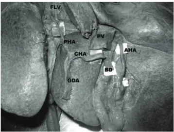
During routine dissection in the Department of Anatomy, Kasturba Medical College, Manipal, a 43-year-old male cadaver revealed the superior mesen-teric artery (SMA) having its origin right next to the celiac trunk (CT). On further dissection, we noticed that the CT was giving rise to all three of its normal branches, which followed their usual course, except for the proper hepatic artery. Even the common hepatic artery (CHA) had its normal origin and course from the CT running towards the right side. It continued upwards as the proper hepatic, after giving off the gastroduodenal artery (GDA). It entered the liver at the fissure for ligamentum venosum (FLV) and gave two branches to the left lobe and one to the right lobe (Figure 1).
1 . Department of Anatomy, Centre for Basic Sciences, Kasturba Medical College, Manipal University, Manipal, Karnataka, India. 2 . Department of Anatomy, KMC International Centre, Manipal University, Manipal, Karnataka, India.
3 . Department of Anatomy, Melaka Manipal Medical College, Manipal University, Manipal, Karnataka, India. Manuscript received Jul 06 2007, accepted for publication Jan 28 2008.
J Vasc Bras. 2008;7(1):84-86.
Copyright © 2008 by Sociedade Brasileira de Angiologia e de Cirurgia Vascular
However, an accessory hepatic artery was seen aris-ing from the SMA. This artery was arisaris-ing from the con-vexity in the proximal 2 cm of the SMA (Figure 2). It was running obliquely upwards and to the right, lying anterior to the inferior vena cava (IVC). At the porta hepatis, it was lying to the right of the bile duct.
Discussion
Knowing anomalous origin of hepatic arteries is important for successful hepatobiliary and liver trans-plant surgeries. A number of abnormalities occurring in hepatic artery anatomy are reported by various authors.8,11-14Abdullah et al. studied 932 cases of liver transplantation and reported that normal hepatic artery distribution was found in 635 cases (68.1%). Variations of hepatic artery were detected in 297 subjects (31.9%) and were divided into three groups, describing 48 (52%) CHA anomalies, 236 (25.3%) left hepatic artery (LHA) or right hepatic artery (RHA) anomalies, and 13 (1.4%) rare variations.1
Gruttadauria et al. studied 701 patients and encoun-tered hepatic artery anomalies in 42%. In his study, the most common anomaly was a replaced/accessory RHA arising from the SMA (15%); they also reported one case
of a LHA arising from a proper hepatic artery with a RHA arising from a GDA.4
A study performed by Covey et al. on 600 digital sub-traction angiographies of the hepatic arterial tree reported that 61.3% of patients had standard arterial anatomy. Approximately 15% of these individuals were found to have variant right hepatic arterial supplies, with accessory right hepatic arteries seen in 2.5% of patients. Of the accessory right hepatic arteries seen, 73% arose from the SMA, with one each arising from the left gas-tric artery, CT, right phrenic artery and GDA.2
Jones & Hardy also reported abnormalities in 43% of 180 cadaveric dissections, with 48% of these having multiple anomalies present. In 75% of cases the RHA arose from the proper hepatic artery, in 17% from the SMA and in 6% from the GDA.5
In the current study, the site of entry of the proper hepatic artery into the liver is at the FLV. To the best of
AHA = accessory hepatic artery; BD = bile duct; CHA = common hepatic artery; FLV = fissure for ligamentum venosum; GDA = gastroduodenal artery; PHA = proper hepatic artery; PV = portal vein.
Figure 1- Proper hepatic artery entering the fissure for liga-mentum venosum and accessory hepatic artery
entering the porta hepatis AHA = accessory hepatic artery; CT = celiac trunk; LGA = left gastric artery; PHA = proper hepatic artery; SMA = superior mesenteric artery.
Figure 2- Hepatic and accessory hepatic arteries arising from the celiac trunk and the superior mesenteric arter-ies, respectively
our knowledge, there are no articles in the literature com-menting on the site of entry. The accessory RHA aris-ing from the SMA is in accordance with previous studies.1,2,4,5,8Among the variations of accessory hepatic artery, this type is the most commonly found. Our great-est limitation was that we were unable to find out how much of the liver tissue is being supplied by each of these arteries as we do not have the infrastructure for such studies.
All these hepatic artery variations must be appropri-ately managed during split liver transplantation to ensure a complete vascular and biliary supply to both grafts.15
When questions arise during bench surgical prepa-ration of the graft, assistance of an angiogram can be helpful to better understand variant anatomy before fur-ther dissection is carried out. Despite its apparent rar-ity, this variant aspect, as well as others, should be known by transplant surgeons.
Acknowledgements
We are grateful to Daphne Pereira, Binod Kumar Tamang, Vishal Kumar, and Soubhagya Ranjan Nayak for their valuable contributions during the revision of the article.
References
1. Abdullah SS, Mabrut JY, Garbit V, et al.Anatomical varia-tions of the hepatic artery: study of 932 cases in liver trans-plantation. Surg Radiol Anat. 2006;28:468-73.
2. Covey AM, Brody LA, Maluccio MA, Getrajdman GI, Brown KT.Variant hepatic arterial anatomy revisited: digi-tal subtraction angiography performed in 600 patients. Radi-ology. 2002;224:542-7.
3. De Santis M, Ariosi P, Calo GF, Romagnoli R.Hepatic arte-rial vascular anatomy and its variants. Radiol Med (Torino). 2000;100:145-51.
4. Gruttadauria S, Foglieni CS, Doria C, Luca A, Lauro A, Marino IR.The hepatic artery in liver transplantation and surgery: vascular anomalies in 701 cases. Clin Transplant. 2001;15:359-63.
5. Jones RM, Hardy KJ.The hepatic artery: a reminder of sur-gical anatomy. J R Coll Surg Edinb. 2001;46:168-70. 6. Peschaud F, El Hajjam M, Malafosse R, et al.A common
hepatic artery passing in front of the portal vein. Surg Radiol Anat. 2006;28:202-5.
7. Volpe CM, Peterson S, Hoover EL, Doerr RJ.Justification for visceral angiography prior to pancreaticoduodenectomy. Am Surg. 1998;64:758-61.
8. Michels NA. Blood supply and anatomy of the upper abdominal organs. Philadelphia: JB Lippincott; 1955. 9. Arnold MM, Kreel L, Lo YF, Law H.Are the hepatic
arter-ies “end arterarter-ies”?Invest Radiol. 1991;26:337-42.
10. Weiglein AH.Variations and topography of the arteries in the lesser omentum in humans. Clin Anat. 1996;9:143-50. 11. Haller A. Icones Anatomicae in quibus praecipae partes
cor-poris humani delineate proponuntur et arteriarum potissi-mum historia continetur. Gottingen. Vandenhoeck. 1756:VIII 270.
12. Tiedemann F. Tabularum arteriarum corporus humani. In: Koerpers, Carlsruhe, M¸ller CF, eds. Abbildungen der Pulsad-ern des menschlichen; 1822. p. 1-250.
13. Adachi B. Arterien system der Japaner. Kyoto: Kerkyusha; 1928. Band II 46-60.
14. Flint ER. Abnormalities of the right hepatic, cystic and gas-tro duodenal arteries and of the bile ducts. Brit J Surg. 1923;10:509-19.
15. Streitparth F, Pech M, Figolska S, et al.Living related liver transplantation: preoperative magnetic resonance imaging for assessment of hepatic vasculature of donor candidates. Acta Radiol. 2007;48:20-6.
Correspondence: Bhagath Kumar Potu
Department of Anatomy, Center for Basic Sciences Kasturba Medical College
576104 - Manipal, Karnataka - India Tel.: +91 820-2922327
E-mail: [email protected]