Reactivity of the isolated
perfused rat tail vascular bed
Departamento de Ciências Fisiológicas, Centro Biomédico,
Universidade Federal do Espírito Santo, 29040-095 Vitória, ES, Brasil A.S. França, L.V. Rossoni,
S.M.C. Amaral and D.V. Vassallo
Abstract
Isolated segments of the perfused rat tail artery display a high basal tone when compared to other isolated arteries such as the mesenteric and are suitable for the assay of vasopressor agents. However, the perfusion of this artery in the entire tail has not yet been used for functional studies. The main purpose of the present study was to identify some aspects of the vascular reactivity of the rat tail vascular bed and validate this method to measure vascular reactivity. The tail severed from the body was perfused with Krebs solution containing different Ca2+ concentrations at different flow rates. Rats were anes-thetized with sodium pentobarbital (65 mg/kg) and heparinized (500 U). The tail artery was dissected near the tail insertion, cannulated and perfused with Krebs solution plus 30 µM EDTA at 36oC and 2.5 ml/ min and the procedures were started after equilibration of the perfu-sion pressure. In the first group a dose-response curve to phenyleph-rine (PE) (0.5, 1, 2 and 5 µg, bolus injection) was obtained at different flow rates (1.5, 2.5 and 3.5 ml/min). The mean perfusion pressure increased with flow as well as PE vasopressor responses. In a second group the flow was changed (1.5, 2, 2.5, 3 and 3.5 ml/min) at different Ca2+ concentrations (0.62, 1.25, 2.5 and 3.75 mM) in the Krebs solution. Increasing Ca2+ concentrations did not alter the flow-pres-sure relationship. In the third group a similar protocol was performed but the rat tail vascular bed was perfused with Krebs solution contain-ing PE (0.1 µg/ml). There was an enhancement of the effect of PE with increasing external Ca2+ and flow. PE vasopressor responses increased after endothelial damage with air and CHAPS, suggesting an endothe-lial modulation of the tone of the rat tail vascular bed. These experi-ments validate the perfusion of the rat tail vascular bed as a method to investigate vascular reactivity.
Correspondence
D.V. Vassallo PPGCF/CBM/UFES Av. Marechal Campos, 1468 29040-095 Vitória, ES Brasil
Research supported by FINEP and CNPq.
Received September 13, 1995 Accepted April 23, 1997
Key words
•Rat tail vascular bed •Phenylephrine •Calcium •Flow •Endothelium •Vascular tone •Perfusion pressure
Introduction
The rat tail artery has been used as a model to study several functional aspects of vascular smooth muscle (VSM). Perfusion of isolated segments of this artery has shown that it is suitable for the assay of vasopressor agents (1-5), in contrast to other vascular beds such as the mesenteric bed, where the
response to vasopressor agents is small (1,6,7). Previous reports have also shown that this artery, when isolated and perfused under constant flow, develops a high basal tone (1) compared to other isolated vascular beds (6,7).
entire isolated rat tail vascular bed. Since the tail artery is a suitable preparation for analysis of vasoconstrictor agents and, as previously suggested, has a high intrinsic tone, the whole rat tail vascular bed severed from the body was perfused in vitro under constant flow to establish some aspects of its reactivity. Also, the experiments were used to validate the perfusion of this prepa-ration as a method to investigate vascular reactivity. Specifically the present study evaluated the rat tail vascular bed response to changes of flow and external Ca2+
con-centration both under control conditions and during continuous infusion of phenyleph-rine (PE). Endothelial participation in vas-cular tone modulation was also evaluated by comparing PE-induced contractions obtained before and after endothelial lesion.
Material and Methods
Rat tail arteries obtained from 30 male Wistar rats (EPM strain) weighing 250-350 g were used in this study. Care and use of laboratory animals were in accordance with NIH guidelines. All rats had free access to water and rat chow. Six to ten rats were used for each experimental group. For the perfu-sion experiments the rats were anesthetized with 65 mg/kg sodium pentobarbital ip and received 500 units of heparin ip. After 10 min, a 1-cm strip of the tail artery was dissected and cannulated near the base of the tail using stretched PE-10 tubing. The rat tail arteries were perfused with Krebs-Henseleit (KH) bicarbonate buffer contain-ing 27.2 mM NaHCO3, 119 mM NaCl, 1
mM NaH2PO4, 1.2 mM MgSO4, 1.25 mM
CaCl2.2H2O, 5 mM KCl, 11 mM glucose
and 0.03 mM EDTA, pH 7.4, bubbled with 5% CO2-95% O2 at 36 ± 0.5oC using a
peristaltic pump (Milan, Colombo, Paraná, Brazil) at a constant flow of 2.5 ml/min, except when flow rate changes were evalu-ated. The tail was then severed from the body and placed in a tissue bath containing
the same Krebs buffer at the same tempera-ture. The experimental protocol was initi-ated after a 30- to 45-min equilibration pe-riod with the tails perfused at 2.5 ml/min. The rat tail perfusion pressure was measured with a TP-200T-Nihon-Kohden pressure transducer (connected to an MP-100 FUNBEC preamplifier) inserted between the pump and the arterial cannula and recorded continu-ously with a polygraphic (ANAMED, AM-820) recorder. Randomized bolus doses of phenylephrine (Sigma) (0.5, 1, 2 and 5 µg in 0.1 ml) were injected into the rubber tube just before the pump and pressor effects were measured. Consecutive doses of PE were administered at 7-10-min intervals and always injected after the perfusion pressure returned to basal values. Since a constant flow was maintained the changes in perfu-sion pressure represented changes in vascu-lar resistance.
Experimental protocol
Four protocols were used. In the first protocol (N = 8) the effects of flow changes on the rat tail perfusion pressure and pressor responses to PE were studied. Step changes in flow rate were made (1.5, 2.5 and 3.5 ml/ min) and the perfusion pressure was evalu-ated after an equilibration period of 5 min. Pressor responses to PE bolus injections (0.5 µg, 1 µg, 2 µg and 5 µg) were studied at each flow. In the second protocol (N = 6) the effects of flow increments (1.5, 2, 2.5, 3 and 3.5 ml/min) on the rat tail perfusion pressure were studied at several external Ca2+
min) before and after endothelial damage. Endothelium was damaged by perfusing air and CHAPS buffer (3,3-cholamidopropyl-dimethylammonio-1-propanesulfonate; Sigma) at a flow rate of 3.5 ml/min. Air was perfused for 2 min followed by perfusion with 5 ml KH bicarbonate buffer (1.25 mM Ca2+, 2.5 ml/min) containing 0.015%
CHAPS. Endothelial damage was confirmed in 4 preparations used only for histological analysis (hematoxylin/eosin staining of 5-µm transversal and longitudinal paraffin sec-tions of tail arteries fixed in Bouin). In the other 7 preparations acetylcholine infusion (5 µg in 0.01 ml) was used in preparations pre-contracted with 0.1 µg/ml PE. Acetyl-choline promoted changes in perfusion pres-sure which was reduced after endothelial damage (∆ reduction in perfusion pressure: 41.3 ± 10.9 mmHg before and 8.29 ± 3.68 mmHg after endothelial damage, P<0.05, Student t-test).
Statistical analysis was done using re-peated-measures ANOVA and the Tukey test was used when statistical significance was attained. The results are reported as mean ± SEM and were considered to be significant at the 5% level.
Results
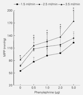
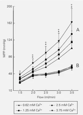
The results of the first protocol are shown in Figure 1. The perfusion pressure increased with flow rate. PE bolus injections produced dose-dependent vasopressor responses that were larger at higher flow rates. The pres-sure-flow curves were displaced upwards (Figure 1) indicating that for the same dose of PE the vasopressor effects were larger at higher flow rates but maintained similar pres-sure-flow relationships with no difference among the effects of different flows (two-way ANOVA, interaction P>0.05). In Figure 2, the lower group of curves (B) shows the changes in mean perfusion pressure (MPP) induced by increasing flow at various Ca2+
concentrations in the KH solution. The
in-crease in perfusion pressure induced by the higher flow was not changed despite the increase of Ca2+ in the KH perfusion
solu-tion.
The third protocol was performed by pre-contracting the preparations with 0.1 µg/ml PE. When perfusing the preparations with 1.25 mM Ca2+ at a flow rate of 2.5 ml/min
the continuous infusion of 0.1 µg/ml PE almost doubled the perfusion pressure, pro-ducing an increase of 95 ± 0.25%. Varying the external Ca2+ at several flow rates
(Fig-ure 2, upper group of curves, A) caused the pressure-flow curves to be displaced up-wards, but with greater MPP changes at higher Ca2+ concentrations and flow.
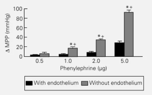
The last protocol was performed by in-ducing PE vasopressor responses before and after perfusion with air and CHAPS buffer. Histological studies showed that the endo-thelial layer was damaged by the treatment. After endothelial damage the perfusion pres-sure increased (49.7 ± 1.98 mmHg for con-trols and 71.6 ± 3.84 mmHg after endothelial damage in 10 preparations). Figure 3 shows that after endothelial damage PE vasopres-sor responses were enhanced, suggesting that in this preparation the endothelial layer con-tinuously modulates the VSM tone probably producing vasodilator agents.
Figure 1 - Effects of 3 different flow rates on the vasopressor responses to phenylephrine (PE) in the rat tail vascular bed per-fused with Krebs solution (con-stant flow, Ca2+ = 1.25 mM). Each point represents the means ± SEM for N = 8. *P<0.01 compared to 1.5 ml/ min (Tukey test). Two-way ANOVA was used for compari-sons among PE responses at dif-ferent flow volumes. +P<0.01 compared to zero PE at each flow (Tukey test). The interac-tion between the PE and flow effects was not significant (P = 0.99), suggesting that the three curves are only displaced up-wards as the flow increases without changes in their slopes. MPP, Mean perfusion pressure.
MPP (mmHg)
200
170
140
110
80
50
20
2.5 ml/min 3.5 ml/min
0 0.5 1.0 2.0 5.0 Phenylephrine (µg)
+
*
+
*
+
*
+
*
*
* *
*
Discussion
The rat tail artery is an important model for the study of several aspects of vascular function such as perfusion experiments (1-5) and analysis of the Na+-pump activity in
preparations obtained from hypertensive ani-mals (8). All of those studies were performed using small isolated segments of this artery (1-5,8). Although important contributions were made by those studies, the participa-tion of an entire vascular bed, such as the mesenteric bed, with vessels of different diameters and behavior is not possible. We therefore decided to use the entire rat tail vascular bed, with the tail severed from the body, to establish some aspects of its vascu-lar reactivity in response to changes in perfu-sion pressure in a constant flow model. The rat tail vascular bed developed a large perfu-sion pressure in response to the 2.5 ml/min flow compared to other vascular beds (1-3). The perfusion pressure increased as a func-tion of flow and, as previously suggested for isolated segments of the tail artery (1-5), is suitable for the assay of vasopressor agents.
The arteries responded to PE with dose-dependent pressor responses. Also, PE pres-sor responses were larger with increasing flow. So, as previously described for the small segments of the rat tail artery (1-5), we also observed an important intrinsic tone and a large vasopressor response to PE in the rat tail vascular bed.
The flow increase did not generate sar-colemmal Ca2+ influx into the VSM. This
conclusion is based on the fact that external Ca2+ increment did not displace the
pres-sure-flow curves. If the increasing flow pro-duced a sarcolemmal Ca2+ influx, the
pres-sure-flow relationships should be displaced upwards at higher Ca2+ concentration.
However, if the sarcolemmal Ca2+ influx
into the VSM was elicited with PE under similar conditions (Figure 2), the flow-induced pressure change was potentiated. This fact can be confirmed by comparing the values of the perfusion pressures in the pressure-flow curves in Figure 2. Al-though our results did not explain the under-lying mechanism, they suggest that PE po-tentiates the pressure increase produced by flow.
The fourth protocol investigated the ef-fects of PE after endothelial damage. Flow has been described as a stimulus for the endothelial cells to liberate vasodilator sub-stances (9-11). On the other hand, the in-crease in pressure stretches the endothelial layer, inducing the release of endothelium-derived contraction factor (EDCF) (9,10,12). In our protocol, the increase in pressure may release endothelial EDCF that acts synergis-tically with PE by potentiating the pressure increment produced by flow. Results ob-tained after endothelial damage showed an increase of the PE vasopressor response to-gether with an increase of the intrinsic basal tone. These findings suggest that the endo-thelium modulates the intrinsic basal tone and the VSM pressor responses of the rat tail vascular bed. These mechanisms confirm previous reports showing that the
endotheli-Figure 2 - Effects of increasing flow on the mean perfusion pres-sure (MPP) of the rat tail vascular bed at four different Ca2+ con-centrations. Group B (lower set of curves) was perfused with phenylephrine (PE)-free Krebs solution and the other group (A, upper set of curves) was pre-contracted with 0.1 µg/ml PE. Each point represents the means ± SEM for N = 6 rat tails. *P<0.01 compared to 1.5 ml/min (two-way ANOVA). Symbols are valid for all curves obtained at different Ca2+ concentrations. Observe that changes in extra-cellular Ca2+ concentrations did not alter the flow-perfusion pres-sure relationship in PE-free Krebs solution. In preparations pre-contracted with PE two-way ANOVA indicated a significant increase in MPP as a function of increasing external Ca2+ concen-tration at each flow (+P<0.03), and a significant difference in the interactions among groups (P<0.04), suggesting that the re-lationships between PE and flow at different Ca2+ concentrations are different; the curves show a different slope.
MPP (mmHg)
200
162
124
86
10 48
1.5 2.0 2.5 3.0 3.5 Flow (ml/min)
0.62 mM Ca2+
1.25 mM Ca2+
2.5 mM Ca2+
3.75 mM Ca2+
+
*
+
*
+
*
+
*
+
* * *
*
A
al cells produce vasodilator substances, such as endothelium-derived relaxing factor (EDRF) and endothelium-derived hyperpo-larizing factor (EDHF) (11). EDRF has been identified as nitric oxide (13,14) produced in response to several stimuli acting on the endothelial cell including flow. Endothelial disruption then reduces the production of vasodilator substances, permitting the in-crease of basal tone and enhancing the vaso-constrictor effects of PE.
In conclusion, our results suggest that the entire rat tail vascular bed perfused under constant pressure has a higher intrinsic basal tone than other isolated perfused vascular beds. The preparation shows a suitable vaso-pressor response to PE and its mean perfu-sion pressure is increased when the flow is enhanced. An increase in extracellular Ca2+
does not alter the pressure-flow relationship if the VSM is not stimulated to contract. If
Figure 3 - Effects of increasing doses of phenylephrine (PE) on the increase in mean perfusion pressure (MPP) of the rat tail vascular bed before and after endothelial damage with air and CHAPS buffer. Preparations were perfused at a flow rate of 2.5 ml/min with Krebs solution containing 1.25 mM Ca2+. Each column represents the means ± SEM for N = 10. +P<0.01 for comparisons between PE ef-fects before and after endothe-lial damage (ANOVA). *P<0.01 compared to 0.5 µg PE (two-way ANOVA). The interaction between the two conditions was significant (P<0.0001), sug-gesting an enhancement of PE effects after endothelial dam-age.
PE is perfused continuously it potentiates the flow effects. Also, in this vascular bed the endothelial layer continuously modu-lates the VSM tone by producing vasodilator substances; endothelial damage increased the intrinsic basal tone and enhanced PE-induced vasopressor responses. Taken to-gether, these findings validate the perfusion of the entire rat tail vascular bed as a method to investigate vascular reactivity.
∆
MPP (mmHg)
100
80
60
40
20
0
0.5 1.0 2.0 5.0 Phenylephrine (µg)
With endothelium Without endothelium
*+
*+
*+
References
1. Nicholas TE (1969). A perfused tail artery preparation from the rat. Journal of Phar-macy and Pharmacology, 21: 826-832. 2. Longhurst PA, Rice PJ, Taylor DA &
Fleming WW (1988). Sensitivity of caudal arteries and the mesenteric vascular bed to norepinephrine in DOCA-salt hyperten-sion. Hypertension, 12: 133-142. 3. Thorin-Trescases N, Oster L, Atkinson J &
Capdeville C (1990). Norepinephrine and serotonin increase the vasoconstrictor re-sponse of the perfused rat tail artery to changes in cytosolic Ca2+. European
Jour-nal of Pharmacology, 179: 469-471. 4. Capdeville-Atkinson C, Oster L,
Thorin-Trescases N, Robert A, Boutinet S & Atkinson J (1993). Intracellular free Ca2+ and vasoconstriction determined simulta-neously in the perfused rat tail artery. American Journal of Physiology, 265: C1689-C1702.
5. Chen XL & Rembold CM (1995). Phenyl-ephrine contracts rat tail artery by one electromechanical and three pharmaco-mechanical mechanisms. American Jour-nal of Physiology, 268: H74-H81. 6. Vasquez EC, Bissoli NS, Moyses MR &
Cabral AM (1988). Contractile reactivity of the perfused mesenteric vascular bed from sinoaortic denervated rats to norepi-nephrine, metoxamine and verapamil. BrazilianJournal of Medical and Biological Research, 21: 629-632.
7. Cabral AM, Musso MN, Bissoli MN, Carvalhinho FB & Vasquez EC (1992). Chlorthalidone reduces vascular hyperre-sponsiveness in DOCA-salt hypertensive rats. Clinical and Experimental Hyperten-sion, A14: 667-683.
8. Songu-Mize E, Bealer SL & Caldwell RW (1982). Effect of AV3V lesions on devel-opment of DOCA-salt hypertension and vascular Na+-pump activity. Hypertension, 4: 574-580.
9. Bevan JA & Laher I (1991). Pressure and flow-dependent vascular tone. FASEB Journal, 5: 2267-2273.
10. Bevan JA (1993). Flow regulation of vas-cular tone. Its sensitivity to changes in sodium and calcium. Hypertension, 22: 273-281.
11. Shepherd JT & Katusic ZS (1991). Endo-thelium-derived vasoactive factors: I. En-dothelium-dependent relaxation. Hyper-tension, 18 (Suppl III): III.76-III.85. 12. Katusic ZS & Shepherd JT (1991).
Endo-thelium-derived vasoactive factors: II. En-dothelium-dependent contraction. Hyper-tension, 18 (Suppl III): III.86-III.92. 13. Moncada S, Palmer RMJ & Annie Higgs E
(1988). The discovery of nitric oxide as the endogenous nitrovasodilator. Hyper-tension, 12: 365-372.