UNIVERSIDADE FEDERAL DO CEARÁ
FACULDADE DE FARMÁCIA, ODONTOLOGIA E ENFERMAGEM PROGRAMA DE PÓS-GRADUAÇÃO EM ODONTOLOGIA
MESTRADO EM ODONTOLOGIA
TALITA ARRAIS DANIEL MENDES
ASSOCIAÇÃO ENTRE A TÉCNICA ÚMIDA ETANÓLICA E DIFERENTES AGENTES DE BIOMODIFICAÇÃO DE COLÁGENO NO PROCEDIMENTO
ADESIVO EM DENTINA
TALITA ARRAIS DANIEL MENDES
ASSOCIAÇÃO ENTRE A TÉCNICA ÚMIDA ETANÓLICA E DIFERENTES AGENTES DE BIOMODIFICAÇÃO DE COLÁGENO NO PROCEDIMENTO
ADESIVO EM DENTINA
Dissertação apresentada ao Programa de Pós-Graduação em Odontologia da Universidade Federal do Ceará, como requisito parcial à obtenção do título de mestre em Odontologia. Área de concentração: Clínica Odontológica. Orientador: Prof. Dr. Juliano Sartori Mendonça.
TALITA ARRAIS DANIEL MENDES
ASSOCIAÇÃO ENTRE A TÉCNICA ÚMIDA ETANÓLICA E DIFERENTES AGENTES DE BIOMODIFICAÇÃO DE COLÁGENO NO PROCEDIMENTO
ADESIVO EM DENTINA
Dissertação apresentada ao Programa de Pós-Graduação em Odontologia da Universidade Federal do Ceará, como requisito parcial à obtenção do título de mestre em Odontologia. Área de concentração: Clínica Odontológica. Aprovada em: ___/___/______.
BANCA EXAMINADORA
________________________________________ Prof. Dr. Juliano Sartori Mendonça (Orientador)
Universidade Federal do Ceará (UFC)
_________________________________________ Prof. Dr. Sérgio Lima Santiago
Universidade Federal do Ceará (UFC)
_________________________________________ Prof. Dr. Jiovanne Rabelo Neri
A Deus.
AGRADECIMENTOS
A Deus por ser o significado de tudo na minha vida, que foi e é o meu “porto seguro” em todos os momentos.
Aos meus pais, Zilná e Eriberto, que são a minha base, grande exemplo de seres humanos batalhadores e honestos. Sempre acreditaram em mim e me deram todo o suporte para que eu pudesse finalizar mais essa etapa, sem vocês eu não teria conseguido, minha admitação e amor por vocês não se mede.
Aos professores que compõe a banca examinadora, Prof. Dr Jiovanne Neri e Prof. Dr. Sérgio Santiago, pela disponibilidade e todas as valiosas considerações realizadas.
Ao meu orientador, Prof. Dr Juliano Mendonça, que sempre foi um professor por quem tive grande admiração desde a graduação. Em sua sabedoria, me fez desenvolver a capacidade de começar pensar como pesquisadora, sempre buscou tirar as minhas dúvidas e acreditou nas minhas ideias, muito obrigada.
Ao professor, Sérgio Santiago, por sempre estar presente desde o ínicio dessa “jornada científica”, me ajudando, apoiando e acredianto em ideias minhas. Sou grata a todas oportunidadades que me foram concedidas.
À minha irmã, Sarah Arrais, que por muitas vezes escutou as minhas dificuldades nesse período e com todo o seu dom de acalmar o fazia. Obrigada por ser essa melhor-amiga que sempre está comigo e acredita muito em mim, sou grata por todo o companheirismo que você tem por mim.
Ao meu namorado, companheiro de pesquisa e de vida, Marcelo Sidou, pessoa a quem mais ouviu as minhas lamentações, e com toda sua calmaria conseguia me acalmar e me mostrar uma saída.
Aos meus sobrinhos, Gabriel e João Miguel, por darem sentido a minha vida e por quem eu busco ser melhor, todos os dias.
Aos meus avós, Telma, José e Mirian, e minha tia Zildene, por todo o apoio e amor que me foi concedido, o meu muito obrigada.
A todos os meu familiares, que torceram muito por mim e me apoiaram em cada vitória e derrota.
Ernanda Sales, Bernardo Aguiar, Luciana Arcanjo, César Filho, Adeilson Alves, Pedro Henrique Acioly, Iana Sá, por todos os momentos, bons e ruins, divididos, e todo o companheirismo e tocida, muito obrigada!
Ao técnico do laboratório de pesquisa, David Queiroz, por toda a ajuda concedida sempre que precisei.
A minha equipe de pesquisa, Samuel Chillaverd, Maria Clara Estellita, Ana Laura Mota, Paula Motoyama, Emanuel Filho, Antonino, sem os quais o trabalho não teria acontecido, obrigada por serem tão companheiros e me ajudarem em tudo, vocês são a melhor equipe que alguém poderia ter.
À CAPES, pelo apoio financeiro com a manutenção da bolsa de auxílio.
A Universidade Federal do Ceará, que foi minha “segunda casa” desde a época da graduação, nessa instituição consegui adquirir muitas experiências e aprendizado.
“Nenhuma pessoa, por mais inteligente que seja, é melhor que o grupo.”
RESUMO
associação entre a técnica TUE e uso de agentes biomodificadores irão melhorar as propriedades mecânicas do colágeno, no entanto não mostrou influenciar no procedimento adesivo.
ABSTRACT
of biomedical agents will improve the mechanical properties of the collagen, but it has not been shown to influence the adhesive procedure.
SUMÁRIO
1 INTRODUÇÃO GERAL... 13
2 PROPOSIÇÃO ... 16
3 CAPÍTULO ... 17
Abstract ... 18
1 Introduction ... 19
2 Materials and methods ... 20
3 Results... 24
4 Discussion ... 24
5 Conclusion... 27
6 References... 27
4 CONCLUSÃO GERAL ... 37
5 REFERÊNCIAS... 38
ANEXO A... 41
13
1 INTRODUÇÃO GERAL
A demanda por estética tem sido bastante elevada nos últimos anos, consequentemente o uso de compósitos resinosos em restaurações dentárias vem sendo bastante empregado. Os sistemas adesivos convencionais (SACs) aderem ao dente por meio de uma ação micromecânica, que ocorre através da infiltração de monômeros resinosos em fibrilas colágenas expostas ou micropororidades geradas no esmalte por um prévio condicionamento da superfície, com ácido fosfórico em diferentes concentrações entre 35- 37%, seguido de aplicação do primer e adesivo (NIU et al., 2014). O processo de união mais crítico ocorre no substrato dentinário, devido a sua característica morfofuncional. Esse substrato corresponde à maioria do tecido dentário, apresentando uma maior quantidade de conteúdo orgânico e umidade quando comparado ao esmalte, onde a união de restaurações de resina composta já é bem estabelecida e duradoura (FRASSETO et al., 2016).
A região onde ocorre a união do sistema restaurador adesvio à dentina é denominada de camada híbrida (CH) (NAKABAYASHI; KOJIMA; MASUHARA, 1982), sendo a principal responsável pela retenção do mesmo ao dente, entretanto a CH é susceptível a ação de degradação (SCHEFFEL et al., 2014; PHANSALKER et al., 2015; AYAR et al., 2016), que pode ocorrer por vários meios como ação de enzimas endógenas, como metaloproteinases de matriz (MMPs) e cisteíno-catepsinas (CTPs), ação de hidrólise, ocasionada por presença de água em excesso nessa interface, bem como degradação de monômeros por lixiviação (PASHLEY et al., 2011; MARAVIC et al., 2017).
14
Quando a dentina desmineralizada não apresenta água suficiente, as fibras colágenas poderão entrar em colapso, formando ligações de hidrogênio entre si, comprometendo, dessa maneira, a infiltração adequada dos monômeros presentes nos sistemas adesivos (CARVALHO et al., 2005; AGGARWAL et al., 2016). Assim, faz-se necessário manter a umidade desse substrato através da técnica úmida, que é a mais utilizada atualmente (; AYAR et al., 2016; YANG et al., 2016; ZHOU et al., 2016).
Como alternativa à técnica úmida (convencional) surgiu a técnica úmida etanólica [TUE] (PASHLEY et al., 2007), que consiste na substituição da água livre, presente na dentina, por um solvente isento de água, o etanol, por meio de uma desidratação química, mantendo a conformação espacial do colágeno (YANG et al., 2016), gerando dessa forma um maior espaço interfibrilar, por meio da diminuição do diâmetro das fibrilas, e permitindo a penetração adequada dos monômeros hidrofóbicos presentes nos sistemas adesivos (CHIBA et al., 2016; GÖSTEMEYER et al., 2016; ZHOU et al., 2016).
O etanol se constitui em um solvente superior à água, em relação à infiltração de monômeros dimetacrilatos no substrato dentinário, levando a um melhor desempenho na união efetiva desses ao dente (BEDRAN-RUSSO et al., 2011). Além da TUE, existem várias outras manobras que auxiliam na otimização das propriedades mecânicas e durabilidade, em longo prazo da interface adesiva, sendo que dentre elas, destaca-se a aplicação de agentes biomodificadores no substrato dentinário (ZHOU et al., 2016).
Esses agentes biomodificadores ou agentes de ligações cruzadas (ALC) irão interagir com o colágeno constituinte da estrutura de dentina a fim de formar ligações com os seus aminoácidos, resultando em formações de ligações químicas interfibrilares e intrafibrilares, aumentando a biomecânica desse substrato e reduzindo significativamente a função enzimática das MMPs e CTPs (BEDRAN-RUSSO et al., 2014; VIDAL et al., 2016).
15
constituintes do colágeno (GU et al., 2011; MAZZONI et al., 2014). O GA irá formar ligações covalentes entre dois grupos amina de hidroxilisinas com seus grupos aldeídos, a fim de formar compostos de piridíneo, sendo que essa ligação apresenta uma grande estabilidade, porém é um agente que possui uma alta citotoxicidade, sendo necessária uma técnica operatória mais minunciosa para sua utilização na odontologia restauradora (BEDRAN-RUSSO et al., 2008). Existem também, produtos de origem natural, como as proantocianidinas (PAC), que são polifenóis derivadas da semente de uva (vitis vinifera). Apesar de seu mecanismo de ação ainda não estar bem esclarecido, acredita-se que sua interação com o colágeno vai ocorrer através o grupamento fenol presente em sua molécula que irá reagir com a amina presente nos aminoácidos constituintes do colágeno, afim de formar ligação de hidrogênio (BEDRAN-RUSSO et al., 2014).
Dessa forma, os ALC irão melhorar a estabilidade do colágeno, bem como suas propriedades mecânicas, porém não são capazes de remover a água em excesso presente no substrato dentinário, responsável por dificultar tanto a infiltração quanto a polimerização dos monômeros (VENIGALLA et al., 2016; VIDAL et al., 2016).
16
2 PROPOSIÇÃO
O presente trabalho teve como objetivos:
2.1 Objetivo geral
Avaliar se a associação entre a técnica úmida etanólica com diferentes agentes de biomodificação de colágeno, aplicados em diferentes tempos, é eficiente na biomodificação do colágeno e se esses tratamentos influenciam na união de um sistema restaurador adesivo à dentina.
2.2 Objetivos específicos
• Analisar o potencial biomodificador de colágeno dos agentes de ligações cruzadas incorporados ao etanol absoluto nos diferentes tempos de aplicação em dentina, por 1 hora e 1 minuto;
• Investigar se há uma influência desses primers, com um minuto de aplicação, utilizados em
dentina no grau de conversão do sistema adesivo no procedimento restaurador;
17
3 CAPÍTULO
Esta dissertação está baseada no Artigo 46 do Regimento Interno do Programa de Pós-Graduação em Odontologia da Universidade Federal do Ceará que regulamenta o formato alternativo para dissertações de Mestrado e teses de Doutorado, e permite a inserção de artigos científicos de autoria ou coautoria do candidato. Por se tratar de estudos envolvendo seres humanos, ou parte deles, o projeto de pesquisa foi submetido à apreciação do Comitê de Ética em Pesquisa da Universidade Federal do Ceará, tendo sido aprovado. Assim sendo, esta dissertação é composta de um artigo científico que será submetido ao periódico Dental Materials, conforme descrito abaixo:
ASSOCIATION OF ETHANOL WET BONDING AND DIFFERENT COLLAGEN BIOMODIFICATION AGENTS ON THE PHYSICAL CHEMICAL PROPERTIES AT
AN ETCH-AND-RINSE ADHESIVE TO DENTIN
18
ASSOCIATION OF ETHANOL WET BONDING AND DIFFERENT COLLAGEN BIOMODIFICATION AGENTS ON THE PHYSICAL CHEMICAL PROPERTIES AT
AN ETCH-AND-RINSE ADHESIVE TO DENTIN
ABSTRACT
Objective: To evaluate the association between Ethanol Wet Bonding (EWB) and collagen cross-linking agents (CLA) on the mechanical properties of collagen and on the physical chemical proprieties of an etch-and-rise adhesive to dentin.
Methods: demineralized dentin bars (n=10) were immersed, for 1 min or 1 h, according to the respective groups: distilled water control (WC); absolute ethanol (EA); absolute ethanol with the incorporation of proanthocyanidins 6,5% (PAC+AE); absolute ethanol with the incorporation of 1-ethyl-3- (3-dimethylaminopropyl) carbodiimide 0.3M (EDC+EA); absolute ethanol with the incorporation of 5% glutaraldehyde (GA+AE). All as solutions used as dentin primer per 1-min of application, for 3-point bending test was performed to evaluate the modulus of elasticity [ME] (n = 10) and mass change [MC] (n = 10) of collagen treated with the respective solutions, at different times of application (1min and 1h). The in situ conversion rate [GC] (n = 3), microtensile bond strength testing [μTBS] (n = 6) and nanoleakage evaluation [NE] (n = 3) of the teeth restored with the respective treatments were analyzed.
Results: After 1 min and 1 h of application, it was observed that WC group showed the lowest ME (P<0,05). The groups EDC+AE, GA+AE and AE presented a better ME with one hour of application, since the PAC+AE and WC were statistically similar. Treatments did not influence GC and μTBS. In the nanoleakage evaluation the AE presented "gaps" in its interface, as well as a great amount of silver.
19
1 INTRODUCTION
One of the major shortcomings that approach restorative Dentistry is the failure of resinous restorations generated by nanoinfiltration of the adhesive interface. The main means that promote this degradation are the hydrolysis and activity of protein hydrolases enzymes, matrix metalloproteinases (MMPs) and cysteine cathepsins (CTPs), which disrupt the collagen through peptide bonds.1,2
The hydrolysis is facilitated by incomplete impregnation of resinous monomers in bare collagen fibrils by acid etching in dentin, in etch-and-rinse adhesive systems, and also by the presence of hydrophilic monomers present in the adhesive systems.3,5 In order to reduce this degradation, alternatives, such as the use of the Ethanol Wet Bonding (EWB), have been studied. EWB consists of the chemical dehydration of the wet dentin, by means of successive ethanol applications, in order to replace the rinse water,5 responsible for the preservation of the spatial conformation of the collagen. This technique has been shown to be effective in increasing the immediate bond strength, as well as an improvement over time compared to the conventional wet technique.2,3
The presence of ethanol makes the dentine propitious so that dimethacrylate monomers adequately infiltrate, since most of the monomers are soluble in that solvent. In addition, their presence will promote a transverse constriction of the collagen fibril, increasing the interfibrillars spaces3,4,5,6 and some studies speculate that there will be a complete replacement of the residual water in a way that will prevent the action of endogenous hydrolases.2,7
Collagen crosslinking agents (CLA) have been studied in the present context of restorative dentistry. They consist of chemical compounds of natural or synthetic origin that will biomodify the dentin structure, improving the mechanical properties of the collagen network, as well as reducing rates of biodegradation of the extracellular matrix. The interaction with collagen occurs most often through the chemical bonds of the CLA between proline and hydroxyproline present in the molecular structure of collagen, such bonds can occur at both the interfibrillar and intrafibrillary levels.8
20
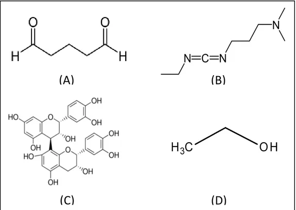
type I collagen, but its mechanism of action is not well established.8,9 Carbodiimide (EDC) will act effectively as CLA (Fig. 1), and show an effective action in the inhibition of MMPs, requiring a short period of application for its effectiveness.10 In addition, glutaraldehyde (GA) is a synthetic CLA (Fig. 1) that will react with the free amino acids of the triple helix structure of the collagen, improving the biomechanical properties of collagen, although it presents a high cytotoxicity when compared to the others.11
The present study aims to evaluate the biomodifying capacity of EDC, PAC and GA in dentin collagen when incorporated into absolute ethanol by means of the evaluation of the biomodifier potential, and the influence of this treatment in the adhesive procedure to dentin by means of microtensile bond strength test and nanoinfiltration analysis of the adhesive interface. The hypothesis of the study are that (1) biomodifiers diluted in absolute ethanol will not improve the modulus of elasticity of the demineralized dentin, as well as the change mass at different times of application, (2) this association will not influence the in situ degree of conversion of the adhesive system; and (3) the association of EWB with CLA will not interfere in the efficiency of union of an adhesive restorative system to dentin (bond strength and nanoleakage).
2 MATERIALS AND METHODS
The study was approved by the Research Ethics Committee of the Institution (Protocol number 2.338.233). After the patient’s informed consent was obtained, the teeth were collected under a research protocol in accordance with the Guidelines and Standards of the National Health Council (Resolution No. 554/2017).
21
2.1 Dentin bioactivity Assays 2.1.2 Sample preparation
Dentin bars with 1.7 X 0.5 X 6.0 mm in dimension were prepared. The specimens were then completely demineralized in 10% phosphoric acid solution over a period of 5 hours at room temperature and under constant stirring. Dentin bars were randomly distributed in the different groups (n = 10) and maintained in their respective solutions (WC, AE, EDC+AE, PAC+AE and GA+AE) for periods of 1 min 12 or 1 h at room temperature.13
2.1.2 Modulus of elasticity
The modulus of elasticity (ME) of the demineralized dentin bars was evaluated at the beginning and after immersion at different times in their respective solutions. This ME was determined in a 3-point flexural test with a 5.0N load cell mounted on a universal mechanical testing machine (Instron, Canton, Massachusetts, EUA) at a cross-head speed of 0.5mm/min. The data were expressed in Megapascal (MPa), and the change of the ME will be calculated between the ratio of the final value (after the dentin biomodification in the respective groups) and the baseline values.13
2.1.3 Mass change
The masses were measured before (M1) and after (M2) the biomodification process of the demineralized dentin bars with an analytical balance (Shimadzu, Kyoto, Japan) precision of five decimal places (0.00001g). The samples were dehydrated in a vacuum desiccator for 24 hours at room temperature. The mass change assessment (MM%) was determined as the percentage gain or loss of each sample based on the following formula:
22
2.2 Bonding procedures
Third molars humans, free of caries, were used for bonding procedures, the occlusal enamel and the roots of each tooth were removed with a diamond disk (Isomet 4000; Buehler, Lake Bluff, USA) at low speed to expose the flat surface of medium dentin, then the smear layer was made with granulation 600 silica carbide, under constant irrigation for 30s. All medium dentin blocks were conditioned with 37% phosphoric acid in gel (Condac 37%, FGM, Joinville, Brazil) for 15s, then washed abundantly for twice the time. The excess water was removed with absorbent paper and a solution of 10μL of absolute ethanol, incorporated or not with the biomodifying agents at their respective concentrations,13 was applied to the test groups, making 4 exchanges, totalizing a period 60 seconds. Then, excess of ethanol was removed from the specimen with the absorbent paper by applying the bond resin of the conventional adhesive (Scotchbond Multipropose Adhesive - 3M ESPE) actively for 30s. After application, the adhesive layer was light-cured for 40 seconds by an LED light curing at 1100mW/cm2 (DB-685, Dabi Atlante, Ribeirão Preto, Brazil). The adhesive procedure in the negative control group followed the protocol of the conventional wet technique. Then, a resin composite plateau (Filtek Z350 XT-3M ESPE) (Table1) of approximately 4mm was made, with each increment of 1 millimeter photopolymerized for 40s. The procedure was used for test DC, µTSB and NE.
2.3 Degree of conversion in situ
For the analysis of micro-Raman spectroscopy, three additional teeth were restored for each group. These teeth were cut logitudinally to the medium (mesial-distal) in order to generate slices of 2,0 mm.
For the adhesive interface analysis, micro-Raman spectra of the adhesive-dentin interface were obtained from each hemi-tooth, and were recorded by a Raman Horiba (Xplora, HORIBA Jobin Yvon, Paris, France) spectrometer. A monochromatic argon laser beam set at a wavelength of 638nm with an output level of 20mW was used as the source of excitation.
23
The values were inserted in the following formula:
% DC= 1- x 100
For this purpose, a reading was performed on unpolymerized adhesive in order to obtain the necessary values for filling the formula.15
2.4 Microtensile bond strength testing
Resin-bonded teeth were sectioned in small resin–dentine sticks (cross-sectional area of approximately 1.0mm2) suitable for the microtensile bond strength. After the restorative procedure the specimens were stored in distilled water in an oven for 24 h at 37 °C. Six teeth (n=6) were evaluated for each group. The sticks from the most peripheral area presenting residual enamel were excluded from the test. The exact cross-sectional area of each tested stick was measured with a high precision digital calliper. The sticks were glued to a jig with a cyanoacrylate gel (Super Bonder gel; Octite Henkel, Rocky Hill, CT, USA) and tested in universal testing machine (EMIC, model DL 2000. São José dos Pinhais, Brazil) with a 500N load cell (cross-head speed: 0.5 mm/min). The µTBS results were calculated and expressed in MPa.
2.5 Nanoleakage evaluation
24
2.6 Statistical analysis
Sigma Plot was the program used for the analyzes. For ME test the data were transformed into Log n and submitted to the Shapiro-Wilk normality test and then submitted to analysis of variance (ANOVA two-way), followed by a Tukey post-hoc test. In the MC test the data were submitted to Kruskal-Wallis non-parametric analysis. The DC in situ and µTSB testing test data were submitted to the normality test followed by analysis of variance ANOVA one- way. All tests were performed with a significance level of 5%.
3 RESULTS
When the modulus of elasticity values were analyzed, all tested groups showed a statistical improvement over the negative control (p<0,001), and no differences were observed between the other tested groups in both application periods (p>0,05). However, when analyzed individually, the EDC+AE, GA+AE and AE groups were shown to be more effective after 1 hour of immersion (p<0,05). In the PAC+AE group, the effect was independent of the immersion time (Table 2).
When the mass change was analyzed, there was not statistical difference between the different groups tested in both immersion times (p >0.05). When the individual treatments were analyzed, there werw no differences between the different application times (Fig. 2).
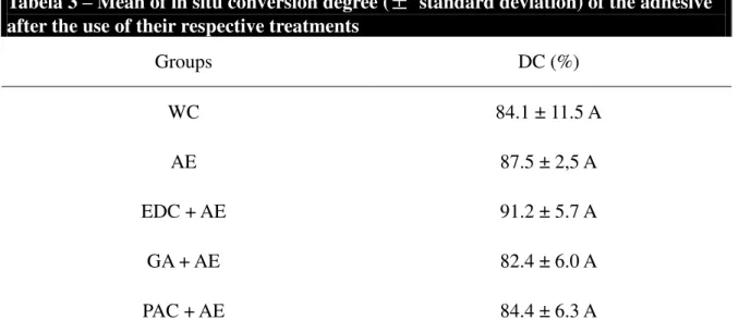
The treatments performed did not influence the degree of conversion of the adhesive when analyzed in situ (p=0.529, F=0,729), as described in Table 3. No statistically significant differences was observed in microtensile bond strength for the tested groups (p=0.194; F=1.647), as shown in Table 4.
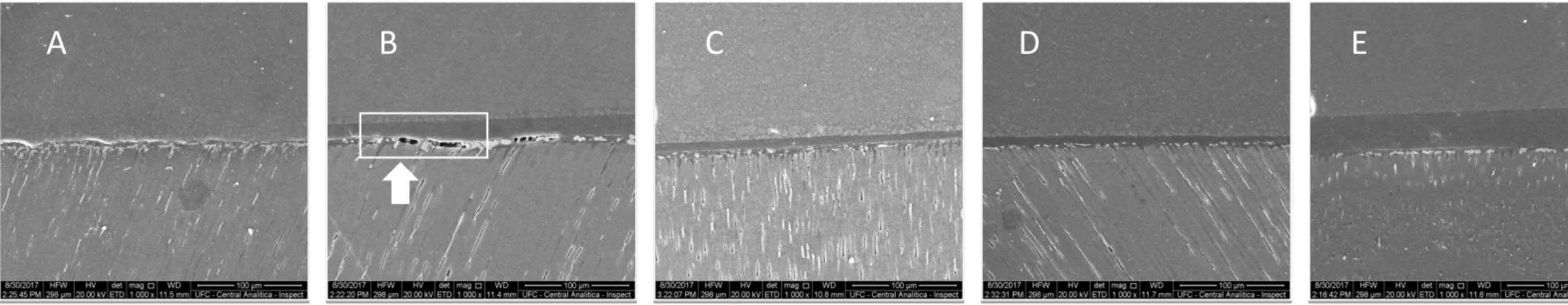
In a qualitative analysis, it was observed presence of silver near the adhesive layer in all tested groups, but in the AE group, large areas of crack formation were observed in this region (Fig. 3).
4 DISCUSSION
25
degradation, being used separately in previous studies, however this association as a technique or dentin primer has not been observed until then.
The bare collagen treatments with the respective cross-links solutions influenced the ME of the demineralized dentin, this data does not accepting the first hypothesis of the study (Table 2). It can be analyzed that in the dentin treatments, all the test groups presented better ME values than the WC, in the different times of application, proving the effectiveness of the EWB in increasing the mechanical properties of the dentin. The only group, among the treatments, that the application time did not influence the modulus of elasticity was PAC+AE. This can occur due to its effectiveness already proven in the period of 1 minute of application, 9 being this polyphenol an effective adjuvant incorporated to the EWB technique in the treatment of demineralized dentin. The PAC may have a good efficacy due to its mechanism of action with collagen, which is more versatile when compared to other cross-linkings agents. This interaction is not yet well elucidated in the literature, however it is believed that it can occur in several different ways, such as through hydrogen bonding, hydrophobic interactions between the aromatic ring of the PAC and proline, and the reaction by means of ester-like bonds between the glutamic and aspartic acid with the amine group, present in the lysine, forming a stable covalent bond.17
The PAC group at 6.5% concentration showed effectiveness in the time of application of 1 minute becoming quite relevant in clinical practice regarding the increase of the biomechanical properties of collagen, evidencing its applicability in restorative dentistry, having already been shown that there is a significant improvement in the modulus of elasticity, 8 however, some data already describe a greater effectiveness in the improvement of these properties of the collagen, when diluted in water instead of ethanol. 11, 13, 17
The 1-minute CLA application protocol for the dentin biomodification test 12 is the one that best simulates the clinical use, however, it showed less satisfactory ME results for the EDC+AE, GA+AE and AE groups, when compared to the time of application of 1-hour.11,13 However it is known that one hour of use of these pre-treatments becomes impractical for day-to-day dental practice, it is observed. However, that after application of one minute of the respective primers the groups were statistically best when compared to the WC.
cross-26
linking agents in these primers, generating no significant change in mass change.20,21
The dentin treatments did not influence the degree of in situ conversion of the adhesive used in the restorative procedure, not rejecting the second hypothesis of the study. This is due to the fact that the ethanol present in the hybrid layer for a longer period of 60 seconds, due to the operative technique used, will generate a better volatilization of the water present in the dentin, bringing the constituent monomers closer together, and helping to improve the adequate polymerization of the same.22 In addition, the biomodification agents incorporated in this organic solvent did not seem to influence the polymerization of the constituent monomers of the used adhesive system, contrasting the study of Leme-Kraus et al. (2017), that affirms that the PACs will interfere significantly on the degree of conversion of hydrophobic dimethacrylate monomers into wet dentin. Such adequate polymerization of the adhesive layer will depend directly on the degree of hydrophilicity of the adhesive monomers, 20 as in the present study, used only the bond, which has a significant amount of hydrophobic monomers in its composition, may have influenced the improvement of the polymerization of the adhesive layer.
It can be observed that there was no statistical difference in microtensile bond strengh (µTBS) in the different treatments used in dentin after the period of 24 hours corroborating with one of the thrid hypotheses of the study. Although the highest values of bond strength were in the PAC+AE group, this difference was not significant in relation to the other treatments. Studies evaluated, in an immediate analysis, using different CLAs, such as EDC and PAC, these values of dentin bonding strength did not present statistical difference immediately, and only after 6 months of evaluation that difference was accentuated, with a more effective EDC.23,24 In addition, studies show that the application of EWB for up to 30 seconds is equivalent to its use for 130 seconds, as well as the use of the conventional technique, 20,25 corroborating with the present study that uses a period of one minute of application, 14 in order to make the clinically feasible technique.
27
once again diluted in water, because it is able to interact efficiently with a dynamic dentin, favoring an infiltration of adhesives with hydrophilic monomers and reducing permeability of the adhesive layer, thus as PACs are most effective in conventional wetting technique.8 The crack formation in group AE (Fig. 3) may occur due to incomplete impregnation of dimethacrylate monomers due to the interfibrillar space that was not sufficient for its penetration. 26
5 CONCLUSION
It is concluded that the association between the EWB technique and the use of biomodifiers will improve the mechanical properties of the collagen, but it has not been shown to influence the adhesive procedure. The treatments showed better efficacy in increasing the modulus of elasticity with one hour of application, except for PAC + AE and NC. This association did not influence DC in situ and µTBS.
REFERENCES
[1] Pashley DH, Tay FR, Breschi L, Tjäderhane L, Carvalho RM, Carrilho M, Tezvergil-mutluay A. State of the art etch-and-rinse adhesives. Dent Mater, 2011; 27: 1-16.
[2] Jee SE, Zhou J, Tan J, Breschi, Tay RF, Grégoire G, Pashley DH, Jang SS. Investigation of ethanol infiltration into demineralized dentin collagen fibrils using molecular dynamics simulations. Acta Biomater, 2016; 36: 175-185.
[3] Ayar MK. A review of ethanol wet‑bonding: Principles and techniques. Eur J Dent, 2016; 10:155- 159.
28
[5] Grégoire G, Sharrock P, Delannée M, Deslisle. Depletion of water molecules during ethanol wet-bonding with etch and rinse dental adhesives. Mater Sci Eng C Mater Biol Appl, 2013; 33: 21-27.
[6] Li MZ, Wang JR, Liu H, Wang X, Gan K, Liu JX, Niu DL, Song XQ. Effects of light curing modes and ethanol-wet bonding on dentin bonding properties. J Zhejiang Univ Sci B, 2017. 17: 703-711.
[7] Ahn J, Jung K, Son AS, Hur B, Kwon YH, Park JK. Effect of additional etching and ethanol-wet bonding on the dentin bond strength of one-step self-etch adhesives. Restor Dent Endod, 2014; 40: 68-74
[8] Leme-Kraus AA, Aydin B, Vidal CMP, Phansalkar RM, Nam JW, McAlpine J, Pauli GF, Chen S, Bedran-Russo AK. Biostability of the Proanthocyanidins-Dentin Complex and Adhesion Studies. J Dent Res, 2017; 96: 406-412.
[9] Bedran-russo AK, Pauli GF, Chen SN, Mcalpine J, Castellan CS, Phansalkar RS, Aguiar TR, Vidal CMP; Napotilano J.G.; Nam JW, Leme AA. Dentin biomodification: strategies, renewable resources and clinical applications. Dent Mater, 2014; 30: 62-76.
[10] Mazzoni A, Apolonio FM, Saboia VPA, Santi S, Angeloni V, Checchi V, Curci R, Lenarda RD, Tay FR, Pashley DH, Breschi L. Carbodiimide Inactivation of MMPs and Effect on Dentin Bonding. J Dent Res, 2014; 93: 263-268.
[11] Bedran-Russo AK, Pashley DH, Agee K, Drummond JL, Miescke KJ. Changes in stiffness of desmineralized dentin following appliccation of collagen cosslinkers. J Biomed Meter Res B Appl Biomater, 2008; 86: 330-334.
[12] Moreira MA, Souza NO, Sousa RS, Freitas DQ, Lemos MS, De Paula DM, Maia FJN,
Lomonaco D, Mazzeto SE, Feitosa VP. Efficacy of new natural biomodification agentsfrom Anacardiaceae extracts on dentin collagencross-linking. Dent Mater, 2017; 33: 1103-1109.
29
Pauli GF, Bedran-russo AK. Dentin Biomodification Potential Depends on Polyphenol Source. J Dent Res, 2014; 93: 417-422.
[14] Scheffel DLS, Hebling J, Schefell RH, Agee KA, Cadenaro M, Turco G, Breschi L, Mazzoni A, Costa CAS, Pashley DH. Stabilization of dentin matrix after cross-linking treatments, in vitro. Dent Mater, 2014; 30: 227-233.
[15] Yang H, Li K, Yan H, Wang Y, Huang C. High-performance therapeutic quercetin-doped adhesive for adhesive-dentin interfaces. Sci Rep, 2017; 7: 81-89.
[16] Tay F.R.; Pashley D.H. Biomimetic remineralization of resin-bonded acid-ecthed dentin. J Dent Res, 2009; 88:719-724.
[17] Vidal CMP, Zhu W, Manhoar S, Aydin B, Keiderling TA, Messersmith PB, Bedran-Russo. Collagen-collagen interactions mediated by plant-derivedproanthocyanidins: A spectroscopic and atomic force microscopy study. Acta Biomater, 2016; 41: 110-118.
[18] Castellan CS, Pereira PNR, Viana G, Chen SN, Pauli GF, Bedran-Russo AK. Solubility study of phytochemical cross-linking agents on dentin stiffness. J Dent, 2010; 38: 431-436.
[19] Epasinge DJ, Burrow MF, Yiu CKY. Effect of proanthocyanidin on ultrastructure and mmineralization of dentin collagen. Arch Oral Biol, 2017; 84: 29-36.
[20] Sadek FT, Mazzoni, Breschi L, Tay FR, Braga RR. Six-month evaluation of adhesives interface created by a hydrophobic adhesive to acid-etched ethanol-wet bonded dentine with simplified dehydration protocols. J Dent, 2010; 38: 276-283.
[21] Becker TD, Agee KA, Joyce AP, Rueggeberg FA, Borke JL, Waller JL, Tay FR, Pashley DH. Infiltration/evaporation-induced shrinkage of demineralized dentin by solvated model adhesives. J Biomed Mater Res B Appl Biomater,2007; 80: 156-165.
30
Influenced by Extended Air-Activated or Passive Solvent Volatilization Modes. Oper Dent, 2012; 37 (3): 246-252.
[23] Shafiei F, Firouzmandi M, Zamanpour M. The effect of two cross-linking agents on dentin bond strength of resin-modified glass ionomer. Eur J Dent, 2017; 11: 486-490.
[24] Zhang Z, Beitzel D, Madj H, Mutluay M, Tezvergil-Mutluay A, Tay FR, Pashley DH, Arola D. Effect of carbodiimide on the fatigue crack growth resistance of resin-dentin bonds. Dent Mater, 2016; 32: 211-222.
[25] Aggawal V, Singla M, Sharma R, Miglani S, Bhasin SS. Effects of simplified ethanol-wet bonding technique on immediate bond strength with normal versus caries-affected dentin. J Conserv Dent, 2017; 19: 419-423.
31
Fig. 1: Images of the structural chemical formulas of the biomodification agents used, GA (A), EDC (B), PAC (C) and AE (D).
(A)
(B)
32
Table 1. Composite and adhesive resin compositions and application protocols.
Composition Application procedure
Adper™ Scotchbond™ Multi-Purpose Adhesive (3M ESPE, St Paul, USA)
Primer: HEMA, polyalkenoic acid copolymer, water; Bond: Bis-GMA; HEMA
CQ
Active application for 30 seconds, followed by photopolymerization
for 40 seconds
Filtek TM Z350 XT, A3 (3M ESPE, St Paul, USA)
UDMA, Bis-GMA, TEGDMA, Bis-EMA,
silica (20 nm nanogglomerated/aggrega
ted), zirconia silica (4-11 nm
nanogglomerated/aggrega ted), clusters, zirconia/ silica aggregated particles
(20 nm silica particles combined with 4-11nm
zirconia 3)
Incremental placement (in 1mm levels or less) and curing of
composite restorations is recommended to minimise polymerisation shrinkage. Curing
each increment separately
*HEMA – hydroxy ethyl methacrylate; GMA - bisphenol-A glycidyl methacrylate; Bis-EMA – ethoxylated bisphenol-A glycidyl methacrylate; TEGDMA – triethylene glycol-imethacrylate;
Table 2: Means of Elasticity modulus (MPa) [± standard deviation] of completely demineralized dentin specimens before and after the application of different trataments solutions for 1min or 1 h.
Groups 1 minute 1 hour
WC 1.2 ± 1.4 B, a 1.2 ± 0.6 B, a
AE 1.6 ± 1.6 A, b 4.8 ± 1.5 A, a
EDC + AE 3.8 ± 3.0 A, b 10.1 ± 14.7 A, a
GA + AE 1.7 ± 1.4 A, b 5.3 ± 4.0 A, a
PAC + AE 4.5 ± 4.3 A, a 7.4 ± 6.9 A, a
33
34
Tabela 3 – Mean of in situ conversion degree (± standard deviation) of the adhesive
after the use of their respective treatments
Groups DC (%)
WC 84.1 ± 11.5 A
AE 87.5 ± 2,5 A
EDC + AE 91.2 ± 5.7 A
GA + AE 82.4 ± 6.0 A
PAC + AE 84.4 ± 6.3 A
* uppercase letters comparing between columns and lowercase letters between rows
Table 4 –Microtensile bond strength testing (MPa) immediate of bonding procedure
Groups µTSB
WC 24.5 ± 12.4 A
AE 26.9 ± 5.0 A
EDC + AE 20.9 ± 7.9 A
GA + AE 19.2 ± 10.6 A
PAC + AE 32.3 ± 11.6 A
35
Fig. 3: Evaluation of the dentin / adhesive union interface after the use of dentin pre-treatments (A) WC, (B) AE, (C) EDC+AE, (D) GA+AE and (E) PAC+AE, with nanoleakage being performed and analyzed by SEM in an increase of 1000X, where a great amount of silver infiltrate in the WC group and, mainly EA, is observed, presenting slits in that union interface.
B C D E
36
4 CONCLUSÃO GERAL
37
REFERÊNCIAS
AGGAWAL, V.; SINGLA, M.; SHARMA, R.; MIGLANI, S.; BHASIN, S.S. Effects of simplified ethanol-wet bonding technique on immediate bond strength with normal versus caries-affected dentin. J Conserv Dent. v. 19, n. 5, p. 419-423, Novembro 2017.
AYAR, M.K. A review of ethanol wet‑bonding: Principles and techniques. Eur J Dent. v.10, n. 1,p. 155- 159, Janeiro 2016.
BEDRAN-RUSSO, A.K.; PASHLEY, D.H.; AGEE, K.; DRUMMOND, J.L.; MIESCKE, K.J. Changes in stiffness of desmineralized dentin following appliccation of collagen cosslinkers. J Biomed Meter Res B Appl Biomater. v. 86, n. 2, p. 330-334, Agosto 2008.
BEDRAN-RUSSO, A.K.; VIDAL, C.M.P.; SANTOS, P.H.; CASTELLAN, C..S. Long-term effect of carbodiimide on dentin matrix and resindentin bonds. J Biomed Mater Res B Appl Biomater. v. 94, n. 1, p. 250-255, Julho 2011.
BEDRAN-RUSSO, A.K.; PAULI, G.F.; CHEN, S.N.; MCALPINE, J.; CASTELLAN, C.S.; PHANSALKAR, R.S.; AGUIAR, T.R.; VIDAL, C.M.P.; NAPOTILANO, J.G.; NAM, J.W.; LEME, A.A. Dentin biomodification: strategies, renewable resources and clinical applications. Dent Mater, v.30, n. 1, p. 62-76, Janeiro 2014.
CARVALHO, R.M.; CHERSONI, S., FRANKENBERGER, R., PASHLEY, D.H., PRATI, C.; TAY, F.R. A challenge to the conventional wisdom that simultaneous etching and resin infiltration
always occurs in self-etch adhesives. Biomaterials. v. 26, p. 1035–1042, Março 2005.
CHIBA, A.; ZHOU, J.; NAKAJIMA, M.; TAN, J.; TAGAMI, J.; SCHEFFEL, D.L.S.; HEBLING, J.; AGEE, K.A.; BRESCHI, L.; GREGOIRE, G.; JANG, S.S.; TAY, F.R.; PASHLEY, D.H. The effects of ethanol on the size-exclusion characteristics of type I dentin collagen to adhesive resin monomers. Acta Biomater. v. 33, p. 235- 241, Março 2016.
38
n.2, p.41-53, Fevereiro 2016.
GÖSTEMEYER, G.; SCHWENDICKE, F. Inhibition of hybrid layer degradation by cavity pretreatment: Meta- and trial sequential analysis. J Dent. v.49, p. 14-21, Junho 2016.
GU, L.; KIM, Y.K.; LIU, Y.; TAKAHASHI, K.; ARUN, S.; WIMMER, C.E.; OSORIO R.; LING J.; LOONEY S.W.; PASHLEY D.H.; TAY F.R. Immobilization of a phosphonated analog of matrix phosphoproteins within cross-linked collagen as a templating mechanism for biomimetic mineralization. Acta Biomater. v. 7, p. 268-277, Janeiro 2011.
MARAVIC, T.; MAZZONI, A.; COMBA ,A.; SCOTTI, N.; CHECCHI, V.; BRESCHI, L. How Stable is Dentin As a Substrate for Bonding? Curr Oral Health Rep. v. 4, n. 3, p. 248-257, Setembro 2017.
MAZZONI, A.; APOLONIO, F.M.; SABOIA, V.P.A.; SANTI, S.; ANGELONI, V.; CHECCHI, V.; CURCI, R.; LENARDA, R.D.; TAY, F.R.; PASHLEY, D.H.; BRESCHI, L. Carbodiimide
Inactivation of MMPs and Effect on Dentin Bonding. J Dent Res. v. 93, n. 3, p. 263-268, Março 2014.
NAKABAYASHI, N.; KOJIMA, K.; MASUHARA, E. The promotion of adhesion by the
infiltration of monomers into tooth substrates. J Biomed Mater Res. v. 16, n.3, p. 265-273, Maio 1982.
NIU, L.; ZHANG, W.; PASHLEY, D.H.; BRESCHI, L.; MAO, J.; CHEN, J.; TAY, F.R. Biomimetic remineralization of dentin. Dent Mater. v.30, n. 1, p. 77-96, Janeiro 2014.
PHANSALKAR, R.S.; NAM, J.W.; CHEN, S.N.; MCALPINE, J.B.; NAPOLITANO, J.G.; LEME, A.; VIDAL, C.M.P.; AGUIAR, T.; BEDRAN-RUSSO, A.K.; PAULI, G.F. A galloylated dimeric proanthocyanidin from grape seed exhibits dentin biomodification potential. Fitoterapia. v.101, n. 2, p.168-178, Dezembro 2015.
39
resins using a macromodel of the hybrid layer. Am J Dent, v.20, n. 1, p. 7-20, fevereiro 2007.
PASHLEY, D.H.; TAY, F.R.; BRESCHI, L.; TJÄDERHANE, L.; CARVALHO, R.M.;
CARRILHO, M.; TEZVERGIL-MUTLUAY, A. State of the art etch-and-rinse adhesives. Dent Mater, v.27, n.1, p.1-16, Janeiro 2011.
SCHEFFEL, D.L.S.; HEBLING, J.; SCHEFELL, R.H.; AGEE, K.A.; CADENARO, M.; TURCO, G.; BRESCHI, L.; MAZZONI, A.; COSTA, C.A.S.; PASHLEY, D.H. Stabilization of dentin matrix aftercross-linking treatments, in vitro. Dent Mater, v. 30, n. 2, p. 227-233, Fevereiro 2014.
SCHEFFEL, D.L.S.; SACONO, N.T.; RIBEIRO, A.P.D.; SOARES, D.G.; BASSO, F.G.; PASHLEY, D.; COSTA, C.A.S.; HEBLING, J. Immediate human pulp response to ethanol-wet bonding technique. J Dent, v. 43, n. 5, p. 537-545, Maio 2015.
VENIGALLA, B.S.; JYOTHI, P.; KAMISHETTY, S.; REDDY, S.; CHERUKUPALLI, R.C.; REDDY, D.A. Resin bond strength to water versus ethanol-saturated human dentin pretreated with three different cross-linking agents. J of conserv dent, v.19, n. 6, p. 555-559, novembro 2016.
VIDAL, C.M.P.; ZHU, W.; MANOHAR, S.; AYDIN, B.; KEIDERLING, A.T.; MESSERSMITH, P.B.; BEDRAN-RUSSO, A.K. Collagen-collagen interactions mediated by plant-derived
proanthocyanidins: A spectroscopic and atomic force microscopy study. Acta Biomaterialia, v. 41, p. 110-118, Setembro 2016.
YANG, H.; GUO, J.; DENG, D.; CHEN, Z.; HUANG, C. Effect of adjunctive application of epigallocatechin-3-gallate and ethanol–wet bonding on adhesive–dentin bonds. J Dent, v. 44, p. 44-49, Janeiro 2016.
40
![Table 2: Means of Elasticity modulus (MPa) [± standard deviation] of completely demineralized dentin specimens before and after the application of different trataments solutions for 1min or 1 h](https://thumb-eu.123doks.com/thumbv2/123dok_br/15654639.621131/33.892.122.750.144.653/elasticity-deviation-completely-demineralized-specimens-application-different-trataments.webp)