A rare case of supernumerary fused and malrotated kidney
_______________________________________________
Volkan Sen
1, Ibrahim Halil Bozkurt
1, Tarik Yonguc
1, Ozgu Aydogdu
1, Ismail Basmaci
11 Izmir Bozyaka Training and Research Hospital – Urology, Izmir, Turkey
_______________________________________________________________________________________
561
RADIOLOGY PAGE
MANUSCRIPT
The supernumerary kidney is an accessory organ with its own blood supply and collecting system. It is a very rare type of congenital renal anomaly with fewer than 100 cases reported since firstly described at 1656 (1). Embryological basis of supernumerary kidney is connected to the ab-normal division of the nephrogenic cord into two metanephric blastemas which will form two kid-neys (2). It may be either completely separate or only loosely attached to the major kidney on the ipsilateral side. This anomaly is usually asymp-tomatic but may rarely become sympasymp-tomatic in early adulthood (3). The mean age at diagnosis is 36 years. The most common presenting symptoms are pain, fever and a palpable abdominal mass. Ultrasonography, CT and MR urography may be needed to identify the anomaly.
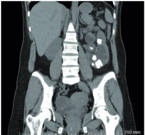
We aimed to present a rare case of super-numerary fused and malrotated kidney. Thirty--seven year-old woman was admitted to our clinic with left flank pain. CT scan demonstrated multi-ple calculi in the left kidney and supernumerary fused kidney (Figures 1 and 2). The third kidney was below the right kidney, malrotated and had its own blood supply (Figure-3).
Grieshammer and colleagues showed that mutant mice lacking either SLIT2 or its receptor ROBO2 develop supernumerary ureteric buds that are correlated with abnormal maintenance of Gdnf expression in anterior metanephric mesenchyme (4). The SLIT2/ROBO2 intercellular signaling sys-tem restricts, directly or indirectly, in extent of the Gdnf expression and plays a critical role in
Vol. 43 (3): 561-562, May - June, 2017
doi: 10.1590/S1677-5538.IBJU.2015.0420
Figure 1 - Computed tomography scan with multiple calculus in left kidney.
IBJU| RADIOLOGY PAGE
562
precisely positioning the side of kidney induction. The rare location and malrotation of supernume-rary kidney of our case could be explained by this hypothesis. Percutaneous nephrolithotomy was performed to the left kidney and no problem was observed in follow-up.
CONFLICT OF INTEREST
None declared.
ARTICLE INFO
Int Braz J Urol. 2017; 43: 561-2
_____________________ Submitted for publication: July 25, 2015
_____________________ Accepted after revision: March 25, 2016
_____________________ Published as Ahead of Print: November 02, 2016
REFERENCES
1. Sureka B, Mittal MK, Mittal A, Sinha M, Thukral BB. Supernumerary kidneys—a rare anatomic variant. Surg Radiol Anat. 2014;36:199-202.
2. Tada Y, Kokado Y, Hashinaka Y, Kadowaki T, Takasugi Y, Shin T, et al. Free supernumerary kidney: a case report and review. J Urol. 1981;126:231-2.
3. CARLSON HE. Supernumerary kidney: a summary of 51 reported cases. J Urol. 1950;64:224-9.
4. Grieshammer U, Le Ma, Plump AS, Wang F, Tessier-Lavigne M, Martin GR. SLIT2-mediated ROBO2 signaling restricts kidney induction to a single site. Dev Cell. 2004;6:709-17.
_______________________ Correspondence address: Volkan Sen, MD Department of Urology Dokuz Eylul University School of Medicine – Urology Izmir, 35340, Turkey Telephone: + 90 553 389-6859 E-mail: [email protected]