OR
IGI
N
A
L
R
E
S
E
A
R
C
H
Motor imagery in the treatment of acute lateral ankle
sprains in soccer athletes: a pilot study
Imagética motora no tratamento da entorse lateral de tornozelo em atletas de futebol de
campo: um estudo piloto
Imagética motora en el tratamiento de esguinces de tobillo lateral en los atletas de fútbol de
campo: un estudio piloto
Guilherme S. Nunes1, Marcos de Noronha1,2, Vanderlei A. de Carvalho Jr.1
1Physiotherapy Department, Health and Sport Science Center, Universidade do Estado de Santa Catarina – Florianópolis (SC), Brazil. 2Department of Community and Allied Health, La Trobe University – Bendigo (VIC), Australia.
282
ABSTRACT | Ankle sprain is a common injury in soccer athletes and has a high relapse rate. Motor imagery (MI) may be an alternative treatment to diminish the neuromuscular consequences after the injury. Thus, this study aimed to verify the preliminary results of the efectiveness of MI in the rehabilitation of soccer athletes with acute ankle sprain. Twenty young athletes of the male sex participated in the study. They were randomly divided into two groups: intervention (IG) and control (CG). Participants underwent conventional rehabilitation (cryotherapy, electrotherapy and kinesiotherapy) for ankle sprain, but only the IG performed imagery exercises to try to recognize the ankle-foot igures, projected by a computer, from various perspectives and angles. The ranges of motion (ROM) were measured for dorsilexion and plantar postural control, edema and functional stability. After treatment, no diference between groups were observed regarding dorsilexion ROM (p=0.23), plantar lexion ROM (p=0.50), Star Excursion Balance Test (SEBT) in the anterior direction (p=0.70), SEBT in the posterolateral direction (p=0.29), SEBT in the posteromedial direction (p=0.79), perimetry in “8” (p=0.50) and the Cumberland Ankle Instability Tool (CAIT) questionnaire for functional instability (p = 0.70). The MI was not an efective method for ankle sprains treatment in ield soccer athletes to improve ROM, dynamic balance, edema and functional stability. However, this is a pilot study and further investigations are required.
Keywords | Ankle Injuries/rehabilitation; Imagery (Psychotherapy); Functional Laterality; Postural Balance.
Mailing address: Guilherme S Nunes - Rua Pascoal Simone, 358, Coqueiros – CEP 88080-350 – Florianópolis, SC – Brazil. E-mail: [email protected] – Phone: +55 48 3321-8660
Presentation: July 2014 – Accepted for publication: Sept. 2015 – Financing sources: none– Conlicts of interest: Nothing to declare.
Human Research Ethics Committee, Universidade do Estado de Santa Catarina (UDESC), number 88/2011. Brazilian Registry of Clinical Trials number RBR-2tthrr.
Descritores | Traumatismos do Tornozelo/reabilitação; Imagens (Psicoterapia); Lateralidade Funcional; Equilíbrio Postural.
RESUMEN | El esguince de tobillo es una lesión común en los atletas de fútbol y presenta una alta tasa de recaídas. La imagética motora (IM) puede ser un tratamiento alternativo para disminuir las consecuencias neuromusculares presentadas post-lesión. Por lo tanto, el objetivo de este estudio fue veriicar los resultados preliminares de la efectividad de la IM en la rehabilitación de atletas de fútbol con esguince agudo de tobillo. Participaron 20 atletas jóvenes del sexo masculino, divididos de manera aleatoria en dos grupos: intervención (GI) y control (GC). Los participantes experimentaron un proceso de rehabilitación convencional (crioterapia, electroterapia y cinesioterapia) para esguince de tobillo, sin embargo sólo el GI realizaba ejercicio de imagética al tratar de reconocer las iguras del tobillo-pie, proyectados en una computadora, en
diferentes perspectivas y ángulos de orientación. Se midió las amplitudes de movimiento (ADM) de lexión dorsal y plantar, control postural, edema y estabilidad funcional. Después del tratamiento no se observó ninguna diferencia entre los grupos en relación a la ADM de lexión dorsal (p=0,23), ADM de lexión plantar (p=0,50), Star Excursion Balance Test (SEBT) en dirección anterior (p=0,70), SEBT en la dirección posterolateral (p=0,29), SEBT en la dirección posteromedial (p=0,79), perimetría en “8” (p=0,50) y cuestionario CAIT-P a la inestabilidad funcional (p=0,70). La IM no fue un método eicaz en el tratamiento de esguinces de tobillo en atletas de fútbol de campo para la mejora de ADM, equilibrio dinámico, edema y estabilidad funcional. Sin embargo este es un estudio piloto y mayores investigaciones son necesarias.
Palabras clave | Traumatismos del Tobillo/rehabilitación; Imágenes (Psicoterapia); Lateralidad Funcional; Balance Postural.
INTRODUCTION
he ankle is considered one of the most susceptible body parts to injury during sports activity. About 30% of sports injuries involving contact, jumping and running are ankle injuries, and ankle sprains account for 77% of ankle lesions1. Repetitive episodes of ankle sprains cause
chronic injury in the proprioceptive and sensory-motor function, by generating a deicit in the neuromuscular relex response time2. In 20% of the cases, repeated
ankle sprain events can result in instability of the ankle joints3,4. his can lead to disturbances and reduction
of nerve information concerning the ability to resist unexpected displacements and to maintain functional stability of the joint.
Studies have investigated more efective methods for treating ankle sprains. Among the possible treatments already investigated, muscle strengthening and proprioceptive exercises can be mentioned as efective techniques for the treatment of ankle sprains5,6. Recently,
motor imagery (MI) has been investigated as an alternative treatment for injuries related to peripheral joints7-9. It is
an exercise of mental origin that possibly stimulates the reorganization of lost motor function due to a deicit in the integration of motor processes that occurs in the cortex after the injury, seeking an unconscious action of the involved members10-12. he use of MI in the rehabilitation
of upper limbs motor function in individuals after a stroke
is supported by evidences13,14. hus, we expect MI to be
of some beneit in the rehabilitation of musculoskeletal injuries, such as in ankle sprains, since these lesions also relate to changes in motor control15.
Some studies have investigated the efects of MI in the treatment of musculoskeletal injuries. Stenekes et al.9, used
MI as a form of rehabilitation during the immobilization period for patients who underwent surgery to repair injury to the lexor tendon in the wrist. he MI positively inluenced the recovery time of the lexor tendon after surgery. Other studies have attempted to assess the efectiveness of MI in the treatment of ankle sprains7,8. MI, in these studies,
was used by asking participants to mentally review all physical therapy performed at the end of each session. Athletes from diferent types of sports were included and positive results in rehabilitation regarding increased muscular resistance were obtained7. However, the quality
soccer athletes with acute ankle sprains could contribute to a more efective rehabilitation.
METHODOLOGY
his was a randomized clinical study, controlled by the Brazilian registration of clinical trials number
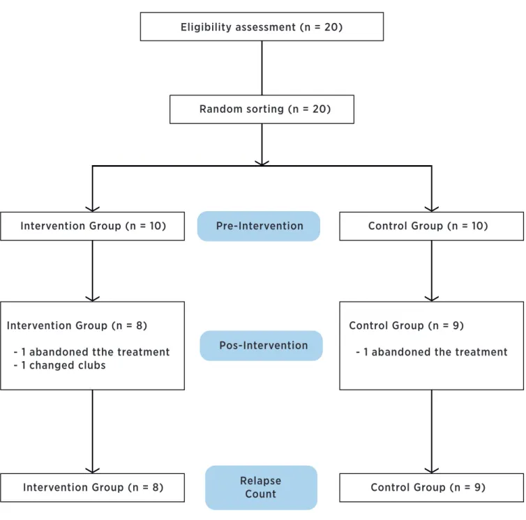
RBR-2tthrr. Participants were randomly allocated to the intervention group (IG) or control group (CG). Both groups received the standard treatment for rehabilitation of ankle sprains, but the IG also received MI stimulus. Randomization was done by a researcher not involved in the recruitment or evaluation of participants, using sealed and opaque envelopes (Figure 1).
Eligibility assessment (n = 20)
Random sorting (n = 20)
Intervention Group (n = 10) Pre-Intervention
Pos-Intervention
Relapse Count Intervention Group (n = 8)
Intervention Group (n = 8)
- 1 abandoned tthe treatment - 1 changed clubs
Control Group (n = 9)
- 1 abandoned the treatment
Control Group (n = 10)
Control Group (n = 9)
Participants
Twenty athletes, male, ield soccer players in the lower soccer clubs from Florianópolis participated in the study. Participants trained regularly at least ive days per week prior to injury. To be included in the study, participants had to be aged between 16-20 years, have sufered recent lateral ankle sprain that incapacitated their sport performance, and had their allocation carried out within 72 hours after the sprain. Ankle sprain was diagnosed when the participant had at least one of these symptoms: pain, swelling, bruising or inability to perform physical activities for more than a day16. Participants who had ankle sprain associated
with lower limb fracture, athletes undergoing invasive treatment or those who had their ankles immobilized after the sprain were excluded from the study. his research was approved by the Human Research Ethics Committee of the State University of Santa Catarina under the protocol number 88/2011. he consent was obtained from all participants or their legal guardians before procedures were performed.
Procedure
he evaluations and interventions were carried out within the premises of the club’s physiotherapy department. First, athletes with acute ankle injury were taken to the medical department of the club, where they were evaluated by a doctor specialized in sports medicine, responsible for diagnosing the degree of the ankle sprain17. Afterwards, the athletes were evaluated
by a physical therapist regarding the inclusion and exclusion criteria. he athletes were included in the study regardless of whether the afected ankle was or not in the dominant leg. Once they were included and consented to participate, participants were assessed by a blind investigator for the allocation of groups; and thus the treatment was initiated. After the treatment was completed and the patients were discharged from physical therapy, participants underwent the same battery of tests initially performed by the blind investigator during the allocation of the groups. After treatment, participants were observed for six months for any recurrence of ankle sprains. Monitoring of relapses was conducted via e-mail, every fortnight. When participants failed to respond to e-mails for one month, contact was made via telephone. Following this telephone contact, e-mail contact was resumed.
Evaluations
Range of movement in ankle plantar flexion
he participant was put into a supine position with the knee lexed and the sole of the foot in full contact with the stretcher. In this position, the participant was instructed to extend the knee and slide the foot on the rigid stretcher, keeping the heel, the 5th and 1st
metatarsal head in contact with the stretcher. With the help of a measuring tape, the vertical distance between the highest point of the knee and the stretcher was measured, as well as the horizontal distance from the heel to the point of projection measured between the knee and the stretcher (intersection point) forming a 90° angle. he maximum ROM for plantar ankle lexion was calculated by trigonometry, using the measured distances18. his measure had an intraclass correlation
coeicient (ICC) between 0.88-0.9218.
Range of movement in ankle dorsal flexion
he participant was placed standing in a squatting exercise position, facing a wall. In this position, he was instructed to squat, dorsal lexing the ankle to the maximum position of dorsilexion, without loss of contact between the heel and the ground, while being able to touch the knee to the wall. he subtalar joint position was controlled by maintaining alignment of the foot perpendicular to the wall with the aid of measuring tapes ixed to the ground and the wall. From this position, the horizontal distance between the posterior region of the heel wall, and the vertical distance between the knee point of contact with the wall and the loor were measured. he maximum ROM for ankle dorsilexion was calculated by trigonometry, with the use of the measured distances19,20. his measure
has an ICC between 0.97-0.9819.
Star Excursion Balance Test (SEBT)
Performed to assess dynamic balance21. he participant
performed the test standing in a single-leg stance, barefoot, on the center of three measuring tapes arranged on the loor. Measuring tapes stretched out from the center going three diferent directions: anterior (SEBT Ant), posterolateral (SEBT PL) and posteromedial (SEBT MP). Between the two posterior measuring tapes we had an angle of 90°, and between the posterior tapes and the anterior tape we had an angle of 135°22. he position of the
reach each of the lines as far as possible and the distance they were able to reach was recorded. he procedure was repeated three times for each direction. Before the test, participants were familiarized with it. Between familiarization and each repeat testing, an interval to rest was allowed until participants felt comfortable to repeat testing. For test analysis, the distance achieved was standardized by dividing the distance obtained by the length of the lower limb multiplied by 100. he length of the lower limb was measured using a measuring tape and the distance between the anterior superior iliac spine and the medial malleolus was measured22. his measure has an
ICC between 0.84-0.9223.
Ankle perimetry
he patient in a supine position and with the use of a measuring tape had the swelling in their ankle sized. Initially, the measuring tape was placed on the tendon of the anterior tibia, taken to the middle of the navicular tuberosity, then placed into the base of the 5th
metatarsal bone for crossing the sole of the foot and inally directed to the medial malleolus. After that, it was taken posteriorly, crossing the Achilles tendon, through the lateral malleolus to reach the starting point. hus, the tape was placed around the ankle in an “8” shape24. hree measurements were performed and the
average of these three measures was used for analysis. his measure has an ICC between 0.98-0.9924.
CAIT Questionnaire
Used to evaluate the functional instability showed by participants. CAIT questionnaire is a tool composed of 9 questions with a modiied Likert scale that generates a score between 0 and 30, with high reliability (ICC = 0.95) and discriminative validity25.
Intervention
After the initial assessment, the groups received the same physical therapy treatment, following the conventional physical therapy practices for the treatment of lateral ankle sprain5,6. All participants received the
same treatment protocol, adapted for each individual in terms of intensity: (1) cryotherapy: 20 minutes in the irst two sessions and repeated at the end of each session should inlammatory signs be exacerbated; (2) electrotherapy: TENS, ultrasound, or laser used in the irst few sessions according to the disability reported by patients; (3) kinesiotherapy, initiated according
to the exercise capacity of the patient, with intensity and volume being increased as the patient evolves: (a) stretching of the muscles in the lower limbs to maintain muscle length, (b) joint mobilization of the ankle, irstly passive, limited by the pain of the participant, and then evolving for active mobilization with circular movements of the ankle and squatting exercises, (c) sensorimotor training, starting with exercises of body support transfer, evolving into balance exercises, irst with bipedal support advancing to single leg support and balance with eyes closed, (d) strengthening of the ankle muscles performed with elastic bands at moderate intensities, (e) return to sport activities in the inal phase of rehabilitation. he sessions occurred ive times a week, lasting an average of two hours.
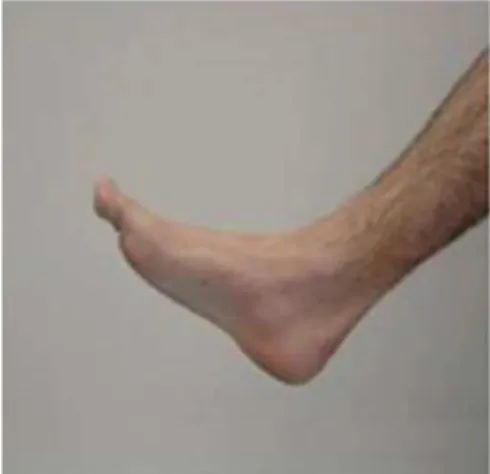
he MI group, at the end of each treatment, was taken to a room attached to the physiotherapy department where each participant sat in front of a computer, in a calm environment, without external disturbance or assistance from others. On the computer screen, 40 diferent ankle-foot images were randomly projected (20 left and 20 right), in diferent angles and with diferent ankle-foot positions (Figure 2). he participant was asked to press the right arrow or the left arrow of the computer keyboard when he was able to identify the image of the foot to be the right or left limb. he right arrow should be pressed when the participant identiies a right foot and the left arrow when he identiies a left foot. he maximum time that the image remained projected on the screen was 4 seconds, after which the image changed, should the participant fail to identify it. Before the MI exercise, participants received explanations on the use of the program, and were shown some images of the ankles, from the right and left foot. At the end of the 40 images, the software speciied the time used to identify each foot and the number of right guesses12.
Statistical Analysis
Descriptive statistics were used to calculate the averages, standard deviations and conidence intervals of the diferences in the data collected. Mann-Whitney test was used to compare the efect of MI on the measured variables among groups. Analyses were performed using SPSS software version 17.0 (SPSS Inc., Chicago, IL, USA). he size of the efect of the diferences between the groups was also measured and considered for the classiication of the efect size: 0.2= small; 0.5 = moderate; 0.8 = large26,27. he
intention-to-treat principle was used for analysis. Participants were asked to perform revaluation even without completing the treatment, though none accepted the ofer to return (Figure 1).
RESULTS
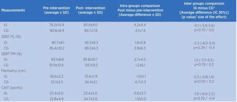
Of the 20 participants, two dropped out of treatment and one transferred clubs during the treatment (Figure 1). Characterization data can be found in Table 1. As for the pre-intervention measurements, the groups were similar and not signiicantly diferent. he groups were not signiicantly diferent after treatments regarding dorsilexion ROM, plantar lexion ROM, SEBT Ant, SEBT PL, SEBT PM, perimetry and CAIT (Table 2). he greatest efect was observed in ROM measurements (efect size =0.5 – Table 2). Of the participants who completed the study and the follow-up period of relapses, one IG participant had a recurrent ankle sprain.
Table 1. Participants’ characterization data
Measurements Intervention Group Control Group
Number of participants 10 10
Age (years) 17.2±1.6 17.4±1.8 Height (m) 1.76±0.09 1.73±0.10 Body weight (Kg) 68.6±10.5 67.7±8.3 History of sprain ankles (% n) 40 50
Degree of ankle sprain Degree I 9 7
Degree II 1 3
Degree III 0 0
Field position Goalkeeper 1 0
Central-back 3 1
Wingback 1 2
Midield 3 4
Striker 2 3
Number of visits 6.3±3.9 8.2±4.4
Data presented in averages ± Standard deviation (SD)
Table 2. Group average and the average diference intra and inter groups
Measurements Pre-intervention
(average ± SD)
Post- intervention (average ± SD)
Intra groups comparison Post minus pre-intervention
(Average diference ± SD)
Inter groups comparison IG minus CG* [Average diference (IC 95%)]
(p value/ size of the efect)
Dorsal lexion (degrees)
IG 53.1±4.5 54.3±2.9 1.2±2.8 2.2 (-1.8-6.2)
p=0.23 / 0.5
CG 54.3±5.0 53.3±4.1 -1.0±5.3
Plantar lexion (degrees)
IG 29.2±7.0 31.4±7.6 2.2±2.1 -1.8 (-1.7-5.3)
p=0.50 / -0.5
CG 27.7±4.2 31.7±5.1 4.0±4.9
SEBT Ant (%)
DISCUSSION
Preliminary results of this pilot study indicate that MI may have no efect on increasing ankle ROM, postural control, edema, and functional stability in ankle sprains treatments for athletes.
he focus of the ankle sprain post-rehabilitation should be to restore joint function and also prevent possible deicits caused by sprains, which may lead to relapse and chronical instabilities5,6. he main factors
of rehabilitation include the restoration of ROM, since dorsilexion ROM restriction is a major deicit after ankle sprains28, and permanent restrictions of this
ROM could be a leading factor for the occurrence of new sprains29. Another concern is the sensory motor
system integrity, which can be afected by disuse during rehabilitation or also by central changes2,3. hus, the
MI exercise could be an auxiliary tool in the recovery process after ankle sprains.
During the mental process of imagining the position of the ankle and foot during image identiication, constant joint mobilization can occur, thus promoting an improvement in ankle ROM. In addition, the movements caused by MI could promote an increase in muscle activation as shown in the study by Christakou et al.7, and thus assist in the restoration
of ROM. Preliminary results of this study do not support such an assumption. Christakou and Zervas’s8
study, like this study, treated athletes with ankle
sprains and observed no diferences between the MI treated and the control groups regarding ankle ROM. Perhaps the micro movements caused in the ankle by the imaginary process is not efective in adding or excelling the stimuli already conducted during conventional treatment, which includes stretching and invigoration of the muscles involved in movements and the dynamic of ankle stabilization. Another argument is that the mental process may have no efect on physical aspects such as ankle ROM. However, the moderate size efect values for ranges of movements may indicate that, with the continuity of the study, MI may end up having a signiicant inluence on ankle movements.
he same hypothesis of efectiveness could be considered for edema reduction. he micro movements could cause continuous muscular contractions, increased metabolism and copy metabolic exercises performed to reduce edema. he position at which the participants performed the MI exercise may have been a hindrance to the conduction of the interstitial luid by lymphatic vessels. As participants performed the exercise in a sitting position, gravity may have hindered the lymphatic return to thicker lymph vessels, lymph nodes and lymph ducts, for subsequent reabsorption of lymphatic luid. In the study of Christakou and Zervas8, which evaluated
ankle edema through volumetry, MI also showed no edema reduction.
Measurements Pre-intervention
(average ± SD)
Post- intervention (average ± SD)
Intra groups comparison Post minus pre-intervention
(Average diference ± SD)
Inter groups comparison IG minus CG* [Average diference (IC 95%)]
(p value/ size of the efect)
IG 79.2±10.9 83.4±9.0 4.2±4.4 -0.1 (-5.8-5.6)
p=0.70 / 0.0
CG 80.6±8.9 84.7±7.8 4.1±7.4
SEBT PL (%)
IG 90.7±8.1 92.3±8.3 1.6±5.8 -2.3 (-8.0-3.4)
p=0.29 / -0.4
CG 85.4±10.2 89.3±6.3 3.9±6.3
SEBT PM (%)
IG 93.1±8.8 95.8±10.7 2.7±4.5 1.5 (-3.5-6.5)
p=0.79 / 0.3
CG 91.9±10.6 93.1±9.3 1.2±6.1
Perimetry (cm)
IG 56.6±2.2 55.6±1.9 -1.0±1.1 0.3 (-0.8-1.4)
p=0.50 / 0.3
CG 55.1±2.5 54.4±2.1 -0.7±1.3
CAIT (points)
IG 23.4±5.0 23.4±5.0 0.0±3.7 -1.9 (-6.0-2.2)
p=0.70 / -0.4
CG 22.8±4.9 24.7±2.8 1.9±5.0
*Calculations using the diference post minus pre-intervention in each group. IG: intervention group; CG: control group; SEBT Ant: Star Excursion Balance Test Anterior; SEBT PS: Star Excursion Balance Test Posterolateral; SEBT PM: Star Excursion Balance Test Posteromedial; CAIT: Cumberland Ankle Instability Tool
he main purpose for using MI in rehabilitation processes is to stimulate the central nervous system to obtain a stimulus to the reorganization of lost motor function, either caused by central or peripherical injuries9-12. In this study, the task of identifying foot
and ankle laterality caused the participant to imagine the position of his own foot. his imagination process of the body itself could help in improving body image, facilitating the control of movements and disturbances around the ankle and improving postural control as a whole. However, MI showed no efect on balance in this study. he stimulation caused by MI may not have been suicient to cause an improvement in postural control, especially because IG participants required few physical therapy session before being discharged, an average of about six sessions (Table 1). A longer MI stimuli time may be needed to cause postural control improvements. However, the study by Christakou et al.7,
which included a larger number of MI interventions (12 sessions), also failed to observe any diference between the groups regarding postural control measured by the Biodex Stability System.
hus, it seems the addition of MI to the treatment does not add any beneit to the conventional treatment, regarding structural and functional aspects, as evaluated by the CAIT questionnaire. Perhaps the MI method used in this research is the cause of the lack of positive results. he participants had to identify static images of feet and ankles, mental accommodation may have occurred during treatment and its beneits remained limited. However, the lack of positive results in this study should be considered with caution since this is a pilot study and results are only preliminary.
Some points can be decisive in these preliminary results, such as the low number of sessions performed. his may be due to the participants being athletes and young people, who complete the rehabilitation process faster than most. Another point is the amount of degree I ankle sprains. his degree of damage cause less structural and functional alterations and, thus, the efects of MI could be limited in this degree of injury. Another item was the lack of control over the motor imagination condition of the patients prior to treatment. Participants may have started treatment in diferent conditions, as almost half of the participants had a history of ankle sprains and their bodily and mental outlook are diferent from those who sufered a irst sprain.
On the other hand, with continuing research and the collection of the sample size suggested by the preliminary results, more consistent data regarding the evidence of the use of MI in ankle sprains treatment may be provided. Continuing the research will also allow collection of more concrete data on the monitoring of recurrent ankle sprains after treatment. Other studies using MI for longer periods of time can verify their efect on proprioception, postural control, pain and strength of individuals post-rehabilitation for ankle sprains, or to verify its efects as a method of prevention.
CONCLUSION
From the results of this study, we can conclude that MI was not an efective method for treating ankle sprains in ield soccer athletes to improve ROM, dynamic balance, edema and functional stability. he experimental model proposed in this pilot study proved to be feasible and may bring concrete evidence regarding the use of MI as a therapeutic tool in the rehabilitation of athletes after ankle sprains.
ACKNOWLEDGEMENTS
he authors would like to thank the physical therapist Bruno Seara Polidoro and to Avaí Futebol Clube for the assistance in performing this study.
REFERENCES
1. Kemler E, van de Port I, Backx F, van Dijk CN. A systematic review on the treatment of acute ankle sprain: brace versus other functional treatment types. Sports Med. 2011;41(3):185-97.
2. Munn J, Sullivan SJ, Schneiders AG. Evidence of sensorimotor deicits in functional ankle instability: a systematic review with meta-analysis. J Sci Med Sport. 2010;13(1):2-12.
3. de Vries Jasper S, Krips Rover, Sierevelt Inger N, Blankevoort Leendert, van Dijk C N. Interventions for treating chronic ankle instability. Cochrane Database Syst Rev. 2014;12: CD004124.
4. Gehring D, Faschian K, Lauber B, Lohrer H, Nauck T, Gollhofer A. Mechanical instability destabilises the ankle joint directly in the ankle-sprain mechanism. Br J Sports Med. 2014;48(5):377-82.
ligament injuries: a systematic review. Arch Orthop Trauma Surg. 2013;133(8):1129-41.
6. Bleakley CM, O’Connor SR, Tully MA, Rocke LG, MacAuley DC, Bradbury I, et al. Efect of accelerated rehabilitation on function after ankle sprain: randomized controlled trial. BMJ. 2010;340(4):1964.
7. Christakou A, Zervas Y, Lavallee D. The adjunctive role of imagery on the functional rehabilitation of a grade II ankle sprain. Hum Movement Sci. 2007;26(1):141-54.
8. Christakou A, Zervas Y. The efectiveness of imagery on pain, edema, and range of motion in athletes with a grade II ankle sprain. Phys Ther Sport. 2007;8(3):130-40.
9. Stenekes MW, Geertzen JH, Nicolai J-PA, De Jong BM, Mulder T. Efects of motor imagery on hand function during immobilization after lexor tendon repair. Arch Phys Med Rehabil. 2009;90(4):553-9.
10. Tamir R, Dickstein R, Huberman M. Integration of motor imagery and physical practice in group treatment applied to subjects with Parkinson’s disease. Neurorehabil Neural Repair. 2007;21(1):68-75.
11. Hudson ML, McCormick K, Zalucki N, Moseley GL. Expectation of pain replicates the efect of pain in a hand laterality recognition task: bias in information processing toward the painful side? Eur J Pain. 2006;10(3):219-24.
12. Moseley GL, Barnett C. Motor imagery for peripheral injury. Arch Phys Med Rehabil. 2009;90(8):1443-4.
13. Hong IK, Choi JB, Lee JH. Cortical changes after mental imagery training combined with electromyography-triggered electrical stimulation in patients with chronic stroke. Stroke. 2012;43(9):2506-9.
14. Kho AY, Liu KPY, Chung RCK. Meta-analysis on the efect of mental imagery on motor recovery of the hemiplegic upper extremity function. Aust Occup Ther J. 2014;61(2):38-48. 15. Bastien M, Mofet H, Bouyer LJ, Perron M, Hébert LJ, Leblond
J. Alteration in global motor strategy following lateral ankle sprain. BMC Musculoskelet Disord. 2014;15:436.
16. De Noronha M, França LC, Haupenthal A, Nunes GS. Intrinsic predictive factors for ankle sprain in active university students: A prospective study. Scand J Med Sci Sports. 2013;23(5):541-7.
17. Malliaropoulos N, Papacostas E, Papalada A, Maffulli N. Acute lateral ankle sprains in track and field
athletes: an expanded classification. Foot Ankle Clin. 2006;11(3):497-507.
18. Nunes G, Bayer G, Costa L, de Noronha M. Intraobserver and interobserver reliability of a method to measure ankle plantar-lexion range of motion in the hook-lying position. (Technical Report). J Sport Rehabil. 2012;(4):1-4.
19. Bennell KL, Talbot RC, Wajswelner H, Techovanich W, Kelly DH, Hall AJ. Intra-rater and inter-rater reliability of a weight-bearing lunge measure of ankle dorsilexion. Aust J Physiother. 1998;44(3):175-80.
20. Chisholm MD, Birmingham TB, Brown J, MacDermid J, Chesworth BM. Reliability and validity of a weight-bearing measure of ankle dorsilexion range of motion. Physiother Can. 2012;64(4):347-55.
21. Olmsted-Kramer LC. Simplifying the star excursion balance test: analyses of subjects with and without chronic ankle instability. J Orthop Sports Phys Ther. 2006;36(3):131-7. 22. Gribble PA, Hertel J. Considerations for normalizing measures
of the star excursion balance test. Meas Phys Educ Exerc Sci. 2003;7(2):89-100.
23. Munro AG, Herrington LC. Between-session reliability of the star excursion balance test. Phys Ther Sport. 2010;11(4):128-32. 24. Petersen EJ, Irish SM, Lyons CL, Miklaski SF, Bryan JM,
Henderson NE, et al. Reliability of water volumetry and the igure of eight method on subjects with ankle joint swelling. J Orthop Sports Phys Ther. 1999;29(10):609-15.
25. De Noronha M, Refshauge KM, Kilbreath SL, Figueiredo VG. Cross-cultural adaptation of the Brazilian-Portuguese version of the Cumberland Ankle Instability Tool (CAIT). Disabil Rehabil. 2008;30(26):1959-65.
26. Becker L. Efect Size Calculators. http://www.uccs. edu/~lbecker. Acessado em 30 de julho de 2014.
27. Husted JA, Cook RJ, Farewell VT, Gladman DD. Methods for assessing responsiveness: a critical review and recommendations. J Clin Epidemiol. 2000;53(5):459-68. 28. Terada M, Pietrosimone BG, Gribble PA. Therapeutic
interventions for increasing ankle dorsilexion after ankle sprain: a systematic review. J Athl Train. 2013;48(5):696-709. 29. De Noronha M, Refshauge KM, Herbert RD, Kilbreath SL.