112
EVALUATION OF THE PASSIVE RESISTIVE TORQUE
IN FEMALE ATHLETES WITH ANKLE SPRAIN
Márcia Barbanera1
Rubens Correa Araujo2
Tulio Diniz Fernandes3
Arnaldo Jose Hernandez3
1. Laboratory of Biomechanics– São Judas Tadeu University –São Paulo – SP, Brazil
2. Physiotherapy Clinic – Taubaté University – SP, Brazil
3. Medicine School of the University of São Paulo – Institute of Orthopedics and Traumatology – Clinics Hospital (FMUSP IOT HC) – São Paulo – SP, Brazil.
Mailing address:
Rua Dias Leme, 134, ap. 23, Mooca 03118-040 – São Paulo – SP, Brasil E-mail: marciabarbanera@superig. com.br
ABSTRACT
Introduction: The ankle sprain is one of the most common injuries in athletes. Direct evaluation of the ligament laxity can be obtained through the objective measurement of extreme passive inversion and eversion movements, but there are few studies on the use of the evaluation of the passive re-sistive torque of the ankle to assess the capsule and ligaments resistance. Objective: The aim of this study was to compare the inversion and eversion passive torque in athletes with and without ankle sprains history. Method: 32 female basketball and volleyball athletes (16.06 ± 0.8 years old; 67.63 ± 8.17 kg; 177.8 ± 6.47 cm) participated in this study. Their ankles were divided into two groups: control group (29), composed of symptom-free ankles, and ankle sprain group, composed of ankles which have suffered injury (29). The resistive torque at maximum passive ankle movement was measured by the isokinetic dynamometer and the muscular activity by electromyography system. The athletes performed 2 repetitions of inversion and eversion movement at 5, 10 and 20°/s and the same protocol only at maximum inversion movement. Results: The resistive passive torque during the inversion and eversion was lower in the ankle sprain group. This group also showed lower torques at the maximum inversion movement. No differences were observed between inversion and eversion movement. Conclusions: Ankle sprain leads to lower passive torque, indicating reduction of the resistance of the lateral ankle ligaments and mechanical laxity.
Keywords: joint instability, sports, ligaments.
INTRODUCTION
Ankle sprains are one of the commonest injuries among athletes1-5, corresponding to 20% of all musculoskeletal
inju-ries6and over 30% of all sports5. The majority of ankle sprains
occurs in the inversion movement, especially with foot in plantar flexion, straining the anterior talo fibular ligament1,4,7,8.
The lateral ligamentous complex of the ankle is the most commonly injured site. It is composed of the anterior talo fibu-lar, calcaneo fibularand posterior talo fibular ligaments and the mostly injured ligament in the lateral ankle sprain is the anterior talofibular one1,7,8.
Although there is massive scientific research the recurrence of ankle sprains remains high2,4-6. The injury recurrence presents
records above 70% among athletes7,9,10.Ankle sprains can be
pre-vented; however, prevention of these injuries will only be possible if the risk factors were identified2,10,11. The possible risk factors
include: alterations in the foot position8,12, deficit in
propriocep-tion2,4,10,11,13,14, laxity in the lateral ligamentous complex4,15,16 and
muscular weakness4,9,13.
Better understanding on the sprain injury mechanism will pos-sibly help the health professional, including physiotherapists, to plan an objective and efficient treatment. Consequently, the patients will benefit from better planned rehabilitation.
After the sprain occurrence, the ankle ligaments and the articu-lar capsule may become lax and increase articuarticu-lar instability 7,12,13,15,
which favors the recurrence of sprains in inversion. The drawer examination and talar inclination techniques are used to identify ankle articular instability7,12,15. However, the use of these
techni-ques has been techni-questioned, since they involve manual tests and only provide subjective information on the ligamentous laxity. The stress radiographic exam in inversion has been traditionally used to quantify the ligament laxity through evaluation of the talus dislocation, but it is an indirect techinque7,12,15.
Objective evaluation of ligamentous laxity may be performed through the accurate measurement of the passive resistance of the capsule ligamentuss tructures generated by the passive movements of ankle maximal inversion and eversion16,17.
Un-fortunately, there are few studies on the evaluation of the ankle passive resistive torque. Birmingham et al.17evaluated the passive
torque in the ankle maximal inversion with an instrument which caused stress on the ankle lateral compartment and did not find differences in the passive torque of subjects with ankle unilateral sprain history, suggesting that these subjects did not present any ligamentous laxity.
The evaluation of the capsule and ligaments resistance may be obtained with the use of an isokinetic dynamometer, considering the ankle resistance measured by the passive torque16. Since ankle sprains
can be caused by capsule and ligaments laxity, these structures are able to support only light tension during the extreme movements of the tibiotalar joint7,12,18; therefore, the passive torque may be
decrea-sed at the end of the ankle inversion and eversion range of motion.
ORIGINAL ARTICLE LOCOMOTOR APPARATUS IN
113
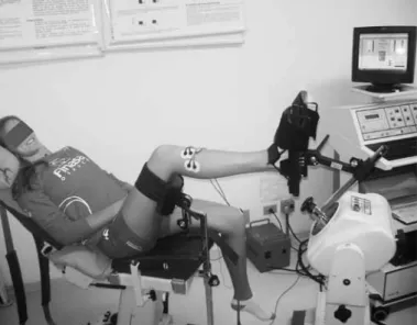
Figure 1. Positioning of the athlete in the isokinetic dynamometer for evaluation of the inversion and eversion passive movements with ankle at neutral position (at 90º in relation to the leg).
Thus, individuals without ankle sprain history will possibly present higher passive torque than individuals who have suffered sprains.
The aim of this study was to compare the inversion and eversion passive torque in athletes with and without ankle sprain history. We expect to find results which support the hy-pothesis that ankles which have suffered sprains will present lower passive torque.
MATERIAL AND METHODS
Subjects
Thirty-two female basketball and volleyball athletes (16.06 ± 0.8 years; 67.63 ± 8.17kg; 177.8 ± 6.47cm) participated in the study. According to their injury history, their ankles were divided in two groups: Control group (29), composed of ankles with no history of injury, and Sprain group, composed of ankles which have suffered sprains (29). The mean number of ankle sprains suffered by the Sprain group was 1.24 ± 0.57, with mean of 18.62 ± 12.2 months from the first sprain episode.
The athletes without history of fracture of the lower limbs and without retro malleolar pain were included in the study. The athletes who presented only episodes of instability, but without ankle sprain history, or those who underwent ankle surgery, were excluded from the study (4 ankles). The specific inclusion criterion for the Sprain group was previous history of ankle sprain for over six months from the date of collection, with or without instability sensation. Concern-ing the Control group, the athletes who did not present any history of injury in lower limbs were included.
The exclusion criterion adopted was pain during the dynamics tests performance, which corresponded to the exclusion of two ankles. All subjects signed a Free and Clarified Consent Form ap-proved by the Local Ethics Committee [Ethics Committee for Analy-sis of Research Projects-CAPPesq from the Clinical Board of Directors of the Clinics Hospital and Medicine School of the University of São Paulo on 08/02/06 (#1051-05)].
Instruments
The passive resistive torque was evaluated through na iso-kinetic dynamometer (Biodex® System 3, Biodex®Inc, USA). The
electromyographic activity (EMG) of the fibularis longus muscle (FL) and of the tibialis anterior muscle (TA) was also recorded to confirm that the movement was passive. The test was can-celled if increase of the EMG sign amplitude was observed. The electromyography instrument used was the Myosystem® 1400 (Noraxon® Inc, USA). The sample frequency was of 1kHz, and the signof both muscles was simultaneously collected. The skin was sanitized with alcohol (95%) to reduce its electrical resistance. The surface electrodes (disposable, Ag/AgCl, 4mm of height and 9mm of width) were placed on the muscle surface between the motor point and the distal extremity of the muscle, according to the SENIAM recommendations (Surface Electromyography for the Non-Invasive Assessment of Muscles)19,20. The tests were performed
after five minutes of warm-up walk on the treadmill21.
Protocol
The athletes remained with trunk inclination of 40° on the seat of the isokinetic dynamometer, hips and knees flexed at 90°, barefoot and rested on the specific support plaque of the instrument (figure 1) for tests performance. The athletes’ feet
were aligned with the mechanical axis of the instrument and the ankle was at neutral position (at 90º in relation to the leg).
The inversion and eversion passive movements were tes-ted. In this protocol, while the athletes remained relaxed, the instrument performed the foot inversion and eversion move-ment until reaching the physiological limit. The evaluation was performed in two different phases.
In phase 1, the protocol consisted in two repetitions of the inversion and eversion passive movements in three different ve-locities (5, 10 and 20°/s). The maximum range of motion (ROM) limit was adjusted according to the sensation reported by the athletes at the end of the ROM.
In phase 2, we adjusted the ROM without the athlete’s foot and only the maximal inversion movement was tested in order to reach the tissue limit. Subsequently, the protocol described in phase 1 was repeated.The athletes were told to press the instrument trigger when they felt any discomfort or strain on the ankle lateral region during the inversion passive arch. After trigger activation, the mo-vement was immediately interrupted and the torque offered by the passive resistance was recorded.
VARIABLES AND STATISTICS
The mean of the EMG sign amplitude and the mean of the torque peaks were calculated for analysis of the passive torque and EMG.
After the parametric statistics was verified, we tested the effect of the group (Control and Sprain), movement (inversion and ever-sion) and velocity (5, 10 and 20°/s) on the passive torque. Analysis of variance of two and three factors (ANOVA), followed by the Tukey HSD test was performed for analysis of the differences. The signifi-cance level adopted was of5% (α=0.05).
RESULTS
Phase1
Three-way ANOVA was used to verify the group, velocity and movement effect in the passive torque. Only the group factor
F(1.694) = 44.62; p < 0.0001) affected torque, while movement
114
Figure 2. Mean and standard deviation of the passive resistive torque of the Control and Sprain groups, obtained in the 5º/s (p = 0.001*), 10º/s (p = 0.002*) and 20º/s (p = 0.001*) velocities during the inversion and eversion movements (total number of measurements = 696).
* –significant difference ; ANOVA (F = 44.62; p = 0.0001).
Tor
que (N
m)
5º/s 10º/s 20º/s
10
8
6
4
2
0
Velocities
Control Sprain
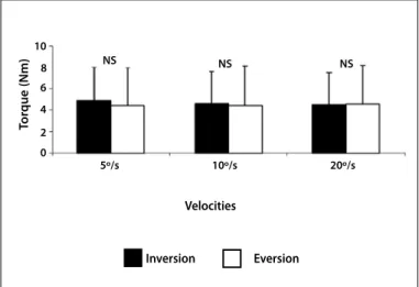
Figure 3. Mean and standard deviation of the passive resistive torque of the inversion and eversion movements, obtained in the 5º/s, 10º/s and 20º/s velocities (total number of measurements = 696).
NS – with no significant difference; ANOVA (F = 0.76; p = 0.38).
Tor
que (N
m)
5º/s 10º/s 20º/s
10
8 6
4
2 0
NS NS NS
Velocities
Inversion Eversion
Figura 4. Mean and standard deviation of the passive resistive torque of the Control and Sprain groups, obtained in the 5º/s (p = 0.01*), 10º/s (p = 0.02*) and 20º/s (p = 0.03*) ve-locities during the maximal inversion movement (total number of measurements = 348). * –significant difference ; ANOVA (F = 12.19; p = 0.0005).
Tor
que (N
m)
5º/s 10º/s 20º/s
20
16
12
8
4
0
Control Sprain Velocities
not. The Tukey HSD post hoc test evidenced that the passive torque was higher in the Control group (p < 0.0001).
The mean and standard deviation of the passive torque of the Control and Sprain groups in the different velocities are presented in figure 2.
Figure 3 presents the mean and standard deviation of the passive torque of the inversion and eversion movements in the different velocities.
Phase2
The two-way ANOVA test was used to verify the group and ve-locity effect in the passive torque. The group factor (F(1.346) = 12.19;
p = 0.0005) affected torque, which did not occur for velocity (F(2.345) = 0.2, p = 0.81). The Tukey HSD post hoct est evidenced that the passive torque was higher in the Control group (p < 0.0005).
The mean and standard deviation of the passive torque of the Control and Sprain groups in the different velocities are presented in figure 4.
DISCUSSION
The majority of studies on ankle injuries is more focused on chronic instability than sprains. The occurrence of sprain recur-rent episodes with recurrecur-rent instability after the first sprain epi-sode may be called chronic ankle instability (CAI)11,18.
Ankle functional and mechanical instability are potential causes of CAI. Ankle functional instability is described by the recurrence of sprains and instability sensation due to neuro-muscular and proprioceptive deficit9,11,13,18. Ankle
mechanica-linstability is characterized by pathological laxity after ligament injury, allowing mobility beyond the physiological limit3,18.
The criteria to differentiate mechanical and functional insta-bility are not well established22, which makes the classification
of the individuals who suffer repetitive sprains difficult and lets each author adopt his/her own classification. Ürgüden et al.23
classified history of traumatic sprain followed by recurrent ankle injuries (two or more times) including pain and edema. Vries et al.11 characterized CAI when the instability symptoms remained
for more than six months. Sefton et al.10 suggested a questionnaire
to evaluate ankle functional instability and defined as criterion: previous history of more than one sprain and recurrent symptoms. Fox et al.24 defined as unstable ankle when the individual suffered
from an ankle lateral sprain and presented at least one episode of instability over the last month.
Many studies have adopted the classification of functional instability when the individuals present history of sprain and instability sensation, perhaps for the difficulty in performing diagnostic tests which confirm the mechanical alterations25, but
mechanical instability is described by instability sensation in one mechanically stable ankle1,16. Injury by sprain in inversion
proba-bly alters the lateral capsule ligamentar structures and modifies mechanical stability of the ankle, making it lax7,12.
The absence of a gold-standard test to differentiate mecha-nical and functional instability through accurate methods makes the appropriate classification of this disorder difficult22.
115
(Sprain), probably for presenting tissue injuries7,12. The athletes who
presented only instability sensation, with no sprain history, were not included in the study since they did not present evidence of tissue injury.
We only assessed the athletes without acute symptoms and who presented their last sprain episode over six months from the date of collection. This criterion was adopted to avoid that the in-flammation could alter the results as well as to guarantee that the alterations found could be directly related to the sprain22.
The inversion and eversion passive movements were tested to evaluate the torque resulting in the end of the movement, and the term used to characterize this procedure was “passive torque”. In that case, torque refers to the passive resistance generated by the capsule ligamentar structures of the ankle.
The electromyographic evaluation indicated that the FL and TA muscles were at rest during the protocol performance. The absence of EMG activity supports our statement that the move-ments were passive and the torque created against the isokinetic dynamometer was offered by the capsule ligamentar tissues.
The passive torque was assessed in two phases. In phase 1, the inversion and eversion movements were adjusted according to the sensation reported by the athletes at the end of the ROM. In or derto confirm the archend, phase 2 was performed. It con-sisted of only the inversion movement, in which the adjustment was performed in the tissue limit to simulate a stress situation of the ankle lateral ligaments.In phase 2, the eversion movement was not considered, since the most frequent injury mechanism occurs in inversion.
In phase 1, the Sprain group presented lower torque peak at the end of the inversion and eversion passive movements (figure 2). This finding suggests that tissue injury caused by ligaments and capsule sprain or rupture has occurred after the trauma7,12.
Decrease of passive resistance of the ankle lateral compart-ment structures, verified by the inversion passive movecompart-ment, was expected due to the previous tissue injury in the Sprain group. However, decrease in passive resistance of the medial compartment was also found. Tourné et al.12 described that
besi-des being the main ankle medial stabilizer, the deltoid also helps in the lateral stability, working in the maintenance of the talus position. When a sprain of high magnitude occurs, consequently changing the talus position, sprain of the ankle lateral ligaments as well as of the deltoid ligament takes place.
Ruptured or lax ligaments and capsule may cause instability,
decreasing hence mechanical resistance16,18. Instability of the
subtalar joint suggests that the ligaments have been damaged after ankle lateral sprain. Such fact may occur due to application of early load and stress on these ligaments, compromising the tissue repair process and leaving them at a more elongated position7.
In phase 2, passive torque was lower in the Sprain group at the end of the inversion passive movement (figure 4). This result corroborates the findings in phase 1, which state that individu-als with sprain history present injury of the capsule ligamentar tissues.
Birmingham et al.17did not find differences in the passive
re-sistance peak in the individuals who suffered sprain and suggest that they do not present ligament laxity, contrary to our findings. The divergence between results may have occurred due to di-fferences in the protocol and instruments. Santos and Liu16 use
daniso kinetic dynamometer to detect passive torque and foun-ded crease in passive laxity of unstable ankles. They conclufoun-ded that decrease of passive torque may be interpreted as increase in articular instability.
A limitation o four study is that all tests were performed in la-boratory environment, which does not allow a reliable reproduc-tion of the musculoskeletal behavior during the sports activities as well as of daily life. Another limitation was the low number of studies about passive torque.
The isokinetic dynamometer was considered a reliable ins-trument in the evaluation of passive torque. However, in order to carry on with our studies, we intend to compare the passive torque data with X-rays of stress in unstable ankles. In case positive correlation is found between these techniques, the radiographic exams will be able to be avoided, since the tests on dynamometer are much safer and offer more protection to the patients. The athletes with ankle sprain history present passive lower torque than the Control group, suggesting capsule and ligaments laxity. Thus, further studies with the use of ankle bandage and or theses should be conducted in order to verify stabilization and decrease of risks of injury.
CONCLUSION
Based on these results, ankle sprain leads to decrease of passive torque, being a possible indication of mechanical instability.
All authors have declared there is not any potential conflict of interests concerning this article.
REFERÊNCIAS
1. Caputo AM, Lee JY, Spritzer CE, Easley ME, DeOrio JK, Nunley II JA, et al. The in vivo kinematics of the tibiotalar joint after lateral ankle instability. Am J Sports Med 2009;37:2241-8.
2. Clark RC, Saxion CE, Cameron KL, Gerber JP. Associations between three clinical assessment tools for postural stability. N Am J Sports Phys Ther 2010;5:122-30.
3. O’Driscoll J, Kerin F, Delahunt E. Effect of a 6-week dynamic neuromuscular training programme on ankle joint function: a case report. Sports Med Arthrosc Rehabil Ther Technol 2011;3:13. 4. Lin CWC, Hiller CE, Bie RA. Evidence-based treatment for ankle injuries: a clinical perspective. J Man
Manip Ther 2010;18:22-8.
5. Waterman BR, Owens BD, Davey S, Zacchilli MA, Belmont Jr PJ. The epidemiology of ankle sprains in the United States. J Bone Joint Surg Am 2010;92:2279-84.
6. Janssen KW, Mechelen W, Verhagen EA. Ankles back in randomized controlled trial (ABrCt): braces versus neuromuscular exercises for the secondary prevention of ankle sprains. BMC Musculoskelet Disord 2011;12:210.
7. Hubbard TJ, Hicks-Little CA. Ankle ligament healing after an acute ankle sprain: an evidence-based approach. J Athl Train 2008;43:523-9.
8. Bonnel F, Toullec E, Mabit C, Tourné Y. Chronic ankle instability: biomechanics and pathomechanics of ligaments injury and associated lesions. Orthop Traumatol Surg Res 2010;96:424-32.
116
10. Sefton JM, Hicks-Little CA, Hubbard TJ, Clemens MG, Yengo CM, Koceja DM, et al. Sensorimotor function as a predictor of chronic ankle instability. Clin Biomech 2009;24:451-8.
11. Vries JS, Kingma I, Blankevoort L, Dijk CN. Difference in balance measures between patients with chronic ankle instability and patients after an acute ankle inversion trauma. Knee Surg Sports Trau-matol Arthrosc 2010;18:601-6.
12. Tourné Y, Besse JL, Mabit C. Chronic ankle instability. Which tests to assess the lesions? Which thera-peutic options? Orthop Traumatol Surg Res 2010;96:433-46.
13. Docherty CL, Arnold BL. Force sense deficits in functionally unstable ankles. J Orthop Res 2008;26:1489-93.
14. Nakasa T, Fukuhara K, Adachi N, Ochi M. The deficit of joint position sense in the chronic unstable ankle as measured by inversion angle replication error. Arch Orthop Trauma Surg 2008;128:445-9.
15. Vries JS, Kerkhoffs GMMJ, Blankevoort L, Dijk CN. Clinical evaluation of a dynamic test for lateral ankle ligament laxity. Knee Surg Sports Traumatol Arthrosc 2010;18:628-33.
16. Santos MJ, Liu W. Possible factors related to functional ankle instability. J Orthop Sports Phys Ther 2008;38:150-7.
17. Birmingham TB, Chesworth BM, Hartsell HD, Stevenson AL, Lapenskie GL, Vandervoort AA. Peak passive resistive torque at maximum inversion range of motion in subjects with recurrent ankle inversion sprains. J Orthop Sports Phys Ther 1997;25:342-8.
18. O’Driscoll J, Delahunt E. Neuromuscular training to enhance sensorimotor and functional deficits in subjects with chronic ankle instability: a systematic review and best evidence synthesis. Sports Med Arthrosc Rehabil Ther Technol 2011;3:19.
19. Hermens HJ, Freriks B, Disselhorst-Klug C, Rau G. Development of recommendations for SENG sensors and sensor placement procedures. J Electromyogr Kinesiol 2000;10:361-74.
20. Merletti R, Botter A, Troiano A, Merlo E, Minetto MA. Technology and instrumentation for detection and conditioning of the surface electromyographic signal: state of the art. Clin Biomech 2009;24:122-34.
21. Kerrigan DC, Franz JR, Keenan GS, Dicharry J, Croce UD, Wilder RP. The effect of running shoes on lower extremity joint torques. PM R 2009;1:1058-63.
22. Docherty CL, Gansneder BM, Arnold BL, Hurwitz SR. Development and reliability of the ankle instability instrument. J Athl Train 2006;41:154-8.
23. Ürgüden M, Kizilay F, Sekban H, Samanci N, Özkaynak S, Özdemir H. Evaluation of the lateral instabil-ity of the ankle by inversion simulation device and assessment of the rehabilitation program. Acta Orthop Traumatol Turc 2010;44:365-77.
24. Fox J, Docherty CL, Schrader J, Applegate T. Eccentric plantar-flexor torque deficits in participants with functional ankle instability. J Athl Train 2008;43:51-4.