Sinus Histiocytosis with Massive Lymphadenopathy
(Rosai-Dorfman Disease): A Case Report and
Literature Review
Rabindra Bhakta Pradhananga
1Kripa Dangol
1Anjan Shrestha
2Dharma Kanta Baskota
11Department of ENT-Head and Neck Surgery, Tribhuvan University
Teaching Hospital, Kathmandu, Nepal
2Department of Pathology, Tribhuvan University Teaching Hospital,
Kathmandu, Nepal
Int Arch Otorhinolaryngol 2014;18:406–408.
Address for correspondence Rabindra Bhakta Pradhananga, MBBS, MS (ENT- Head & Neck Surgery), Department of ENT-Head & Neck Surgery, Tribhuvan University Teaching Hospital, Kathamanduþ977, Nepal (e-mail: [email protected]; [email protected]).
Introduction
Sinus histiocytosis with massive lymphadenopathy (SHML), which is also known as Rosai-Dorfman disease (RDD), is a rare histiocytic proliferative disorder of unknown etiology ini-tially described by Rosai and Dorfman in 1969.1 RDD can occur in any age group but is most commonly seen in children and young adults.2Usually it presents with massive painless lymph node enlargement at any site, with the cervical nodes being common. Histologically, lymph nodes show pericapsu-larfibrosis and dilated sinuses, heavily infiltrated with large histiocytes, lymphocytes, and plasma cells.1,3 Immunohisto-chemically, the sinus histiocytes are strongly positive for S-100 protein.4
Though various modes of treatment like chemotherapy, radiotherapy, and surgical debulking have been tried, all have shown limited efficacy. The course of the disease is somewhat variable, from complete spontaneous remission in some cases to protracted clinical disease for years in other cases. The possibility of involvement of vital organs leading to death is also mentioned in some cases.
Review of Literature with Differential
Diagnosis
RDD commonly presents with massive bilateral and painless cervical lymphadenopathy with fever, night sweating, and weight loss. Extranodal involvement has been documented in Keywords
►
sinus histiocytosis
►
massive
lymphadenopathy
►
Rosai-Dorfman
disease
Abstract
Introduction
Rosai-Dorfman disease (RDD) is a rare histiocytic proliferative disorder of
unknown etiology. Usually it presents with massive painless cervical lymph node
enlargement. Histologically, it shows proliferation of distinctive histiocytic cells that
demonstrate emperipolesis in the background of a mixed in
fl
ammatory in
fi
ltrates.
Immunohistochemically, the cells are positive for markers such as CD68 and S100.
Objective
To report a case of a 12-year-old patient with multiple sites of cervical
lymphadenitis, which was diagnosed as RDD histopathologically as well as
immunohistologically.
Resumed Report
A 12-year-old girl presented with multiple painless sites of cervical
lymphadenitis without any systemic and other ear, nose, and throat manifestations. The
biopsy report of the lymph node showed dilatation of the sinuses,
fi
lled with histiocytes
having foamy cytoplasm. Many of the histiocytes were engul
fi
ng mature lymphocytes.
The sinus histiocytes were strongly positive for S-100 protein.
Conclusion
RDD must be considered in the differential diagnosis of massive or
multiple lymphadenopathies.
received April 10, 2014 accepted June 29, 2014 published online August 25, 2014
DOI http://dx.doi.org/ 10.1055/s-0034-1387163. ISSN 1809-9777.
Copyright © 2014 by Thieme Publicações Ltda, Rio de Janeiro, Brazil
Case Report
25 to 40% of cases, and organs such as skin, respiratory tract, reticuloendothelial system, genitourinary tract, and thyroid are commonly involved.
Ju et al reported a case of RDD in a 26-year-old man with cervical and mediastinal lymphadenopathy with pleural ef-fusion in 2009.5Agarwal et al reviewed seven cases (five nodal and two extranodal) of RDD. Of these,five patients were followed.6However, four had stable disease, and one developed histiocytic sarcoma after a gap of 4 years. Zhu et al found more purely extranodal RDD involving the central nervous system on retrospectively analyzing 13 cases of RDD.7
Clinically, the nodal type of RDD should be differentiated from lymphoma and any other causes of chronic lymphade-nitis, such as tubercular lymphadelymphade-nitis, Kikuchi-Fujimoto disease, among others. Histologically, the disease must be differentiated from Langerhans cell histiocytosis and infec-tious and lymphoproliferative disorders as well as sinus hyperplasia. S-100 positivity can usually distinguish between the latter condition and RDD, whereas in both conditions the histiocytes have a strong macrophage antigen expression.4
Case Report
A 12-year-old girl visited the ENT & Head and Neck Surgery Out Patient Department with presentation of bilateral multi-ple painless swelling in neck of 2 months’duration. She also complained of mild to moderate fever and weakness. Her general physical condition looked normal.
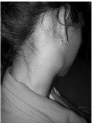
Examination of the neck revealed enlarged lymph node on both sides (►Figs. 1and2). Left side examination noted level
II (34 cm2), level III (21 cm2), and level V (33 cm2)
lymph nodes palpable with scar at a previous biopsy site (taken somewhere else). Similarly, the right side revealed level IB (33 cm2), level III (11 cm2), and level V (43 cm2) lymph nodes. All the lymph nodes were found to befirm, well defined, nontender, and mobile. Examination of ear, nose, oral cavity, oropharynx, and larynx revealed normal
findings.
Routine blood investigations revealed polymorphonuclear leukocytosis and increased erythrocyte sedimentation rate (55 mm/h). Fine Needle Aspiration Cytology (FNAC) was inconclusive. Ultrasonography (USG) of the neck showed multiple enlarged lymph nodes in both sides of the neck, suspicious of lymphoma. USG of the abdomen and pelvis showed normalfindings.
Because a previous biopsy taken elsewhere was inconclu-sive, a revision biopsy was taken and sent for histopathologic examination. Histopathology showed a gross appearance of well-encapsulated brownish tissue measuring 1.81.5 cm2, and cut sections showed homogenous white areas. Micro-scopic examination of the sections of lymph node showed dilatation of the sinuses,filled with histiocytes having foamy cytoplasm. Many of the histiocytes were seen engulfing mature lymphocytes (emperipolesis; ►Figs. 3 and 4). The
histopathologic diagnosis was RDD, which was further
con-firmed by immunohistochemistry. The histiocytic cells were S-100-positive.
The patient was started on oral prednisolone in tapering dose with continuation of low dose for 1 month. Her condi-tion was improved atfirst follow-up after 1 week, and she will be followed further.
Fig. 1 Multiple massive cervical lymphadenopathy on right side.
Fig. 2 Cervical lymphadenopathy with scar on left side of neck.
Discussion
RDD is a rare, idiopathic, histiocytic proliferative disorder. Consequently, many characteristics of this disease remain unknown, restricting early diagnosis and proper treatment. Various theories have been implicated as an etiology; how-ever, there is no strong evidence for any of them.
The presentation of the disease also varies with site of involvement. Commonly it presents with massive cervical lymphadenopathy. But extranodal involvement has also been documented in around 40% of cases, the most common being skin followed by upper respiratory tract and bone.8However, RDD also can occur in a variety of other sites, including the genitourinary system, lower respiratory tract, oral cavity, and soft tissues.9Other reported specific sites include brain, spine, liver, kidney, thyroid, breast, parotid gland, orbit, nasal cavity, and lungs.
The characteristic pathologic feature of this disease is proliferation of distinctive histiocytic cells that demonstrate emperipolesis (the engulfment of lymphocytes and erythro-cytes by histioerythro-cytes) in the background of a mixed infl amma-tory infiltrate, consisting of moderately abundant plasma cells and lymphocytes.3Immunohistochemically, SHML cells express phagocytic markers such as CD68 and S100, but not
the markers for Langerhans (CD1a) or dendritic cells (DRC, CD23, and CNA42).2
Pulsoni et al reviewed case reports of 80 patients who received various modes of treatments.9The study showed all of the treatment modalities including chemotherapy and radiotherapy were ineffective; however, surgical debulking when required was effective. Spontaneous resolution of adenopathies is frequently observed so that clinical observa-tion without treatment is advisable when possible. Systemic corticosteroids are usually helpful in decreasing nodal size and symptoms; however, they can be quite immunosuppres-sive and RDD lesions can recur after a short period of interruption.9Although Oka et al reported an effective pro-longed course of low-dose prednisolone in a case of RDD with skin and lymph node involvement with respiratory obstruc-tion, its role cannot be generalized.10
Final Comments
RDD must be considered in the differential diagnosis of young patients who exhibit massive or multiple lymphadenopa-thies, especially when involvement of the cervical area oc-curs. The disease can be diagnosed on the histopathologically and immunohistochemically.
References
1 Rosai J, Dorfman RF. Sinus histiocytosis with massive lymphade-nopathy. A newly recognized benign clinicopathological entity. Arch Pathol 1969;87(1):63–70
2 Bernácer-Borja M, Blanco-Rodríguez M, Sanchez-Granados JM, Benitez-Fuentes R, Cazorla-Jimenez A, Rivas-Manga C. Sinus his-tiocytosis with massive lymphadenopathy (Rosai-Dorfman dis-ease): clinico-pathological study of three cases. Eur J Pediatr 2006; 165(8):536–539
3 Juskevicius R, Finley JL. Rosai-Dorfman disease of the parotid gland: cytologic and histopathologicfindings with immunohisto-chemical correlation. Arch Pathol Lab Med 2001;125(10): 1348–1350
4 Eisen RN, Buckley PJ, Rosai J. Immunophenotypic characterization of sinus histiocytosis with massive lymphadenopathy (Rosai-Dorfman disease). Semin Diagn Pathol 1990;7(1):74–82
5 Ju J, Kwon YS, Jo KJ, et al. Sinus histiocytosis with massive lymphadenopathy: a case report with pleural effusion and cervical lymphadenopathy. J Korean Med Sci 2009;24(4):760–762 6 Agarwal A, Pathak S, Gujral S. Sinus histiocytosis with massive
lymphadenopathy—a review of seven cases. Indian J Pathol Micro-biol 2006;49(4):509–515
7 Zhu F, Zhang JT, Xing XW, et al. Rosai-Dorfman disease: a retro-spective analysis of 13 cases. Am J Med Sci 2013;345(3):200–210 8 Foucar E, Rosai J, Dorfman R. Sinus histiocytosis with massive lymphadenopathy (Rosai-Dorfman disease): review of the entity. Semin Diagn Pathol 1990;7(1):19–73
9 Pulsoni A, Anghel G, Falcucci P, et al. Treatment of sinus histiocytosis with massive lymphadenopathy (Rosai-Dorfman disease): report of a case and literature review. Am J Hematol 2002;69(1):67–71 10 Oka M, Kamo T, Goto N, et al. Successful treatment of
Rosai-Dorfman disease with low-dose oral corticosteroid. J Dermatol 2009;36(4):237–240
Fig. 3 Section with hematoxylin and eosin stain under 20100 magnification: dilated sinuses with histiocytes.
Fig. 4 Section with hematoxylin and eosin stain under 40100 magnification: characteristic emperipolesis.
International Archives of Otorhinolaryngology Vol. 18 No. 4/2014