Evaluation of agreement between tests
for the diagnosis of leprosy
Avaliação de concordância entre testes para diagnóstico da hanseníase
Alison R. Silva1; Marcos Fabiano A. Queiroz1; Edna A. Y. Ishikawa1; Maria do Perpétuo S. A. Silvestre2; Marilia B. Xavier1
1. Universidade Federal do Pará (UFPA), Pará, Brazil. 2. Instituto Evandro Chagas (IEC), Pará, Brazil.
First submission on 13/10/16; last submission on 27/01/17; accepted for publication on 03/03/17; published on 20/04/17
ABSTRACT
Introduction: Leprosy is a chronic infectious disease caused by the intracellular parasite Mycobacterium leprae. The diagnosis is
essentially clinical, based on symptoms, skin exam, peripheral nerves and epidemiological history. Laboratory tests are carried out to complement the result of clinical diagnosis, or even serving as a conirmatory method. Objective: To investigate the positivity and agreement between skin smear, enzyme-linked immunosorbent assay (ELISA) with synthetic antigen ND-O-BSA, ML Flow test and polymerase chain reaction (PCR) for detection of Mycobacterium leprae in new cases of leprosy. Methods: We conducted a case series study assessing a convenience sample of 39 new cases of leprosy and a control group of 18 household contacts in Belém (PA) and in Igarapé-Açu (PA) from March 2014 to September 2015. Results: The agreement between ELISA, ML Flow and PCR tests combinations showed slight to absent reproducibility (Kappa ≤ 0.24). The results showed greater sensitivity in PCR assay, with higher positivity in multibacillary cases. The ELISA test showed low positivity, even in multibacillary cases, resulting in no reaction to paucibacillary cases and household contacts. Conclusion: The high sensitivity of PCR decreases the agreement with other tests.
Key words: leprosy; enzyme-linked immunosorbent assay; polymerase chain reaction; serology.
INTRODUCTION
Leprosy is an infectious and chronic disease caused by the intracellular bacillus Mycobacterium leprae (M. leprae).
The disease affects the skin and the peripheral system nerves that is part of the human nervous system, causing skin lesions. The clinical manifestations of leprosy depend closely on the interaction between the bacillus and the host immune system: patches initially hypochromic with altered thermal or tactile sensitivity (indeterminate form) can evolve into a pre-inlammatory proile with good immune response and containment of exacerbated bacillary multiplication (tuberculoid form) or to an anti-inlammatory proile with immunodeicient response to contain bacterial multiplication (virchowian form). There are also interpolar forms with variable immunological response (dimorphic form)(1, 2).
In the 1980s, multidrug therapy (MDT) was established as a primary treatment of leprosy, and the prescription for
paucibacillary (PB) was deined for a period of six months, and for multibacillary (MB), for a period of 12 months(3).
In order to help in the clinical diagnosis of leprosy, several complementary exams may be ordered, the pain, tactile and thermal sensitivities tests of the injured skin are the most important ones(4). However, the results of the different diagnostic
tests may vary according to the clinical form of leprosy assessed in the wide range of responses that the patient may manifest.
Lymph smear is one of the most common tests ordered for conirmation of the new cases of leprosy, evaluating under the microscope the biological samples of the lobes of both ears, one of the elbows and a suspected lesion. The result is highly positive (large number of bacilli present) in samples of virchowian patients, variable in dimorphous, and negative in tuberculoids
and indetermined(5).
Tests based on the detection of the humoral immune response seek to identify speciic antibodies to M. leprae, and
J Bras Patol Med Lab, v. 53, n. 2, p. 100-107, April 2017
ORIGINAL ARTICLE
the phenolic glycolipid-I (PGL-I) is most commonly used in the immunoenzymatic assay format. The main serological tests are the enzyme-linked immunosorbent assay (ELISA) – a quantitative in house test – and the commercial ML Flow – a quick and qualitative point of care test. The production of antibodies is related to the number of bacilli circulating in the body, and the positivity of the serological tests follow the results of the skin smear(6, 7).
Molecular biology also allows the identiication of M. leprae,
in order to directly detect the genetic material of the bacillus. The polymerase chain reaction (PCR) is the most widespread method for this purpose, and may use biopsy specimens of skin, blood, urine, lymph, saliva and nasal secretion. However, the positivity in this test, depending on the material used, can only indicate the individual’s exposure to another truly bacilliferous patient, thus, therefore with no real dissemination of the infection(8-10).
The identiication of M. leprae is dificult because of the inability to culture the microorganism in vitro. Alternative
diagnostic methods could be performed in parallel, in order to complement the clinical outcome and provide tools to support epidemiological research. However, these tests may vary in their positivity depending on the clinical form and the particular immune status of the individual analyzed. Thus, the objective of the present study was to investigate the positivity and concordance of diagnostic tests, including ELISA, ML Flow, PCR and skin smear
test for M. leprae detection in new cases of leprosy.
METHODS
Study population and inclusion criteria
The present study evaluated a convenience sample composed of 39 new leprosy cases and a control group of 18 household contacts during the period from March 2014 to January 2015. Patients were attended in two municipalities endemic to the disease (Belém and Igarapé-açu), which belong to the State of Pará, Brazil. Clinical evaluation and collection of biological material were carried out at the Dermatology Outpatient Clinic of the Nucleus of Tropical Medicine of the Universidade Federal do Pará (UFPA), Belém, at the Basic Health Unit of Guamá (Belém), and at the Basic Health Unit of the Vila Santo Antônio do Prata (Igarapé-açu).
Collection of biological material was ordered to patients. In the category of leprosy cases are included individuals of any age, sex or ethnicity, presenting characteristic signs and symptoms for leprosy(4)
and those who, obligatorily, did not initiate MDT. Participants younger than 18 years of age must be accompanied by their
respective legal guardians, who should previously authorize their participation in the survey.
Data regarding the smear results and operational classiication were collected in medical records of the study sites, while epidemiological data were collected through interviews with the individuals participating in the study using a previously established standard questionnaire.
Collection of biological material
4 ml of venous blood were collected from a peripheral vein, using a vacuum collection system in an anticoagulant tube. After centrifugation, the serum was stored in polypropylene microtubes and conditioned at -20ºC until the immunoenzymatic tests were performed.
Prior to the nasal secretion collection, a few drops of sterile saline solution (9%) were applied to each patient’s nostrils in order to moisten the local tissues and facilitate the collection procedure. After approximately 3 minutes, a sterile swab was gently rotated in the anterior segment of the patients’ nasal cavity. This swab was washed in a test tube containing 3 ml of sterile saline solution (9%) and then this suspension was maintained at -20ºC until the time of analysis. The procedure was performed in duplicate and the materials of the same patient were mixed is such a way to form only one sample.
Anti-PGL-I ELISA
The in-house ELISA protocol followed speciications according to the methodology previously described(6), using the synthetic
was added to each plate. For control, a positive reference serum was included in triplicate on each plate. The color reactions on each plate were stopped with 50 µl 2NH2SO4 when the optical density
(OD) reached value 0.6. The optical density was measured in a spectrophotometer using a 492 nm ilter.
All serum samples were tested in duplicate and the ELISA results were expressed as the mean of the absorbance of duplicates. The OD inal value of each serum sample was calculated by subtracting the value of the wells coated only with BSA from the OD value of the wells coated with ND-O-BSA. The cut-off point for positive samples was 0.2 OD.
Extraction of deoxyribonucleic acid (DNA) and PCR
After collection of nasal secretion, the material was centrifuged for further DNA extraction by the Wizard Genomic DNA Puriication Kit® (PROMEGA), according to the protocol
provided by the manufacturer. For PCR, we used groups of primers LP1 –TGCATGTCATGGCCTTGAGG – and LP2 – CACCGATACCAGCGGCAGAA – (RLEP gene of M. leprae, 129 base pairs)(10) and, in parallel, R5 – CACGCTTCCTGTGCTTTGC – and
R6 – TGCGCTAGAAGCTTGCCGTA (M. leprae RLEP, 447 base pairs)(11). Each ampliication reaction consisted of 10 µl inal
volume, containing 0.2 µl of Taq DNA polymerase (Invitrogen), 1.6 µl of deoxyribonucleotide triphosphate (dNTP), 1 µl of 10× buffer, 0.6 µl MgCl2, 0.3 µl of each primer (direct and reverse) and 2 µl of DNA extracted from nasal secretions.
As the positive control for PCR, a skin lesion biopsy sample was extracted from a patient positively diagnosed as MB leprosy, the DNA was isolated as described above and stored for subsequent analyzes. As the negative control, a sample containing only the PCR reagents was used, without addition of DNA.
The PCR reactions were performed on thermocyclers
(GeneMate®) according to the conditions presented below for each
primer pair: Primers Lp1 and Lp2: 95ºC for 5 minutes, 58ºC for 2 minutes and 72ºC for 2 minutes, followed by 35 cycles at 94ºC for 30 seconds, 63.5ºC for 30 seconds, 72ºC for 1 minute and 72ºC for 10 minutes. Primers R5 and R6: 95ºC for 3 minutes, followed by 32 cycles at 94ºC for 1 minute, 53ºC for 1 minute, 72ºC for 1 minute, 55ºC for 1 minute and 72ºC for 1 minute.
The PCR products were fractionated by horizontal electrophoresis on 1% agarose gel, immersed in 1× TAE buffer. The DNA fragments were stained using ethidium bromide (0.5 µg/ml). The result was examined in ultraviolet transilluminator (L.pix Molecular Imaging, Locus Biotecnologia). Samples with negative result were retested for conirmation.
ML FLOW test
It was performed according to the manufacturer’s instructions, using 5 µl of serum for 10 minutes. A sample was considered positive when it was possible to visualize in red both bands test and control zones. A sample was considered negative when only the control zone was visible. Negative samples were retested for conirmation.
Statistical analysis
To evaluate the values of sensitivity, speciicity, accuracy, positive predictive values (PPV) and negative predictive values (NPV) and other pointers of screening diagnostic, individual screening tests were performed in ELISA, PCR and ML Flow tests in relation to the operational classiication. Due to the fact that the PCR test uses two different primer pairs, screening tests were applied regardless of the results of LP1/LP2 and R5/R6.
Statistical analysis of agreement between ELISA, PCR and ML Flow tests were performed using the Kappa agreement test. The PCR assay was considered the most sensitive, and was assigned to the agreement analysis with the other diagnostic tests. The Landis & Koch scale(12) was used to measure the degree
of agreement according to the Kappa value, with the scores divided into: < 0 no agreement; 0.0-0.20 slight; 0.21-0.40 fair; 0.41-0.60 moderate; 0.61-0.80 substantial; 0.81-0.99 almost perfect; 1 perfect. Agreement analyzes were performed between the tests using: 1) total sample of cases (PB and MB); 2) sample of PB cases; 3) sample of MB cases; 4) total sample of household contacts; 5) sample of household contacts of PB; and 6) sample of household contacts of MB. Differences between groups were assessed by chi-square test or G-test, where appropriate. Values
of p ≤ 0.05 were considered statistically signiicant. All statistical
inference was performed on Bioestat 5.0 software(13).
Ethical aspects
The research was submitted to the Human Research Ethics Committee of the Universidade do Estado do Pará (UEPA), according to resolution no. 196/96 of the National Health Council, and was approved according to opinion number 544.914. All participants signed a free and informed consent form, explaining in brief and succinct the objectives of the research project, the progress of their treatment was ensured regardless of their acceptance in participating in the Project, as well as compensation for any damages caused by misuse of the equipment used during
the survey.
RESULTS
Characterization of the sample of leprosy cases
and household contacts
Thirty-nine leprosy patients were evaluated, which, according to the operational classiication, were 58.97% MB (23/39). The majority of cases were male (19/39, 51.28%) and the mean age of the new cases was 40 years. The sample of household contacts was composed of 18 individuals, almost totally female participants (15/18, 83.3%) with a mean age of 21.3 years of age (Table 1).
According to the Madrid Classiication, the dimorphic form was identiied in 48.71% (19/39) of the cases (Table 2). The 18 contacts evaluated in this study were related to seven new cases, because those involved belong to the same family and share the
same home.
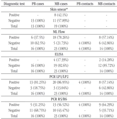
Positivie skin smear, ELISA, ML FLOW and PCR test
The skin smear showed positivity in eight cases (excluding seven patients who did not take the test). The serology for the ML Flow test showed positivity for 61.53% of the new cases (24/39). The ELISA results showed seropositivity only in four samples, both from MB patients. The best PCR results were found in the assay using LP1 and LP2 primers, with 84.61% positivity (33/39). Among the MB patients, 86.95% of the individuals (20/23) presented positivity for PCR using LP1/LP2 primers, while 56.52% (18/23), positivity using R5/R6 primers. Positivity among PB cases was higher when the LP1 and LP2 primers were used. In the household contacts group, the highest positivity was related to the R5/R6 assay, with positivity in 72.2% (13/18)
(Table 3).
When the screening test was used in the cases sample, the ML Flow test showed sensitivity of 80%, with an accuracy of 66.67%. The screening test was applied to the ELISA samples using the total case sample and only to the multibacillary cases, and the sensitivity values were, respectively, 10.26% and 17.39%. The screening test showed sensitivity of 82.35% and accuracy of 55.74% when PCR results of LP1 and LP2 primers were evaluated. The results of the
TABLE 1 − Demographic characteristics in a sample from leprosy patients
and their household contacts, Pará, Brazil, 2014-2015 Demographic
characteristics
Cases of leprosy n (%)
Household contacts
n (%) p value Gender
Male 20 (51.28) 3 (16.7) Chi-square Female 19 (48.71) 15 (83.3) p = 0.0288
Total 39 (100) 18 (100) Age (years)
< 15 - 9 (50) G-test
15-44 20 (51.28) 8 (44.4) p < 0.0001 45-65 13 (33.33) 1 (5.6)
> 65 3 (15.38) -
Total 39 (100) 18 (100) Mean age ± standard
deviation 41.6 ± 18.9 21.3 ± 13.5
TABLE 2 − Clinical characteristics in a sample from leprosy patients and
house-hold contacts distribution by case, Pará, Brazil, 2014-2015
Classiication n (%) Number of cases with
contacts examined
Number of contacts examined
Operational classiication
Paucibacillary 16 (41.02) 3 4 Multibacillary 23 (58.97) 4 14
Total 39 (100) 7 18
Madrid classiication
Indeterminate 8 (20.51) 2 2 Tuberculoid 8 (20.51) 2 2
Dimorph 19 (48.71) 2 12
Virchowian 4 (10.25) 1 2
Total 39 (100) 7 18
TABLE 3 − Skin smear, ML Flow, ELISA and PCR (LP1/LP2 and R5/R6 primers)
results in sample from leprosy patients and household contacts, Pará, Brazil, 2014-2015
Diagnostic test PB cases MB cases PB contacts MB contacts
Skin smear*
Positive - 8 (42.1%) -
-Negative 13 (100%) 11 (57.89%) - -Total 13 (100%) 19 (100%) -
-ML Flow
Positive 6 (37.5%) 18 (78.26%) - 8 (57.14%)
Negative 10 (62.5%) 5 (21.73%) 4 (100%) 6 (42.86%) Total 16 (100%) 23 (100%) 4 (100%) 14 (100%)
ELISA
Positive - 4 (17.39%) - 2 (14.28%)
Negative 16 (100%) 19 (82.6%) - 12 (85.72%) Total 16 (100%) 23 (100%) - 14 (100%)
PCR LP1/LP2
Positive 13 (81.25%) 20 (86.95%) 4 (100%) 8 (57.14%)
Negative 3 (18.75%) 3 (13.04%) - 6 (42.86%) Total 16 (100%) 23 (100%) 4 (100%) 14 (100%)
PCR R5/R6
Positive 5 (31.25%) 13 (56.52%) 4 (100%) 9 (64.29%)
Negative 11 (68.75%) 10 (43.47%) - 5 (35.71%) Total 16 (100%) 23 (100%) 4 (100%) 14 (100%)
same test, when applied to the results of the tests using R5 and R6 primers, showed lower sensitivity and accuracy (46.15% and 48.48%, respectively). For this reason, the LP1/LP2 test results were used in the agreement evaluations with the other diagnostic
tests (Table 4).
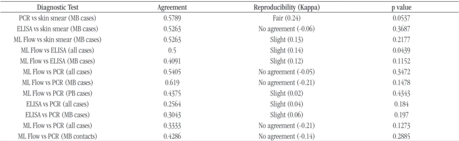
Agreement between skin smear, ELISA, ML FLOW
and PCR tests
Agreement evaluations were performed between ELISA, ML Flow, PCR and skin smear tests (patients whose did not performed the test were excluded from the analysis). The application of the statistical test was not possible in the evaluations using skin smear and ELISA in PB patients due to the lack of positivity of these tests. In MB cases, the Kappa agreement observed in relation to the skin smear presented a variation of fair (in the examinations evaluating the PCR) and absent (in tests evaluating the ELISA). In concordance evaluations between ML Flow and PCR tests,
variations between slight and no agreement were observed. For the agreement analysis between ELISA and PCR tests, the Kappa values obtained for all evaluations were below 1, resulting in reproducibility considered as slight (Table 5).
DISCUSSION
Serologic methods such as ELISA and the ML Flow rapid test are characterized by a slightly higher sensitivity when compared to the usual methods, such as the skin smear test(14). Investigating
circulating antibodies in the bloodstream, serology avoids the usual discomfort to the patient provided by lymph collections or skin biopsies. Because ELISA is a laborious technique, the use of ML Flow would be the most appropriate, given its low cost and time to read and interpret results(15).
Anti-PGL-I IgM antibody titers signiicantly decline between four and eight weeks of multidrug therapy, achieving
TABLE 5 − Agreement between skin smear, ML Flow, ELISA and PCR in sample from leprosy patients and household contacts, Pará, Brazil, 2014-2015
Diagnostic Test Agreement Reproducibility (Kappa) p value
PCR vs skin smear (MB cases) 0.5789 Fair (0.24) 0.0537 ELISA vs skin smear (MB cases) 0.5263 No agreement (-0.06) 0.3687 ML Flow vs skin smear (MB cases) 0.5263 Slight (0.13) 0.2177 ML Flow vs ELISA (all cases) 0.5 Slight (0.14) 0.0439 ML Flow vs ELISA (MB cases) 0.4091 Slight (0.12) 0.1152 ML Flow vs PCR (all cases) 0.5405 No agreement (-0.05) 0.3472 ML Flow vs PCR (MB cases) 0.619 No agreement (-0.21) 0.1478 ML Flow vs PCR (PB cases) 0.4375 Slight (0.02) 0.4343
ELISA vs PCR (all cases) 0.2564 Slight (0.04) 0.184
ELISA vs PCR (MB cases) 0.3043 Slight (0.06) 0.197
ML Flow vs PCR (all cases) 0.3333 No agreement (-0.21) 0.1273 ML Flow vs PCR (MB contacts) 0.4286 No agreement (-0.14) 0.2885
ELISA: enzyme-linked immunosorbent assay; PCR: polymerase chain reaction; MB: multibacillary form; PB: paucibacillary form.
TABLE 4 − Screening test results for ML Flow, ELISA and PCR (LP1/LP2 and R5/R6 primers) in sample from leprosy patients, Pará, Brazil, 2014-2015
Screening test Skin smear ML Flow ELISA (all samples) ELISA (MB samples) PCR LP1/LP2 PCR R5/R6
Sensitivity 42.11% 80% 10.26% 17.39% 82.35% 46.15%
Speciicity 100% 51.85% 96.3% 96.3% 22.22% 51.85%
Type 1 error
(false positive) 0% 48.15% 3.7% 3.7% 77.78% 48.15%
Type 2 error
(false negative) 59.89% 20% 89.74% 82.61% 17.65% 53.85%
Prevalence 30% 52.63% 59.09% 46% 51.74% 59.09%
PPV 100% 64.86% 80% 80% 57.14% 58.06%
NPV 71.05% 70% 42.62% 57.78% 50% 40%
Accuracy 76.09% 66.67% 45.45% 60% 55.74% 48.48%
Likelihood ratio positive 113684204.2 1.63 2.77 4.7 1.06 0.96
Likelihood ratio negative 0.58 0.36 0.93 0.86 0.79 1.04
ELISA: enzyme-linked immunosorbent assay; PCR: polymerase chain reaction; MB: multibacillary form; PPV: positive predictive value; NPV: negative predictive value.
immunological stability after 16 weeks(16, 17), demonstrating that
screening for circulating anti-PGL-I should be performed prior to, during, and after patient’s treatment.
Although PCR has good sensitivity when compared to the other tests, its major disadvantage is based on the impossibility
of evaluating the viability of M. leprae. Although there are
tests subsequent to PCR to determine if the bacillus is alive and active(18, 19), the costs for performing these procedures are
inappropriate for the implementation in the public health network, in addition to the already high costs for the performance of molecular biology techniques. However, the direct identiication of the microorganism in situations in which the other tests tend to be negative (regarding indeterminate and tuberculoid forms, for example, or in pure neuritic leprosy, in which there is no apparent skin lesion), it is extremely valuable for diagnostic conirmation.
Both PCR assays proved to be satisfactory for the detection
of M. leprae DNA. The number of positive results and sensitivity values when using LP1 and LP2 primers was slightly higher when compared with R5 and R6. The difference in the number of base pairs ampliied by each primer pairs may be correlated with the difference in results: LP1 and LP2 primers stand out by amplifying 129 base pairs. This is a small ampliiable DNA fragment, meaning that even if the collected genetic material is fragmented or scarce, the probability of detection of the amplicon in question is greater when compared to assays employing fragments with larger base pairs(10).
Studies evaluating the positivity of PCR in material collected from patients undergoing multidrug therapy demonstrate low sensitivity when compared to the material of treatment-free patients, possibly due to the degradation of M. leprae caused by the use of medication(20, 21). Thus, the quality of the collected material
would be impaired and the number of bacilli collected strongly reduced when compared to a treatment-free patient.
The World Health Organization (WHO) classiies any case of positive skin smear, regardless of the value of the bacilloscopic index or the number of lesions, as MB case(4). Therefore, the
positivity of the skin smear in PB will always be null. Likewise, as previously mentioned, the serology in this group tends to be negative because of the very low number of bacilli in the host.
In MB cases, the agreement evaluations among the skin smear of PCR, ML Flow and ELISA showed fair, slight and absent Kappa values, respectively. It is believed that the slight agreement are related to several factors, such as the high sensitivity value of PCR, the low sensitivity value of ELISA and the number of patients who did not perform the skin smear test, as well as the diversity of clinical forms found in the sample evaluated. In the analysis between the agreement of the ML Flow test and the histopathological test, higher and lower agreement can be found
in virchowian and indeterminate patients, respectively, as well as greater diagnostic discrepancy between the dimorphic forms(22).
Although ELISA and ML Flow tests are based on the identiication of anti-PGL-I, the titration of antibodies required for quantiication by ELISA, demonstrating its positivity, is much higher when compared to ML Flow, which is only qualitative. Thus, low antibody loads may be detectable by the rapid test, resulting in discrepancy of data when evaluating PB cases. A study analyzing the positivity of ELISA and ML Flow tests in a sample of 154 patients and household contact found (excluding patients with indeterminate form) a greater number of positive results for the rapid test in cases with substantial agreement between the two tests and higher antibody titers in communicants of MB patients(23). Another study evaluating the performance of the two
tests in endemic and non-endemic areas for leprosy indicated 70% positivity for the ML Flow test against 53.3% of the ELISA test, with a cut-off point lower than the one used in this study (positivity equal or higher than 0.157)(24).
The positive evaluations between the ML Flow rapid test and the PCR showed reproducibility considered to be weak (slight to absent), with a Kappa value below 1 in all combinations performed. These results are expected due to the signiicant difference found between the evaluations of the two tests, of which PCR showed greater sensitivity and marked positivity in both PB and MB cases. The dificulties of agreement analysis between ELISA and PCR are similar to those found between ML Flow and PCR. The high sensitivity of the molecular biology assay decreases the agreement observed between the two tests.
The choice of the diagnostic test in suspected cases of leprosy should be careful, considering the diversity of responses expressed by the different clinical forms. A combined evaluation of various clinical and laboratory methods of diagnosis should be optimal, although the lack of public health care in many regions affected by the disease would prevent satisfactory patient evaluation. The choice of test should consider the suspected clinical form, the cost of the well-aimed examination, and the time and reliability of the results. Technological innovations should be encouraged in order to continue the development of high sensitivity, providing early diagnosis and effectively disrupting the chain of transmission of the disease.
CONCLUSION
The use of primers capable of amplifying small M. leprae
of the disease, and its correlation with clinical examination and
other tests is essential.
ACKNOWLEDGEMENT
To the researchers and employees of the Laboratory of Molecular Biology and the Ambulatory of the Nucleus of Tropical Medicine of the UFPA, Leprosy Laboratory [Instituto Evandro Chagas (IEC)] and Health Units of Guamá and Vila Santo Antônio do Prata for the support during the research development.
CONFLICT OF INTERESTS
All authors declare no conlict of interest.
FINANCIAL SUPPORT
This study was supported by the Research Program for the Brazilian Uniied Health System [(Programa Pesquisa para o Sistema Único de Saúde (PPSUS)], Brazil, 2014.
RESUMO
Introdução: A hanseníase é uma doença infecciosa crônica causada pelo parasita intracelular Mycobacterium leprae. O diagnóstico é essencialmente clínico, com base em sintomas, exame da pele, nervos periféricos e história epidemiológica. Testes laboratoriais são realizados para complementar o resultado de diagnóstico clínico, ou mesmo servindo como método de conirmação. Objetivo: Investigar a positividade e a concordância da baciloscopia, do ensaio de imunoadsorvente ligado à enzima (ELISA)
com o antígeno sintético ND-O-BSA, do ML Flow e da reação em cadeia da polimerase (PCR) para a detecção de Mycobacterium
leprae em casos novos de hanseníase. Métodos: Foi realizada uma série de casos, avaliando uma amostra de conveniência de 39 novos casos de hanseníase e um grupo-controle de 18 contatos domiciliares em Belém (PA) e Igarapé-Açu (PA) a partir de março
2014 a setembro de 2015. Resultados: A concordância entre as combinações ELISA, ML Flow e PCR mostrou reprodutibilidade
leve a ausente (Kappa ≤ 0,24). Os resultados mostraram maior sensibilidade no ensaio de PCR, com maior positividade em casos multibacilares. O teste ELISA mostrou baixa positividade, mesmo em casos multibacilares, resultando em nenhuma reação nos
casos paucibacilares e contatos domiciliares. Conclusão: A alta sensibilidade da PCR diminui a concordância com outros testes.
Unitermos: hanseníase; ensaio de imunoadsorção enzimática; reação em cadeia da polimerase; sorologia.
REFERENCES
1. Ridley DS, Jopling WH. Classiication of leprosy according to immunity – a ive group system. Int J Lepr Other Mycobact Dis. 1966; 34(3): 225-73. 2. Congresso Internacional de Leprologia 6. Madrid, 1953. Memória. Madrid: Association de La Lepra; 1953.
3. Yawalkar SJ, McDougall AC, Lanquillon J, et al. Once-montly rifampicin plus daily dapsone in initial treatment of lepromatous leprosy. Lancet. 1982; 8283(1): 1192-1202.
4. Brasil. Ministério da Saúde. Guia para o controle da hanseníase. Brasília: Ministério da Saúde; 2009.
5. Brasil. Ministério da Saúde. Guia de procedimentos técnicos – baciloscopia em hanseníase. Brasília: Ministério da Saúde; 2010. 6. Bührer-Sékula S, Smits HL, Gussenhoven GC, Van Ingen CW, Klaster PR. A simple dipstick assay for the detection of antibodies to phenolic glycolipid – I of Mycobacterium leprae. Am J Trop Med Hyg. 1998; 58(2): 133-6.
7. Bührer-Sékula S. Sorologia PGL-I na hanseníase. Rev Soc Bras Med Trop. 2008; 41(Suppl 2): 53-5.
8. Scollard DM, Adams LB, Gillis TP, Krahenbhul JL, Williams DL. The continuings challenges of leprosy. Clin Microbiol. 2006; 19(2): 338-81. 9. Pontes ARB, Almeida MGC, Xavier MB, Quaresma JAS, Yassui EA. Detecção do DNA de Mycobacterium leprae em secreção nasal. Rev Bras Enf. 2008; 61(esp): 734-7.
10. Donoghue HD, Holton J, Spigelman M. PCR primers that can detect low levels of Mycobacterium leprae DNA. J Med Microbiol. 2001; 50: 177-82.
11. Woods SA, Cole ST. A rapid method for the detection of potentially viable Mycobacterium leprae in humans biopsies: a novel application of PCR. FEMS Microbiol Lett. 1989; 65: 305-10.
12. Landis JR, Koch GG. The measurement of observer agreement for categorical data. Biometrics. 1977; 33: 159-74.
13. Ayres M, Ayres MJ, Ayres DL, Santos AS. Bio Estat 5.0 – aplicações estatísticas nas áreas das ciências biológicas e médicas. Sociedade Civil Mamirauá. MCT – CNPq; 2007.
CORRESPONDING AUTHOR
Alison Ramos da Silva
Av. Generalíssimo Deodoro, 92; Umarizal; CEP: 66025-240; Belém-PA, Brasil; e-mail: [email protected].
14. Grossi MAF, Leboeuf MAA, Andrade ARC, Lyon S, Antunes CMF, Bührer-Sékula S. A inluência do teste sorológico ML Flow na classiicação da hanseníase. Rev Soc Bras Med Trop. 2008; 41(Suppl 2): 34-8.
15. Silva RC, Lyon S, Lyon AC, et al. Correlation between ELISA and ML Flow assays applied to 60 brazillians patients affected by leprosy. R Soc Trop Med Hyg. 2010; 104: 546-50.
16. Cho SN, Cellona RV, Villahermosa LG, et al. Detection of phenolic glicolipid-I of Mycobacterium leprae in sera from leprosy patients before and after start of multidrug therapy. Clin Diagn Lab Immunol. 2001; 8(1): 138-42.
17. Zenha EM, Ferreira MA, Foss NT. Use of anti-PGL-1 antibodies to monitor therapy regimes in leprosy patients. Braz J Med Biol Res. 2009; 42: 968-72. 18. Truman RW, Krahenbuhl JL. Viable Mycobacterium leprae as a research reagent. Int J Lepr Other Mycobact Dis. 2001; 69(1): 1-12. 19. Lahiri R, Randhawa B, Krahenbhul JL. Application of a viability-staining method for Mycobacterium leprae derived from the athymic (nu/nu) mouse foot pad. J Med Microbiol. 005; 54: 235-42.
20. Torres P, Camarena JJ, Gomez JR, et al. Comparison of PCR mediated ampliication of DNA and the classical methods of detection of Mycobacterium leprae in different types of clinical samples in leprosy patients and contacts. Lep Rev. 2003; 74(1): 18-30.
21. Antunes DE, Araujo PS, Ferreira GP, et al. Identiication of clinical, epidemiological and laboratory risk of factors for leprosy reactions during and after multidrug therapy. Mem Inst Oswaldo Cruz. 2013; 108(7): 901-8.
22. Teixeira AC, Cruvinel DL, Roma FR, et al. Avaliação da concordância entre exames clínicos e laboratoriais no diagnóstico da hanseníase. Rev Soc Bras Med Trop. 2008; 41(Suppl II): 48-55.
23. Lobato J, Costa MP, Reis EM, et al. Comparison of three immunological tests for leprosy diagnosis and detection of subclinical infection. Lep Rev. 2011; 82: 389-401.