Quality control of the slides by Kato-Katz method
for the parasitological diagnosis of schistosomiasis
infection by Schistosoma mansoni
Controle de qualidade das lâminas pelo método Kato-Katz para o diagnóstico
parasitológico da esquistossomose mansônica
Constança S. Barbosa1; Elainne Christine S. Gomes1; Jeann Marie R. Marcelino2; Karina R. L. J. Cavalcante3;Wheverton Ricardo C. Nascimento1
1. Centro de Pesquisas Aggeu Magalhães (CPqAM), Fundação Oswaldo Cruz (Fiocruz), Pernambuco, Brazil. 2. Programa de Vigilância e Controle da Esquistossomose (PCE), Departamento de Vigilância das Doenças Transmissíveis, Ministério da Saúde, Brasília (DF), Brazil. 3. Coordenação Geral de Laboratórios de Saúde Pública (CGLAB),
Secretaria de Vigilância em Saúde (SVS), Ministério da Saúde, Brasília (DF), Brazil.
First submission on 25/10/16; last submission on 04/01/17; accepted for publication on 12/01/17; published on 20/04/17
ABSTRACT
Introduction: Kato-Katz is a laboratory method recommended by the Brazilian Ministry of Health (BMH) and the World Health
Organization (WHO) as the gold standard for the diagnosis of human infection by Schistosoma mansoni. The method has great clinical and epidemiological relevance because it allows the parasite load quantiication of the infected patient by calculating the number of eggs per gram (EPG) of feces. This classiication may also be used to estimate the intensity of infection in the communities, to measure the impact of disease control measures, as well as to establish quality parameters for reading the slides. Objective: To describe the correct laboratory procedures for the parasitological diagnosis of S. mansoni infection by the Kato-Katz method based on the quality control protocol established by the Laboratory and Reference Service in Schistosomiasis/Centro de Pesquisa Aggeu Magalhães (CPqAM)/Fundação Oswaldo Cruz (Fiocruz)/BMH. Methods: We describe: 1) the technical steps for fecal sample preparation and reading the slides; 2) the
technical limitations; 3) the standard operating procedure (SOP) to be adopted by laboratories; 4) the methodology for the internal and external quality control of the reading slides results; and 5) the tolerance limits accepted for such control. Conclusion: This study provides the laboratory which performs the diagnosis of schistosomiasis using the Kato-Katz method with parameters to implement a diagnostic service that can be evaluated internally and externally. The establishment of a quality protocol enables the comparison of data and the identiication of failures in the operational procedure, which can be corrected by training personnel and taking actions for the problems identiied.
Key words: schistosomiasis; quality control; diagnosis; Schistosoma mansoni; parasitology; routine diagnostic tests.
INTRODUCTION
The Schistosomiasis Laboratory of the Centro de Pesquisas Aggeu Magalhães (CPqAM)/Fundação Oswaldo Cruz (Fiocruz) had the Quality Program implemented in 2002 and, due to the accumulation of knowledge and experience in the ield of research, teaching and service provision, it was recognized, in the same year, as the Regional Reference Service in Schistosomiasis for the Brazilian Ministry of Health (BMH), and was renamed as the Laboratory and Reference Service in Schistosomiasis [Laboratório e Serviço de Referência em Esquistossomose (SRE)] of the CPqAM/ Fiocruz(1). In 2004, it became part of the set of national laboratories
networks of the National System of Public Health Laboratories [Sistema Nacional de Laboratórios de Saúde Pública (SISLAB)], aimed at carrying out epidemiological and environmental surveillance activities in health and sanitary surveillance, acting as a support to the Health Surveillance through Laboratory tests, training of human resources, development of technologies and technical advice to national institutions(2).
The Quality Management System of the SRE is governed by the ABNT NBR ISO 15.189:2015 – Clinical analysis laboratories: special quality and competence requirements, March 2015(3) and
ABNT NBR ISO 9004:2010 – Managing for the sustained success of an Organization: A quality management approach(4). The SRE
Quality Program establishes the control and critical analysis of processes and procedures and is subject to annual audits when its 34 standard operating procedures (SOPs) are rigorously evaluated. The laboratory staff is trained systematically in Biosafety training, for actions to prevent and control the risks of the activities carried out, and in Good Laboratory Practices, to optimize the norms of organization and conditions of laboratory studies and/or ield works.
The SRE is qualiied to issue reports, provide training, and capacitate the health services in the Northeast of Brazil in the following areas: laboratory diagnosis in Malacology and Parasitology, ield epidemiology techniques and workshops on strategies for schistosomiasis control. With regards to the laboratory diagnosis of human infection by S. mansoni, it is offered training in the two main parasitological methods of feces to identify parasite eggs: Hoffmann, Pons & Janer(5) and
Kato-Katz(6). Between these methods, the Kato-Katz is recommended
by the BMH for the diagnosis and control of schistosomiasis, since it allows not only the identiication of infection carriers and the estimation of infection prevalence, but also its intensity by calculating the estimate the number of eggs per gram (EPG) of feces. This tool is of great clinical and epidemiological relevance, since it allows to classify the parasite load in the infected individual as light (1-99 epg); moderate (100-399 epg); and high (> 400 epg)(7). Such classiication can also be used
to estimate the intensity of infection in the communities, to measure the impact of disease control measures and also to establish quality parameters for reading the slides.
In this context, this paper proposes to describe the laboratory procedures required for the effective diagnosis of schistosomiasis using the Kato-Katz method and from a quality control protocol established by the SRE/Fiocruz/BMH.
METHOD
Kato-Katz method
It is a quantitative method useful when one wants to know the parasitic load, that is, the amount of eggs that the individuals are eliminating in the feces. It can be performed from fresh feces or preserved in merthiolate-iodine-formaldehyde (MIF) or formaldehyde, but not from diarrheic feces. In order to perform the technique from preserved stools, they should not be liqueied and the preservative should be discarded at the time of examination.
Because of its simplicity and objectivity, it is the main method of schistosomiasis diagnosis and nearly the only one
currently in use in the routine examinations of health services and research laboratories. It is also the best method to be used in campaigns due to its fast preparation and ease of storage of the
slides for reading.
Materials
Kits Kato-Katz, binocular microscope, masks, apron, gloves
and slides.
Preparation technique
• It is performed from fresh stools (or kept in the refrigerator for up to 48 hours) not diarrheic.
• The S. mansoni eggs are large and characteristic,
dispensing with other resources for their visualization, besides the common microscope.
• The diagnostic kit (Kato-Katz) is ready for preparation; follow the instructions:
- place a small portion of the stool sample to be examined on a piece of toilet paper or newspaper;
- compress this portion of feces with the piece of metal mesh that comes in the kit; only helminth eggs and debris smaller than they will pass through this mesh;
- remove the feces that have passed to the top of the mesh and transfer them with the aid of the collector to the rectangular card of the kit containing, in its center, a hole 6 mm diameter (the feces are placed in that hole). This card will be placed on a common slide of microscopy;
- after illing the hole completely, remove the card carefully
leaving the stool on the glass slide;
- cover the feces with the cellophane cover slipper impregnated with malachite green (kit component), pressing the slide, after inverted it, against a sheet of absorbent paper;
- wait 1 to 2 hours and examine under a microscope (all ields);
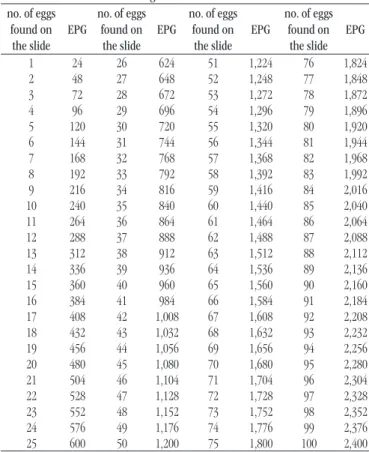
- the number of eggs found in the fecal smear should be multiplied by the constant 24 to obtain the number of eggs per gram of feces (Table 1).
Limitations of the method
The dificulties are related to the absence of eggs in the feces, even in parasitized individuals who, due to certain circumstances, do not release eggs in the feces. This often leads to cases of false negative diagnosis. Among these circumstances, the most common are: 1) absence of eggs in the initial stage of the infection (pre-patent), which usually lasts 4-6 weeks; and in unisexual infections, when there will be no oviposition; 2) absence of eggs soon after the medication, when it is insuficient for healing, but is effective to cause the suspension of the oviposition by the females, temporarily intoxicated and atrophied (during one or more months); and 3) shortage or inconstancy of eggs disposal in light infections and in old cases.
Quality control of Kato-Katz slides: processes and
operational steps
When a laboratory correctly identiies Schistosoma mansoni
eggs, it issues a record that will be decisive for the clinical treatment of the patient, as well as for the epidemiological evaluation and control of schistosomiasis in the communities affected by the disease,
so the importance of qualiied and trained personnel to meet the demand, performing the service in minimum time and effectively. The laboratory, in addition to complying with current legislation, must follow the changes and adapt to the competitive market, using internal quality control and participating in proiciency testing programs. In order to carry out this type of parasitological diagnosis, the laboratory must have a SOP to which all personnel involved must be properly trained following the procedures below:
• pre-analytical process – guarantees the steps: exam order, orientation on collection, preparation and collection of the patient’s material or sample, sample packaging and transport to
the laboratory and registration of the data;
• analytical process – deined as the set of operations to perform the tests according to a certain method in which the Internal Quality Control is applied to the results of the patient sample tests to evaluate if the analytical system is operating within the predeined tolerance limits;
• post-analytical process – are the steps after carrying out the examination: consistency analysis of the results, release of reports, storage of material or sample of the patient, transmission and archiving of results and technical advice in order to guarantee the Traceability, that is, the ability to retrieve the history, application or location of a record using the registered identiications.
The Internal/External Quality Control will evaluate the performance of laboratory tests through intra- or interlaboratory comparisons, which will ensure the quality and reliability of the
test results.
Methodology for internal and external quality
control of slide reading by Kato-Katz method
Double reading
All slides examined under the microscope, regardless of the methodology applied and the microscopist that issued the report, should be properly stored. Each month 10% of the slides should be randomly selected for reexamination by another microscopist from the same laboratory and every six months, this evaluation must be performed by a microscopist external to the institution. This procedure should ensure the internal and external control of the slides analyzed. For both quality controls (internal and external), positive and negative slides should be inserted in this process for reading evaluation. It is essential that the slides are assembled, manipulated and stored properly and within the quality parameters established by national protocols(9, 10) so
that there is no loss of material quality that would making the evaluation process impossible.
TABLE 1 − Egg counting to estimate the intensity of infection by
estimating the calculation of EPG* no. of eggs
found on the slide
EPG
no. of eggs found on
the slide EPG
no. of eggs found on the slide
EPG
no. of eggs found on
the slide EPG
1 24 26 624 51 1,224 76 1,824 2 48 27 648 52 1,248 77 1,848 3 72 28 672 53 1,272 78 1,872 4 96 29 696 54 1,296 79 1,896 5 120 30 720 55 1,320 80 1,920 6 144 31 744 56 1,344 81 1,944 7 168 32 768 57 1,368 82 1,968 8 192 33 792 58 1,392 83 1,992 9 216 34 816 59 1,416 84 2,016 10 240 35 840 60 1,440 85 2,040 11 264 36 864 61 1,464 86 2,064 12 288 37 888 62 1,488 87 2,088 13 312 38 912 63 1,512 88 2,112 14 336 39 936 64 1,536 89 2,136 15 360 40 960 65 1,560 90 2,160 16 384 41 984 66 1,584 91 2,184 17 408 42 1,008 67 1,608 92 2,208 18 432 43 1,032 68 1,632 93 2,232 19 456 44 1,056 69 1,656 94 2,256 20 480 45 1,080 70 1,680 95 2,280 21 504 46 1,104 71 1,704 96 2,304 22 528 47 1,128 72 1,728 97 2,328 23 552 48 1,152 73 1,752 98 2,352 24 576 49 1,176 74 1,776 99 2,376 25 600 50 1,200 75 1,800 100 2,400
It is important to remember that the purpose of quality control is to guarantee the technical quality for laboratory diagnosis of schistosomiasis by strengthening the technical knowledge of microscopists, encouraging responsible attitudes in relation to work. Therefore, quality control should never be confused with inspection, supervision or punitive measure, but as a reinforcement to minimize false results.
Limits of tolerance for the control of the quality of the slides for the diagnosis Kato-Katz
Tem percent of the slides read by each microscopist in the laboratory should be drawn randomly to be examined by another microscopist (internal or external to the laboratory, depending on the quality control) to calculate the concordance rate between the readings. The microscopists from a reference laboratory should obtain 95% reliability under everyday routine conditions, but due to the variability in the presentation of S. mansoni parasite eggs
on Kato-Katz method slide, it is acceptable a concordance rate that may vary up to 25%. This concordance rate was deined based on the minimum range of infection intensity (light infection) determined by the World Health Organization (WHO), consisting of one to four eggs per slide (96 EPG). In this case, for a slightly parasitized individual it is acceptable that the discordance between readings is up to one egg (25%) as shown in Table 2.
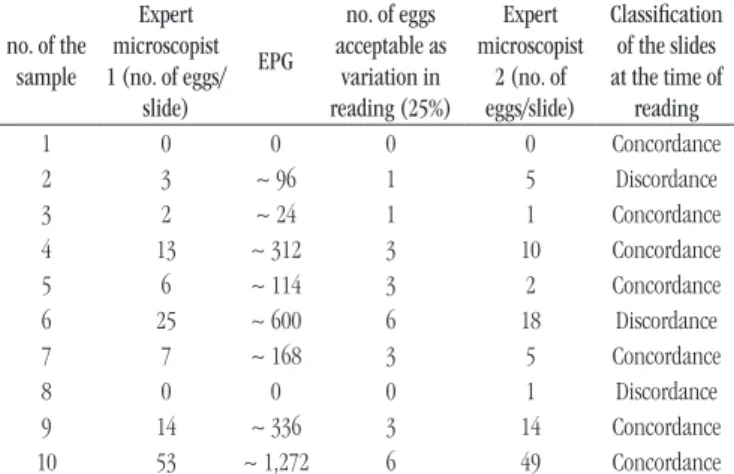
TABLE 3 − Example of a spreadsheet for assessing the concordance and
discordance of the results of Kato-Katz slides readings by two expert microscopists no. of the
sample
Expert microscopist 1 (no. of eggs/
slide)
EPG
no. of eggs acceptable as
variation in reading (25%)
Expert microscopist
2 (no. of eggs/slide)
Classification of the slides at the time of
reading
1 0 0 0 0 Concordance
2 3 ~ 96 1 5 Discordance
3 2 ~ 24 1 1 Concordance
4 13 ~ 312 3 10 Concordance
5 6 ~ 114 3 2 Concordance
6 25 ~ 600 6 18 Discordance
7 7 ~ 168 3 5 Concordance
8 0 0 0 1 Discordance
9 14 ~ 336 3 14 Concordance 10 53 ~ 1,272 6 49 Concordance
EPG: eggs per gram of feces.
TABLE 2 − Acceptable values for S. mansoni egg count variation per slide,
according to the intensity of the infection (EPG) no. of S. mansoni
eggs per slide EPG
Infection intensity
no. of eggs acceptable as variation in reading (25%)
Negative 0 Not infected 0
1-4 1-96 Light 1
5-16 97-384 Moderate 3
17-40 385-960 High 6
> 40 > 960 High 12
EPG: eggs per gram of feces.
To calculate the rate of concordance, the following formula
is used:
Concordance (%) = no. of concordance slides
total of slides reread
There shall be compliance between the positive/negative slides, and the maximum acceptable discordance rate shall be 10%. Once a mismatch above 10% has been detected by the evaluating microscopist, two procedures must be adopted: 1) all slides must be reread; 2) training will be offered to the microscopists who present systematic failures in the evaluation.
Table 3 shows an example of a spreadsheet with the results of Kato-Katz slides reading by two microscopists, in which the reading can be classiied as concordant or discordant from the maximum variation of eggs per slide, according to the intensity of the infection.
In the above example, according to the formula for calculating the concordance rate, the result is 70% (7/10 × 100). In this case, 30% of discordance is veriied, which would be an indicative for rereading of the slides and training of the microscopist.
CONCLUSION
This work is expected that the laboratories that intend to perform the diagnosis of schistosomiasis by the Kato-Katz method may have parameters for the implementation of a diagnostic service that could be evaluated internally and externally. Such parameters are of importance for the reference laboratories for cities and states, since besides issuing diagnostic reports in the development of routine activities, they are often used as a service for the external quality control in relation to the results of slides readings. In conclusion, the implementation of the same quality protocol enables the comparison of data and the identiication of failures in the operational procedure, which may be quickly corrected through training of personnel and implementation of speciic measures for the problems identiied in the process.
CONFLICTS OF INTEREST
CORRESPONDING AUTHOR
Wheverton Ricardo Correia do Nascimento
Laboratório de Referência em Esquistossomose; Centro de Pesquisas Aggeu Magalhães; Fundação Oswaldo Cruz; Avenida Professor Moraes Rego, s/n; campus da Universidade Federal de Pernambuco (UFPE); Cidade Universitária; CEP: 50670-420; Recife-PE, Brasil; Phone/Fax: +55 (81) 2101-2572; e-mail: [email protected].
RESUMO
Introdução: O Kato-Katz é o método laboratorial adotado pelo Ministério da Saúde (MS) e pela Organização Mundial da Saúde (OMS)
como padrão-ouro para o diagnóstico da infecção humana pelo Schistosoma mansoni, sendo uma ferramenta de relevância clínica e epidemiológica, visto que permite classificar a carga parasitária do indivíduo infectado pelo cálculo de ovos por grama de fezes (OPG). Essa classificação pode também ser utilizada para estimar a intensidade da infecção nas comunidades, mensurar o impacto de medidas de controle da doença bem como estabelecer parâmetros de qualidade para a leitura das lâminas. Objetivo: Descrever os procedimentos laboratoriais corretos para o diagnóstico parasitológico da infecção pelo S. mansoni pelo método Kato-Katz a partir do protocolo de controle de qualidade estabelecido pelo Laboratório e pelo Serviço de Referência em Esquistossomose/Centro de Pesquisa Aggeu Magalhães (CPqAM)/Fundação Oswaldo Cruz (Fiocruz)/MS. Método: São descritas: 1) as etapas técnicas para o preparo das amostras de fezes e a leitura das lâminas; 2) as limitações da técnica; 3) o procedimento operacional padrão (POP) a ser adotado pelos laboratórios; 4) a metodologia para o controle de qualidade interno e externo da leitura das lâminas; e 5) os limites de tolerância aceitos para tal controle. Conclusão: Este trabalho instrumentaliza os laboratórios que realizam o diagnóstico da esquistossomose pelo método Kato-Katz com parâmetros para implantar um serviço diagnóstico passível de ser avaliado interna e externamente. O estabelecimento de um protocolo de qualidade viabiliza a comparação de dados e a identificação de falhas no procedimento operacional, que poderão ser corrigidas por meio de capacitação de pessoal e tomada de medidas para os problemas identificados.
Unitermos: esquistossomose; controle de qualidade; diagnóstico; Schistosoma mansoni; parasitologia; testes diagnósticos de rotina.
REFERENCES
1. Fiocruz. Fundação Oswaldo Cruz. Portaria 430/09-PR, de 13 de setembro de 2002. Reconhecimento como serviço regional de referência em esquistossomose pelo Ministério da Saúde. Rio de Janeiro; 2002. 2. Ministério da Saúde (BR). Secretaria de Vigilância em Saúde. Portaria nº 410, de 12 de setembro de 2002. Divulga relação de órgãos/entidades que possuem laboratórios pré-selecionados para integrar a Rede Nacional de Laboratórios de Vigilância Epidemiológica. Brasília: Ministério da Saúde; 2002.
3. ABNT. Associação Brasileira de Normas Técnicas. Laboratórios clínicos – requisitos de qualidade e competência/análises clínicas e diagnóstico in vitro [Internet]. 2015. Available at: https://www.abntcatalogo.com.br/ norma.aspx?ID=329302. [Acessed: June 13, 2016].
4. ABNT. Associação Brasileira de Normas Técnicas. Gestão para o sucesso sustentado de uma organização: uma abordagem da gestão da qualidade. 2010. 47 p. ISBN: 978-85-07-02068-4/2010.
5. Hoffman WA, Pons JA, Janer JL. The sedimentation-concentration method in schistosomiasis mansoni. J Saúde Pública Med Trop. 1934; 9(3): 283-98.
6. Katz N, Chaves A, Pellegrino J. A simple device for quantitative stool thick-smear technique in schistosomiasis mansoni. Rev Med Trop. 1972; 14(6): 397-400.
7. WHO. World Health Organization. Prevention and control of schistosomiasis and soil-transmitted helminthiasis: report of a WHO expert committee. WHO Expert Committee on the Control of Schistosomiasis. (WHO Technical Report Series, n. 912) Geneva; 2002.
8. Barbosa CS, Gomes ECS, org. Guia para vigilância e controle da esquistossomose – prática de laboratório e campo. 1 ed. Recife, PE: Editora Universitária, UFPE; 2008.
9. Ministério da Saúde. Secretaria de Vigilância em Saúde (BR). Departamento de Vigilância em Doenças Transmissíveis. Plano integrado de ações estratégicas de eliminação da hanseníase, ilariose, esquistossomose e oncocercose como problema de saúde pública, tracoma como causa de cegueira e controle das geo-helmintíases: plano de ação 2011-2015. Brasília: Ministério da Saúde; 2012.