http://dx.doi.org/10.1590/jvb.2014.008
Traumatic arteriovenous istula of the
supericial temporal artery
Fístula arteriovenosa traumática de artéria temporal superficial
Otacílio de Camargo Júnior1,2, Márcia Fayad Marcondes de Abreu2, Guilherme Camargo Gonçalves de Abreu2,
Sthefano Atique Gabriel2, Isabella Maria Machado da Silva1
Abstract
Arteriovenous istulae of the supericial temporal artery are rare, and their principal cause is traumas. Complications include pulsatile mass, headache, hemorrhage and deformities that compromise esthetics. Treatment can be performed using conventional surgery or endovascular methods. he authors describe a case of a 44-year-old male patient who developed a large pulsating mass, extending from the preauricular region to the right parietotemporal and frontal regions after a motorcycle accident. he treatment chosen was complete surgical removal of the pulsatile mass and ligature of the vessels feeding the istula.
Keywords: arteriovenous istulae; supericial temporal artery; trauma.
Resumo
As fístulas arteriovenosas de artéria temporal supericial são raras, sendo o trauma sua etiologia principal. Suas complicações incluem massa pulsátil, cefaleia, hemorragia e deformidade estética. O tratamento pode ser realizado por cirurgia convencional ou endovascular. Os autores relatam o caso de um paciente de 44 anos que evoluiu com massa pulsátil extensa desde região pré-auricular até região parietotemporal e frontal direita após acidente motociclístico. Optou-se por remoção cirúrgica completa da massa pulsátil e ligadura dos vasos nutridores da fístula.
Palavras-chave:fístula arteriovenosa; artéria temporal supericial; trauma.
1 Pontifícia Universidade Católica de Campinas – PUC-Campinas, Campinas, SP, Brasil. 2 Hospital e Maternidade Celso Pierro – HMCP, Campinas, SP, Brasil.
Financial support: None.
Conlicts of interest: No conlicts of interest declared concerning the publication of this article. Submitted on: 06.23.13. Accepted on: 07.18.13.
he study was carried out at Hospital e Maternidade Celso Pierro (HMCP)/Pontifícia Universidade Católica de Campinas (PUC-Campinas). he present study was presented at the XI Pan-American Congress on Vascular and Endovascular Surgery, as an oral presentation.
39 J Vasc Bras. 2014 Jan.-Mar.; 13(1):39-42
Brief title: istula of the supericial temporal artery
mass originated. A careful neurological examination detected no abnormalities.
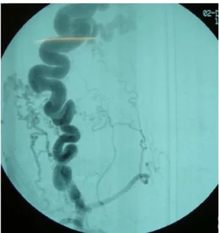
The patient underwent cranial tomography, which found no intracranial involvement. Selective arteriography of the right external carotid artery showed premature opacification of the right
supericial temporal vein, which was dilated and tortuous, and a high low rate arteriovenous istula (Figure 2). The istula was fed by the supericial
temporal artery and drained via the superficial temporal vein into the external jugular vein. There was no collateral circulation from the left external
INTRODUCTION
Arteriovenous istula of the supericial temporal
artery is a rare condition, with an estimated incidence of 0.5% to 2.0% of cases.1,2 In 75% of
patients, etiology is traumatic and these istulae can
be caused by blunt or penetrating traumas or by iatrogenic injuries during diagnostic and therapeutic procedures.1,2
Arteriovenous istulae are connections between
an arterial segment and the venous drainage system, resulting in a tortuous dilated vein and emergence of aneurysmal varicose segments, which can progress to thrombosis, ulceration and rupture.1,2
The objective of this study is to describe a case
of traumatic arteriovenous istula of the temporal supericial artery ha was treated surgically.
CASE DESCRIPTION
An otherwise healthy, 44-year-old male patient with no history of comorbidities was referred to the clinic complaining of a painless pulsating mass that was growing progressively, in a right frontal and preauricular position, and was causing a pulsing buzz in the right ear.
The patient had suffered a motorbike accident 10 years previously that had caused a stellate laceration injury to the right preauricular region. He reported that he had received care at a hospital where laboratory tests and cranial tomography had found no further abnormalities and the stellate laceration wound had been treated by debridement of devitalized tissues and closure with sutures. One year after the accident, the patient had noticed a murmur in the region of the right ear and a swelling in the right parietal region and had sought medical attention, but had not been treated. Fifteen months later he had noted a progressive increase in the murmur and in the volume of the pulsating mass in the frontal, parietal preauricular areas, causing
strong headaches, signiicant cranial discomfort and
unsightly esthetics in addition to preventing the right eye from opening normally.
On physical examination an extensive, painless and immobile pulsating mass was observed to extend from the right preauricular region to the right parietotemporal and frontal region (Figure 1). A thrill was detected on palpation of the mass and auscultation revealed a continuous murmur, with accentuated systolic sounds. Both thrill and murmur
ceased when the right supericial temporal artery was
compressed. After detailed physical examination a small scar was found in the right preauricular region, in the probable location from which the pulsating
Figure 1. Arteriovenous Fistula of the Supericial Temporal Artery.
Figure 2. Selective arteriography of the right external carotid artery, showing premature opaciication of the right supericial temporal vein, which is dilated and tortuous.
Otacílio de Camargo Júnior, Márcia Fayad Marcondes de Abreu et al.
carotid artery to the istula. No communication was
observed between circulation in the internal carotid
artery and the arteriovenous istula of the external
carotid artery.
The decision was taken to employ conventional treatment. The patient was positioned with the head elevated by 20-30 degrees above the level of the heart to improve venous drainage. After general anesthetic, transverse incisions were made in the scalp and the arteriovenous malformations were carefully dissected, then ligated, sectioned and resected (Figure 3). The patient progressed satisfactorily, free from infection or necrosis of the scalp and with evident improvement in esthetic appearance (Figure 4).
DISCUSSION
The superficial temporal artery is vulnerable
to trauma because of its supericial path over the
temporal bone and because of its proximity to cranial sutures. In addition to traumatic etiologies,
arteriovenous istulae of the supericial temporal
artery can also be spontaneous or caused by surgical procedures, such as, capillary implants, external ventricular drainage and craniotomies.3-5 Traumatic
fistulae develop over the course of months or years after the trauma; while spontaneous fístulae may be present at birth, although in the majority of patients they are asymptomatic until puberty.3,4
Traumas, vasomotor disorders, hormonal stimuli
and inlammatory processes worsen the symptoms.3,4
Two mechanisms have been suggested to explain
the formation of traumatic arteriovenous istulae in the scalp. By the irst mechanism, a simultaneous
laceration of the artery and the adjacent vein leads
to formation of the istula.6,7 The second mechanism
begins with rupture of vasa vasorum in the artery wall, proliferation of endothelial cells from the damaged vasa vasorum then forms numerous small vessels, creating vascular communication channels between artery and vein.6,7 In the case of the patient
described here, the small scar in the right preauricular region suggests a direct connection from the artery to
the supericial temporal vein caused by a traumatic
laceration.
Diagnosis of a traumatic istula of the supericial
temporal artery is based on a history of trauma and a detailed physical examination.6-8 Although
angiotomography can provide images in shorter acquisition times and offers reconstruction of images
in iner slices, angiography remains the gold standard
examination, revealing donor and recipient vessels, excluding intracranial components and providing information about the direction and velocity of
blood low.6-8 Some traumatic lesions in supericial
temporal artery topographies may not pulse or may pulse because of transmission from adjacent arteries, meaning that pseudoaneurysms, true aneurysms, arteriovenous malformations, cysts, abscesses, hematoma and aneurysms of the medial meningeal artery with bone erosion are all part of differential
diagnosis of traumatic arteriovenous istulae of the supericial temporal artery.6-8
Treatment for arteriovenous fistulae of the
supericial temporal artery should take account of the elevated blood low through the istula, the complex
vascular anatomy and the esthetic issues involved.9,10
Treatment is indicated to reduce esthetic problems, to prevent hemorrhage and ischemic skin erosion
Figure 3. Intraoperative view.
Figure 4. Postoperative view.
Brief title: istula of the supericial temporal artery
7. Hasturk AE, Erten F, Ayata T. Giant non-traumatic arteriovenous malformation of the scalp. Asian J Neurosurg. 2012;7(1):39-41. http://dx.doi.org/10.4103/1793-5482.95698
8. Mishra SS, Panigrahi S, Parida D, Behera SK. Usefulness of computed tomographic angiography in the management of extracranial scalp arteriovenous malformation. Neurol India. 2012;60(3):357-8. http://dx.doi.org/10.4103/0028-3886.98544
9. Whiteside OJ, Monksield P, Steventon NB, Byrne J, Burton MJ. Endovascular embolization of a traumatic arteriovenous istula of the supericial temporal artery. J Laryngol Otol. 2005;119(4):322-4. http://dx.doi.org/10.1258/0022215054020368
10. Yablonicky KJ, Desai S. A case report of a scalp arteriovenous malformation after trauma. J Emerg Med. 2011;41(5):e117-9. http://dx.doi.org/10.1016/j.jemermed.2009.07.039
Correspondence Otacílio de Camargo Júnior Rua Cândido Gomide, 468 –. Jardim Guanabara CEP 13073-200 – Campinas (SP), Brazil E-mail: [email protected]
Author’s information OCJ é Cirurgião Vascular e Endovascular. Professor Adjunto da PUC-Campinas; Chefe do Serviço de Cirurgia Vascular e Endovascular do
HMCP. MFMA, GCGA, SAG são Médicos do Serviço de Angiologia e Cirurgia Vascular do HMCP. IMMS é Aluna do curso de Medicina da PUC-Campinas.
Author’s contributions Conception and design: OCJ, SAG, GCGA Analysis and interpretation: OCJ , SAG, MFMA Data collection: OCJ, SAG, GCGA, IMMS Writing the article: OCJ , SAG, MFMA, GCGA, IMMS Critical revision of the article: OCJ, SAG, MFMA, GCGA Final approval of the article*: OCJ, SAG, MFMA, GCGA, IMMS Statistical analysis: N/A Overall responsibility: OCJ , SAG
*All authors have read and approved of the inal version of the article submitted to J Vasc Bras. and other symptoms such as headaches and buzzing
sounds.9,10 Treatment options include surgical
excision, ligature of the vessels providing blood supply, transarterial and transvenous embolization and injection of sclerosing agents into the lesion.9,10
Endovascular embolization can be employed as
a deinitive treatment or as an adjuvant to a surgical
procedure, reducing blood loss during removal of
the pulsating mass; but it is not alone suficient for
extensive arteriovenous fistulae, providing only temporary relief from symptoms, involving a high risk of recurrence and conferring a risk of cutaneous necrosis.9,10 Surgical removal of the pulsating mass
and ligature of the vessels feeding the arteriovenous
istulae remains the treatment of choice.9,10
REFERENCES
1. Miekisiak G, Mis M, Sandler A, Druszcz A. Iatrogenic arteriovenous istula of the supericial temporal artery. Oral Maxillofac Surg. 2008;12(4):219-21. http://dx.doi.org/10.1007/s10006-008-0133-5
2. Li F, Zhu S, Liu Y, et al. Traumatic arteriovenous istula of the supericial temporal artery. J Clin Neurosci. 2007;14(6):595-600. http://dx.doi.org/10.1016/j.jocn.2006.04.011
3. Senoglu M, Yasim A, Gokce M, Senoglu N. Nontraumatic scalp arteriovenous istula in an adult: technical report on an illustrative case. Surg Neurol. 2008;70(2):194-7. http://dx.doi.org/10.1016/j. surneu.2007.04.018
4. Leal FS, Miranda CC, Guimarães AC. Traumatic pseudoaneurysm of the sup erf icial temp oral arter y: case rep ort. Arq Neuropsiquiatr. 2005;63(3B):859-61. http://dx.doi.org/10.1590/ S0004-282X2005000500027
5. Bernstein J, Podnos S, Leavitt M. Arteriovenous istula following hair transplantation. Dermatol Surg. 2011;37(6):873-5. http:// dx.doi.org/10.1111/j.1524-4725.2011.02027.x
6. Kelly K, Trites JR, Taylor SM, Bullock M, Hart RD. Arteriovenous malformation of the scalp with cerebral steal. Head Neck. 2009;31(11):1520-3. http://dx.doi.org/10.1002/hed.21032