Evolutionarily Conserved
Joseph J. Gillespie1,2*, Nicole C. Ammerman2, Sheila M. Dreher-Lesnick2, M. Sayeedur Rahman2, Micah J. Worley3, Joao C. Setubal1,4, Bruno S. Sobral1, Abdu F. Azad2
1Virginia Bioinformatics Institute at Virginia Tech, Blacksburg, Virginia, United States of America,2Department of Microbiology and Immunology, University of Maryland School of Medicine, Baltimore, Maryland, United States of America,3Department of Biology, University of Louisville, Louisville, Kentucky, United States of America,
4Department of Computer Science, Virginia Tech, Blacksburg, Virginia, United States of America
Abstract
Background:Bacterial type IV secretion systems (T4SSs) comprise a diverse transporter family functioning in conjugation, competence, and effector molecule (DNA and/or protein) translocation. Thirteen genome sequences from Rickettsia, obligate intracellular symbionts/pathogens of a wide range of eukaryotes, have revealed a reduced T4SS relative to the Agrobacterium tumefaciensarchetype (vir). However, theRickettsiaT4SS has not been functionally characterized for its role in symbiosis/virulence, and none of its substrates are known.
Results: Superimposition of T4SS structural/functional information over previously identified Rickettsia components implicate a functionalRickettsiaT4SS.virB4,virB8andvirB9are duplicated, yet only one copy of each has the conserved features of similar genes in other T4SSs. An extraordinarily duplicated VirB6 gene encodes five hydrophobic proteins conserved only in a short region known to be involved in DNA transfer inA. tumefaciens.virB1,virB2andvirB7are newly identified, revealing aRickettsiaT4SS lacking onlyvirB5relative to thevirarchetype. Phylogeny estimation suggests vertical inheritance of all components, despite gene rearrangements into an archipelago of five islets. Similarities ofRickettsiaVirB7/ VirB9 to ComB7/ComB9 proteins ofe-proteobacteria, as well as phylogenetic affinities to theLegionella lvhT4SS, imply the Rickettsiales ancestor acquired avir-like locus from distantly related bacteria, perhaps while residing in a protozoan host. Modern modifications of these systems likely reflect diversification with various eukaryotic host cells.
Conclusion:We present thervh(Rickettsialesvirhomolog) T4SS, an evolutionary conserved transporter with an unknown role in rickettsial biology. This work lays the foundation for future laboratory characterization of this system, and also identifies theLegionella lvhT4SS as a suitable genetic model.
Citation:Gillespie JJ, Ammerman NC, Dreher-Lesnick SM, Rahman MS, Worley MJ, et al. (2009) An Anomalous Type IV Secretion System inRickettsiaIs Evolutionarily Conserved. PLoS ONE 4(3): e4833. doi:10.1371/journal.pone.0004833
Editor:Stefan Bereswill, Charite´-Universita¨tsmedizin Berlin, Germany
ReceivedJanuary 16, 2009;AcceptedJanuary 28, 2009;PublishedMarch 12, 2009
Copyright:ß2009 Gillespie et al. This is an open-access article distributed under the terms of the Creative Commons Attribution License, which permits unrestricted use, distribution, and reproduction in any medium, provided the original author and source are credited.
Funding:The project described was supported by Award Numbers R01AI017828 and R01AI59118 from the National Institute of Allergy And Infectious Diseases (NIAID) to AFA, and through NIAID contract HHSN266200400035C to BSS. The content is solely the responsibility of the authors and does not necessarily represent the official views of the NIAID or the National Institutes of Health.
Competing Interests:The authors have declared that no competing interests exist.
* E-mail: [email protected]
Introduction
Type IV secretion systems (T4SSs) are multi-component membrane-spanning transporters present in many Gram-negative bacteria, including medically and agriculturally important species. Derived from ancient conjugation machineries [1], T4SSs are presumably used by many species for the exchange of genetic material [2,3,4,5]. However, T4SSs have garnered significant attention for their role in pathogenesis, acting as syringes that inject virulence factors into eukaryotic host cells [6]. These translocated virulence factors, more commonly referred to as ‘effector molecules’ (DNA, protein or nucleoprotein complexes), have a broad range of host-altering functions, such as highjacking of vesicular trafficking [7], cytoskeletal modification [8], ubiqui-tination system exploitation [9], and genome introgression [10,11,12,13,14,15]. T4SSs are found in a variety of etiological agents of human disease, including Bordetella pertussis (whooping
have numerous divergent functions in both Gram-negative bacteria and Gram-positive bacteria, likely arising from multiple progenitors [26].
Much of what is known about the structure and function of T4SSs has been generated from paramount studies on the phytopathogen Agrobacterium tumefaciens [30]. Thus, the virB/virD T4SS ofA. tumefaciensis considered the archetype to which most othervirandvir-like T4SSs (typically referred to as ‘type A’ T4SSs) are compared. InA. tumefaciens, thevirBoperon andvirDlocus on the Ti (tumor inducing) plasmid encode 12 single copy genes (virB1Ti-virB11Ti, virD4Ti) whose products comprise a molecular scaffold that spans the inner and outer membranes (IM and OM, respectively), culminating in a transfer pilus (T pilus) that protrudes from the bacteria [6,31,32,33,34,35].A. tumefaciensuses itsvirT4SS, which is regulated by the VirA/VirG two-component system [36], to introduce a nucleoprotein complex, the T-strand DNA plus VirD2 and VirE2 proteins, into the nuclei of various host plant cells [37]. The byproduct of this interkingdom introgression is crown gall disease, which allows the bacteria to reside and multiply within a modified host niche [38].
The rudimentary functions of most components of the A. tumefaciens vir system have been characterized. VirD4Tiis an IM
ATPase that couples the nucleoprotein complex (and other substrates) to the T4SS channel [39,40]. VirB4Tiand VirB11Ti
are additional IM ATPases that presumably fuel the delivery of substrates to the core periplasmic-spanning channel [32,41,42,43, 44,45,46,47,48,49,50,51,52]. VirB6Ti-VirB10Ti form the core
periplasmic-spanning channel [42,45,46,51,52,53,54,55,56,57,58, 59,60,61], while VirB2Tiand VirB5Tiare the major and minor
constituents of the T pilus, respectively [42,44,46,62,63,64]. VirB1Ti is a member of the specialized lytic transglycosylase
family of proteins implicated in the local degradation of peptidoglycan, allowing the channel to span the periplasm [42,65,66,67]. The C-terminal portion of VirB1Ti (VirB1*) is
cleaved and functions in cell-cell contact and virulence [65].
Despite the vast body of research onvirsystems, the function of VirB3Ti, an exported OM protein presumably involved in host cell
attachment, remains unknown [42,64]. Studies implicating the interaction of Vir components inA. tumefaciens as well as several other bacteria are rapidly growing and, coupled with recent structural studies, the architecture of the Vir scaffold is coming to light (Figure 1B).
A decade has passed since the first genome sequence was published forRickettsia, obligate intracellular bacteria of the class Alphaproteobacteria [68,69,70]. The R. prowazekii str. Madrid E genome sequence (1.1 Mb) exposed an extraordinary trend toward genome reduction via pseudogenization, with major constituents of biosynthetic pathways deleted relative to other bacteria [71]. It also revealed a reduced T4SS as compared to thevirB/virDT4SS ofA. tumefaciens, with only six genes annotated as coding for Vir components (virB4,virB8-virB11,virD4). Interestingly, two of these Vir genes were duplicated (virB4 and virB9, the latter gene annotated astrbG), and the arrangement of the Vir genes was non-canonical relative to most other T4SSs, being scattered in three well-separated locales of the genome. Subsequently sequenced rickettsial genomes have confirmed this atypical nature of the RickettisaT4SS [72,73,74,75,76,77,78,79], and consensus genome annotation revealed a T4SS devoid of only the VirB1, VirB2, VirB5, and VirB7 components [80,81].
Despite intense laboratory effort, a paucity of characterized virulence factors exists forRickettsia[82], with, to date, no genes identified as coding for effectors of the T4SS. This is surprising, as the virulent species of Rickettsia are of great interest both as emerging infectious diseases [83] and for their potential deploy-ment as bioterrorism agents [84,85]. Given the relatively close evolutionary relationship ofAgrobacterium(Rhizobiales) and Rickett-sia(Rickettsiales) within theAlphaproteobacteriatree [86], as well as the availability of 13 genome sequences ofRickettsiaspp. (R. bellii str. RML369-C,R. belliistr. OSU 85 389,R. canadensisstr. McKiel, R. prowazekii str. Madrid E,R. typhi str. Wilmington, R. felis str.
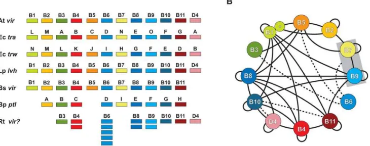
Figure 1. Characteristics of the archetypal bacterial type IV secretion system (type A). (A)Schema depicting several of the type A T4SSs used in this investigation for comparison with theRickettsiaT4SS. Atvir=Agrobacterium tumefaciensTi plasmid; Ectra=Escherichia coliIncN plasmid pKM101; Ectrw=Escherichia coliplasmid R388; Lplvh=Legionella pneumophila; Bsvir=Brucella suis; Bpptl=Bordetella pertussis; Rtvir? =Rickettsia typhi. (B)Map of Vir-Virphysical interactions based on a survey of the literature. Specific studies demonstrating the mapped interactions are discussed in the text and in supportingDocument S1. Solid black lines depict direct physical interactions. Dashed lines depict energetic effects of ATP hydrolysis of VirD4 and VirB11 on VirB10 [100], as well as the stabilization effect of VirB6 on VirB3, VirB5, and VirB7 dimerization [184] and the VirB7/VirB9 heterodimer [131]. VirB1 and VirB1* are modeled accordingly [65] and the VirB7/VirB9 complex is boxed to illustrate interactions with the heterodimer rather than individual components.
URRWXCal2, R. akaristr. Hartford,R. massiliaestr. MTU5, R. rickettsiistr. Sheila Smith CWPP,R. rickettsiistr. Iowa,R. conoriistr. Malish 7,R. sibiricastr. 246, andR. africaestr. ESF-5), it is timely to assess the atypical nature of theRickettsiaT4SS with an attempt to better understand the function (if any) this transporter has in rickettsial pathogenesis. We provide here a bioinformatic analysis of theRickettsiaT4SS, with each component evaluated in light of the latest structural and functional information from studies ofvir and vir-like (e.g.,tra,trw,trb, ptl) T4SSs from other bacteria. We address the nature of Vir gene duplication inRickettsiaand discuss the potential role the vir T4SS plays not only in rickettsial pathogenesis, but also lateral gene transfer (LGT). These results shed new light on the origin and function of the rickettsiae Vir-like genes and lay a much needed foundation for future laboratory assessment of the function of theRickettsiaT4SS.
Results and Discussion
Previously IdentifiedRickettsiaT4SS Components Fifteen highly conserved genes previously annotated in rickettsial genome sequencing projects have similar counterparts in the T4SSs of other bacteria (Table 1). These 15 genes comprise eight of the Vir components well characterized in theA. tumefaciens T4SS that are involved in substrate (DNA and/or protein) presentation (virD4), translocation energetics (virB4,virB11), mating channel structure (virB6, virB8-virB10), and host cell attachment (virB3) [26,37]. Thus, consensus genome annotation supports prior findings that theRickettsiaT4SS is reduced in complexity relative to theA. tumefaciensmodel (missing genes encoding the components VirB1, VirB2, VirB5, and VirB7), with the various components present in three scattered genomic locales in all sequenced Rickettsia genomes (Figure S1). Accordingly, we evaluate the characteristics of each Vir component to propose that the T4SS in Rickettsia is a functional transporter. Important background information regarding these eight Vir components is compiled in Document S1.
Substrate presentation (VirD4). TheRickettsiaVirD4 gene is conserved across 13 genomes (8.1% div.), and the corresponding amino acid sequence (4.2% div.) is highly conserved when compared across divergent pathogenic bacterial type IV coupling proteins (T4CPs), particularly within the five conserved motifs of the two large C-terminal domains (CTDs) (Figure S2). Unpublished data from our laboratories also suggests thatRickettsia VirD4 is capable of binding putative effectors of the T4SS using a bacterial 2-hybrid assay.
Translocation energetics (VirB4 and VirB11). Two VirB4 genes are present in all sequencedRickettsiagenomes (virB4aand virB4b), and both encoded proteins are large and similar in size and composition to VirB4 and VirB4-like proteins from other bacteria (Figure 2). In most Rickettsia genomes, virB4a is clustered with virB3and thevirB6duplicates, whilevirB4bis not located near any other Vir genes (Figure S1). virB4a is more conserved across Rickettsiagenomes thanvirB4bat the nucleotide (6.7% versus 9.2%) and amino acid (3.7% versus 9.9%) level, with more indels in VirB4b relative to VirB4a and other bacteria. In the protein alignment, 11 variable residues are VirB4a-specific, whereas 43 variable residues are unique to VirB4b, illustrating more conservation of VirB4a with non-Rickettsia proteins. Strikingly, seven of the VirB4b-specific substitutions alter critical residues in the NTP-binding cleft, which is essential for T-DNA export inA. tumefaciens[41], casting doubt on the ability of VirB4b to function as a T4SS ATPase.
The Rickettsia VirB11 gene is conserved across 13 genomes (9.7% div.) and the corresponding amino acid sequence (4.8% div.)
is highly conserved when compared to VirB11 and VirB11-like proteins from other bacteria, particularly within the four conserved motifs of the CTD (Figure S3). Not surprisingly, the least conserved region in our alignment maps to the major difference between the two VirB11 crystals, a domain swap in the N-terminal domain (NTD)-CTD linker region of Brucella suis relative toH. pylori[87]. Like most other VirB11 and VirB11-like proteins, Rickettsia VirB4 contains the domain swap and likely forms an additional helix,aC2, in the linker B region that allows for additional interactions at the subunit-subunit interface. Finally, a search within all rickettsial genomes for the recently identified regulator ofH. pyloriVirB11, HP1451 [88], proved unsuccessful.
Mating channel structure (VirB6, VirB8-VirB10). An extraordinary five copies of the VirB6 gene (virB6a-e) are found in most Rickettsia genomes (Figure 3A), with exception to R. massiliae, which is missingvirB6e[72]. Additionally,virB6dis split in theR. belliistr. OSU 85 389 genome [81]. These findings, coupled with blast results (Table 1), suggest that these VirB6 genes are the most variable components of theRickettsia virsystem, particularly when the regions flanking the VirB6/TrbL domains are considered. Individual full length VirB6 genes are conserved across 13 genomes and range in nt divergence from 10.3% (12.4% aa) to 13.2% (15.2% aa); however, the five duplicates are extraordinarily different from one another, averaging ,80% aa divergence in the VirB6/TrbL domains alone (alignment of full length proteins was confounded by variability in the flanking sequences). This lack of conservation, coupled with conflicting
Table 1.Relative Conservation ofRickettsiaVir Proteins to Vir and Vir-like Proteins from Other Bacteria.
Protein1 L query2 No. hits3 Distribution4 Other5
a b c d e
VirD4 591 500 229 64 36 3 31 137 (6)
VirB4a 805 500 244 60 75 2 76 43 (15)
VirB4b 810 456 218 53 59 2 73 51 (28)
VirB11 334 500 200 109 107 18 8 58 (4)
VirB6a 1061 151 130 2 5 0 1 13 (2)
VirB6b 668 181 147 1 13 0 1 19
VirB6c 967 202 158 5 20 1 1 17 (2)
VirB6d 890 197 172 4 18 0 0 3 (1)
VirB6e 1154 247 174 14 35 1 9 14 (3)
VirB8a 228 101 65 4 11 1 3 17 (3)
VirB8b 242 153 80 10 36 0 16 11 (4)
VirB9a 247 411 203 74 73 3 36 22 (13)
VirB9b 158 342 197 53 63 1 8 20 (11)
VirB10 481 380 193 52 69 4 47 15 (11)
VirB3 95 80 65 1 7 0 6 7
1Consensus annotation. Grouped by function: substrate presentation (VirD4),
translocation energetics (VirB4, VirB11), mating channel (VirB6, VirB8-VirB10), attachment (VirB3).
2Length (aa) ofR. typhiquery sequence. 3
Number of blastp subjects yielding significant alignments (maximum hits set to 500).
4Distribution of subjects across five classes of proteobacteria.
5Non-proteobacterial subjects, with number of plasmid encoded proteins in
parentheses. Note: plasmid encoded Vir and Vir-like proteins were not assigned to a taxonomic class, hence some may be from plasmids found in proteobacteria.
evidence and hypotheses for VirB6 channel structure formation [89,90] (Figure 3B), makes it difficult to predict the functionality of any of the five duplicate VirB6-like proteins inRickettsia. The regions flanking the VirB6/TrbL domains are particularly divergent across the five duplicates (Figure 3A) and are similar only in a few instances to the sister taxon ofRickettsia,Orientia tsutsugamushi(Figure S4). The contribution of additional flanking sequences to the structure and function of these expanded VirB6/TrbL domain-containing ORFs, especially regarding substrate transport to VirB2 and VirB9 in the OM, is worthy of exploration as they are seemingly unique to Rickettsiaceae (Figure 3C). Comparison of the VirB6/ TrbL domains of all five VirB6 proteins to VirB6 and VirB6-like
proteins from other bacteria supports previous observations that conservation within this domain is limited to the transmembrane-spanning (TMS)-cytoplasmic region involved in substrate transfer to VirB8 [89,91]. Of the conserved residues in this region, the invariant Trp is essential [92] and required for polar localization of VirB6 [90]. Little conservation in sequence length and composition exists in the functionally important N-terminal large periplasmic region, and a TMS region prediction program [93] calculated at least one additional TMS region in this location of the domain in most analyzed taxa (Figure 3C).
Two VirB8 genes are present inRickettsiagenomes (virB8aand virB8b), separated by one small ORF (discussed below) and
Figure 2. Characteristics and comparative analysis of VirB4 and VirB4-like amino acid sequences across six divergent bacterial species.Bars above alignment depict interactions between VirB4 and other Vir components as revealed by bacterial two-hybrid screen [208] with color scheme as follows: purple = VirB4-VirB10, yellow = VirB4-VirB8, green = VirB4-VirB1, blue = VirB4-VirB11. Residues colored white depict mutations in one rickettsial sequence in otherwise conserved positions in the alignment. Coordinates for each sequence are shown to the right in each block, with numbers in parentheses depicting flanking residues of the alignment not shown. Conservation color scheme as follows: yellow = identical; green = identical in five of six sequences; orange = identical in four of six sequences or a position comprised of only two residue states. See text for alignment details. Taxon abbreviations and associated NCBI accession numbers are as follows: At =Agrobacterium tumefaciensVirB4, NP_059802; Rta =Rickettsia typhi VirB4a, AAU03521; Rtb =R. typhi VirB4b, AAU04227; Xf =Xylella fastidiosa conjugal transfer protein (VirB4-like), Q9PHJ8; Rhizobium etliVirB4, Q8KIM8;Bartonella tribocorumVirB4, Q8GJ64.(A)Alignment of the C-terminal portion of VirB4 containing the conserved DNA-binding domain. Walker A (WA) and Walker B (WB) boxes are enclosed with red and blue boxes, respectively. The highly conserved Gln predicted to be involved in binding is denoted with an asterisk and underlined, with additional structurally proximal residues forming the purported NTP-binding cleft also underlined [209]. Seven residues within conserved NTP-NTP-binding domains mutated only in Rtb are shaded in black.(B)Schematic of the entire VirB4 protein depicting the interactions with four other Vir components [208]. Alignment depicts conservation outside of the conserved NTP binding domains. The CagE_TrbE_VirB domain (PF03135) plus WA and WB boxes are depicted in black, red and blue respectively.
encoded on opposite strands (Figure S1).virB8aflanksvirB9aon the minus strand whilevirB8bis within a largervircluster (virB8b -virB9b-virB10-virB11-virD4) on the plus strand. virB8b is more conserved acrossRickettsiagenomes thanvirB8a at the nucleotide (9% versus 10%) and amino acid (9.4% versus 13.4%) levels. Both VirB8 proteins are similar in size and composition to other VirB8 and VirB8-like proteins, with many residues within superimposed a-helices and b-strands of the solved crystal structures [94,95]
conserved in both proteins (Figure 4A). However, limited conservation is seen in residues implicated in dimerization [94,95,96] as well as positions previously demonstrated as lethal mutants [96,97], even in an expanded comparison of 200 VirB8 and VirB8-like proteins (Figure 4B). Furthermore, VirB8a has two critical residues mutated within the highly conserved ‘‘NPxG’’ motif (XPxX across the 13 Rickettsia genomes), which is not expected to adopt its critical sharp turn confirmation between
Figure 3. Characteristics of the highly duplicated VirB6-domain-containing proteins ofRickettsia, with comparison to VirB6 and VirB6-like amino acid sequences from six divergent bacterial species. (A)Schema of fiveRickettsia typhiORFs (Rta-e) containing putative VirB6 domains, as predicted by the Conserved Domains Database (NCBI) following blastp searches. All searches retrieved COG3704 (VirB6) and PF04610 (TrbL, TrbL/VirB6) domains, with Rtd also scoring limited similarity with COG0558 (PgsA, phosphatidylglycerophosphate synthase).(B)
Competing topology models forAgrobacterium tumefaciensVirB6 in the IM (named here on the basis of the number of predicted TMS regions). Top: 5TMS model [89] with bar at top depicting regions of VirB6 required for formation of T-strand close contacts with several Vir components, as revealed by transfer DNA immunoprecipitation; bottom: 4TMS model [90]. Color scheme of protein as follows: black = periplasm, blue = IM, red = cytoplasm. TMS regions are labeled with Roman numerals sequentially from N- to C-terminus. Shaded (gray) portion depicts alignment inC.(C)Multiple sequence alignment of the central portion of the VirB6 domain of five (a–e)R. typhiORFs and five VirB6 and VirB6-like sequences from diverse bacteria species. Bars above alignment depict shaded region inB, with competing models illustrated accordingly. Bold-shaded residues depict TMS regions as predicted by TMHMM [93]. The essential Trp residue [90,92] is denoted with an asterisk above the alignment. Coordinates for each sequence are shown to the right in each block, with numbers in parentheses depicting flanking residues of the alignment not shown. Bold numbers for the rickettsial sequences depict regions not included in the VirB6 domain alignment. Conservation color scheme is the same as inFigure 2
legend, except orange = identical in seven of eleven sequences or a position comprised of only two residue states. See text for alignment details. Taxon abbreviations and associated NCBI accession numbers are as follows: Rta =R. typhiplasmid conjugal transfer protein VirB6 (VirB6a), YP_067002; Rtb =R. typhi TrbL/VirB6 plasmid conjugal transfer protein (VirB6b), YP_067001; Rtc =R. typhi plasmid conjugal transfer protein VirB6 (VirB6c), YP_067000; Rtd =R. typhihypothetical protein (VirB6d), YP_066999; Rte =R. typhiplasmid conjugal transfer protein TrbL/VirB6 (VirB6e), YP_066998; At =A. tumefaciensVirB6, AAF77166; Bh =Bartonella henselaeVirB6, AAF00944; Sm =Sinorhizobium melilotiVirB6, NP_435960; Bs =Brucella suisVirB6, NP_699271; St =Salmonella typhimurium(IncN plasmid R46) TraD, NP_511193; Lp =Legionella pneumophilaLvhB6, AAM0824.
helix a4 and strand b4 if altered [95]. This, coupled with one of two essential residues mutated in the NTD (G78Q) relative to identical residues in VirB8b and VirB8Ti, as well
as the extraordinary overall variation in the NTD (Figure 4C), casts doubt on VirB8a as a functional component of the Rickettsia T4SS. However, predicted structural models for both Rickettsia VirB8 proteins are similar to the VirB8Ti crystal
(Figure 4D).
Rickettsiagenomes contain two VirB9 genes (virB9a andvirB9b) encoded immediately downstream of the two VirB8 genes (Figure S1).virB9ais more conserved acrossRickettsiagenomes thanvirB9b at the nucleotide (7.6% versus 8.2%) and amino acid (4.7% versus 10.8%) levels. While the entire NTDs of both proteins are comparable to other VirB9 and VirB9-like proteins from other bacteria (data not shown), the CTD of VirB9b is highly truncated and contains residues only in the first b-strand, B1, of the
Figure 4. Comparative analysis of VirB8 and VirB8-like proteins with emphasis on twoRickettsiaorthologs. (A)Multiple sequence alignment of the C-terminal regions of VirB8 and VirB8-like sequences corresponding to regions of the solved crystal structures of VirB8 fromBrucella suis[95] andAgrobacterium tumefaciens[94]. Secondary structural model is shown above the alignment with arrows (b-strands B1–B4) and bars (a-helices A1–A4) with color scheme as follows: gray = agreement across both models, stippled = disagreement between models, red = alignment too variable to support either model. Structural notation follows the earlier model [95]. Red box encloses the highly conserved ‘‘NPxG’’ motif. Blue dots depict residues involved in VirB8 homo-dimerization [94,95,96], and red dots depict previously determined lethal mutants [96,97]. InB. suisVirB8, residues implicated in interactions with VirB10 (T201) and VirB4 (R230) are underlined. Boxed regions are further described inB. Residues colored white depict mutations in one rickettsial sequence in otherwise conserved positions in the alignment. Coordinates for each sequence are shown to the right in each block, with numbers in parentheses depicting the number of flanking residues of the alignment not shown. Conservation color scheme is the same as inFigure 2legend. See text for alignment details. Taxon abbreviations and associated NCBI accession numbers are as follows: At =A. tumefaciensVirB8, NP_059806; Rta =Rickettsia typhiVirB8a, YP_067240; Rtb =R. typhiVirB8b, YP_067242; Bs =Brucella suisVirB8, AAN33274; Bq =Bartonella quintanaVirB8, AAM43802; Bp =Bordetella pertussisVirB8 homolog (PtlE), G47301.(B)Sequence logos [198,199] illustrating consensus sequences for the three boxed regions inAgenerated across an alignment of 200 VirB and VirB-like bacterial sequences (sequences retrieved from NCBI with blastp usingR. typhiVirB8a as a query). The ‘‘NPxG’’ motif is enclosed in red box.(C)Alignment of the N-terminal sequences of VirB8a and VirB8b fromR. typhi. Identical residues are depicted with asterisks, with red dots as described above inA.(D)Predicted structure of Rta (left) and Rtb (right) using SWISS-MODEL v8.05 [200] with theA. tumefaciensVirB8 as a template (PDB ID code 2cc3A). The critical ‘‘NPxG’’ motif is enclosed in red dashed box.
superimposed NMR structure of E. coli TraO (a VirB9 analog) (Figure 5A). Thus, if at all functional, RickettsiaVirB9b proteins lack the necessary domain to interact with VirB7 and VirB7-like lipoproteins. Interestingly, Rickettsia VirB9a proteins contain the conserved Cys residue implicated in disulphide bridge formation with VirB7 (discussed below), despite a lack of conservation of the Cys residue in most other VirB9 proteins. This Cys residue is also conserved in ComB9 proteins of H. pylori and C. jejuni [98] (Figure 5B), perhaps lending insight into the origin of the RickettsiaT4SS. Finally, despite its highly truncated nature, VirB9b shares mild similarity with VirB9a in the NTD, and both proteins are predicted to contain signal peptides (Figure 5C), with the VirB9b signal peptide of R. typhi recently confirmed to mediate secretion in an E. coli-based alkaline phosphatase gene fusion system [99].
TheRickettsiaVirB10 gene is conserved across 13 genomes (11% div.), and the corresponding amino acid sequence (10.5% div.) is highly conserved when compared across related proteins from divergent pathogenic bacteria, particularly within the periplasmic
CTD for which a crystal has been solved forH. pyloriComB10 [95] (Figure S5). Like many other (but not all) VirB10 and VirB10-like proteins [100], the Rickettsia sequences contain a proline-rich tract in the periplasmic region of the NTD that is predicted to form an a-helical coiled-coil [98], which could mediate oligomerization [95]. Interestingly, theRickettsiasequences contain unique insertions flankinga-helix A3 on both the N- (73 aa) and C- (10 aa) terminal sides. The effects these insertions have on the structure/function of Rickettsia VirB10 remain to be determined.
Attachment (VirB3). Like many other bacteria harboring T4SSs,Rickettsiacontain a VirB3 gene that encodes a small protein (,95 aa) with a predicted signal peptide and a hydrophobic N-terminal region (Figure S6). Many of the hydrophobic residues map to predicted TMS regions, which are characteristic of all sampled VirB3 proteins, and two motifs within the N-terminal region, ‘‘(GAT)L(ST)RP’’ and ‘‘GV’’, comprise the most conserved sequences of these proteins. Such an occurrence of conserved motifs within predicted signal peptides with such close
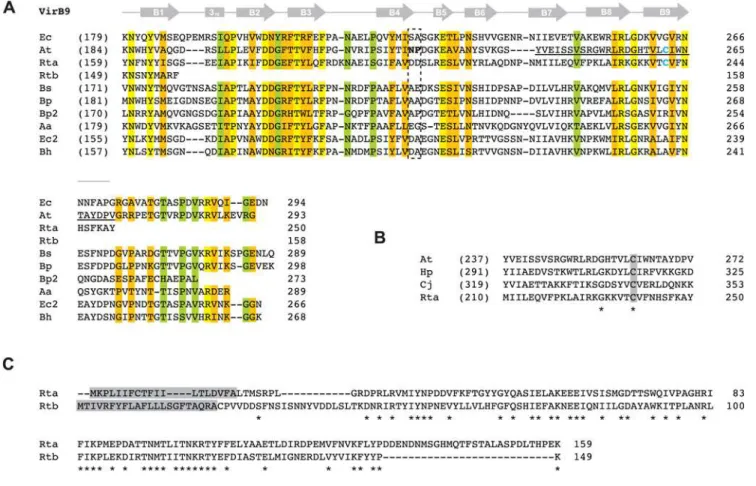
Figure 5. Comparative analysis of VirB9 and VirB9-like proteins with emphasis on twoRickettsiaorthologs.In all panels, coordinates for each sequence are shown to the right in each block, with numbers in parentheses depicting flanking residues of the alignment not shown.(A)
Multiple sequence alignment of ten VirB9 and VirB9-like sequences corresponding to regions of the NMR structure for the VirB9 (TraOCT)/VirB7 (TraN)
interaction inEscherichia coli(IncN plasmid R46) [141]. Secondary structural model is shown above the alignment with arrows (b-strands B1–B9) and a bar (310helix). Dashed box indicates the region demonstrated to protrude extracellularly from the OM. Blue cysteines in At (C262) and Rta (C241)
depict the residues that participate in the disulfide bridge between VirB9Tiand VirB7Ti[53,54,116,135]. Region underlined in VirB9Ti(G242–G271) is
depicted inB. Conservation color scheme is the same as inFigure 2legend, with calculations adjusted for four additional sequences. See text for alignment details. Taxon abbreviations and associated NCBI accession numbers are as follows: Ec =E. coliTraO (IncN plasmid R46), NP_511196; At =Agrobacterium tumefaciensVirB9, NP_396496; Rta =Rickettsia typhiVirB9a, YP_067240; Rtb =R. typhiVirB9b, YP_067243; Bs =Brucella suisVirB9, NP_699268; Bp =Bordetella pertussis VirB9, NP_882293; Bp2 =Bordetella pertussis TraK (plasmid pSB102), NP_361041; Aa =Aggregatibacter actinomycetemcomitansmagB09, NP_067575; Ec2 =E. coliTrwF (plasmid pR388), CAA57030; Bh =Bartonella henselaeTrwF, CAF28337.(B)Illustration of the conserved Cys residue shared between VirB9Ti,R. typhiVirB9a and the ComB9 proteins ofHelicobacter pylori(CAA10656) andCampylobacter
proximity to TMS regions is unusual, and coupled with a growing number ofvirB3/virB4fusions identified from other T4SSs, raises the possibility that VirB3 has more of an IM-periplasmic function [101]. Few other residues are conserved across the sampled taxa. The gene is highly conserved at the nucleotide (6.2%) and amino acid (4%) level acrossRickettsia, suggesting an essential function for this poorly characterized protein.
Bioinformatic prediction of three additional scaffold components
Our previous phylogenomic study on Rickettsia collectively suggested through consensus genome annotation that, relative to theA. tumefaciensarchetype T4SS,Rickettsiagenomes are devoid of Vir scaffold genes encoding VirB1, VirB2, VirB5 and VirB7 [81]. Below, we discuss the various methods used to uncover aRickettsia T4SS that is only lacking a virB5 homolog. We provide background information for these genes, as they may be prone to misidentification via standard genome automation methods for a variety of reasons discussed below.
virB1. VirB1 and related proteins belong to a class of soluble lytic transglycosylases (LTs), enzymes involved in maintaining the integrity of the bacterial cell wall [102]. Specifically, LTs manage the turnover of the murein layer by degrading peptidoglycan, working in concert with murein synthesizing enzymes in a housekeeping fashion [103,104,105]. LTs are part of an ancient glycohydrolase superfamily that includes plant chitinases, bacterial chitosanases, goose and hen g-type lysozymes, and phage T4 lysozymes [106]. The specific class of LTs encompassing VirB1 and VirB1-like proteins also includes proteins from other secretion systems and related enzymes present in bacteriophages [66,67,107,108,109]. This class of ‘‘specialized’’ LTs [110] differs from housekeeping LTs in that peptidoglycan is locally disrupted for specific purposes [107], and in the case of VirB1 and related proteins, this purpose is to accommodate the assembly of the T4SS apparatus across the entire cell envelope [66]. Whereas all other Vir components inA. tumefacienshave been shown to be essential for type IV secretion, VirB1 is not, as deletion/mutation of virB1 greatly reduces virulence but does not eliminate tumor formation [42,111,112,113]. VirB1-like proteins are also non-essential in other systems [66,114], although a VirB1 homolog (HP0523) in thecag T4SS ofH. pylori is critical for both CagA translocation/phosphorylation and interleukin-8 induction in host cells [115]. Aside from self-oligomerization, VirB1 interacts with several other Vir components, namely VirB4 and VirB8-VirB11 [116,117], illustrating its periplasmic role in peptidoglycan degradation. Apart from the LT domain of VirB1Ti, the
C-terminal VirB1* is released by cleavage in the periplasm [65] and acts independent of VirB1Tiin tumorigenesis [118]. Specifically,
VirB1* interacts with VirB2Ti and VirB5Ti to promote T pilus
formation [119]. Despite the conserved consensus sequence ‘‘(YH)Vx(KRQ)V’’ near the cleavage site in VirB1Ti, such
processing has not been demonstrated in any other VirB1 or VirB1-like sequence to date, although a cleaved VirB1 product was reported in the cell lysate ofBrucella abortus[113].
Searches of theRickettsiadatabase [80] for genes annotated as coding for LTs yielded two conserved families. The first family contained genes annotated as either ‘‘soluble lytic murein transglycoslyase’’ or ‘‘hypothetical protein’’. This gene family encodes proteins of an average length 650 aa, consistent with the size of the product of E. coli slt. Considerable variation in annotation existed for the second gene family, ranging from ‘‘hypothetical protein’’ to ‘‘invasion protein’’ to ‘‘soluble lytic transglycosylase’’. These genes encode proteins averaging 290 aa, a size typical of many specialized LTs. Interestingly, one member
of this family,R. prowazekiiORF 457, was previously suggested as a possible candidate for VirB1 in a comparison of several divergent T4SSs [26]. This prediction was never incorporated into genome annotation. A search of the NCBI protein database for ‘‘VirB1’’ yielded 142 results, of which all results containing ‘‘VirB1’’ in the protein annotation were selected for comparison with the smaller rickettsial LT domain containing proteins. These proteins were aligned and a subset of this alignment illustrates the limited conservation in VirB1 and VirB1-like sequences across divergent bacteria (Figure 6). Collectively, the selected sequences are most conserved in the regions corresponding to the active site motifs of the crystal structure ofE. coliSlt [120], which are also conserved in the largerRickettsia‘‘soluble lytic murein transglycoslyase’’ family. The C-terminal region of VirB1* processing is also conserved as previously reported [119], and all predicted VirB1* sequences contain tracts of repeated residues, particularly Pro rich tracts. Across the 13Rickettsiagenomes, these putative VirB1 sequences exhibit the least conservation of all Vir components at the nucleotide (13.9%) and amino acid (16.3%) level (after the variable flanking sequences of VirB6 proteins are excluded, see above). Accordingly, we suggest that Rickettsia genomes contain both housekeeping LTs orthologous toE. coliSlt and related Slts, as well as specialized LTs similar to VirB1, which are likely to play a role in type IV secretion via the localized degradation of peptidogly-can.
virB2. VirB2 and related proteins comprise the major pilin subunit of the T pilus [121,122] and are also essential components of the mating channel [42,62,123,124]. The signal sequences of these proteins are cleaved in the periplasm, followed by additional species-specific processing of the N- and C-termini [92,125,126,127,128,129,130]. InA. tumefaciens, VirB2Tiis made
circular through a cyclization process mediated by an uncharacterized chromosomal-encoded gene product [126,127]. Various laboratory strategies have identified VirB2 in complexes with VirB5 and VirB7 [55,131,132,133,134,135], supporting its role in T pilus formation. The VirB2-VirB5 pilus complex is dependent on the periplasmic interaction between VirB4 and VirB8, which results in the formation of extracellular pili [135]. However, all VirB2 and VirB2-like proteins are incorporated into the IM [2], as evident by at least two predicted TMS regions in all analyzed sequences. Polymers [134] of the protein are critical for substrate transfer in the mating channel [124,136] and possibly span the entire periplasm [101]. Given the recent hypothesis that T4SSs have dual (and independent) biogenesis roles in T pilus formation and substrate transfer [101], it is apparent that VirB2 and VirB2-like proteins are essential for both processes [2].
VirB2 and VirB2-like proteins [137]. Across the 13 Rickettsia genomes, the putative VirB2 sequences are conserved at the nucleotide (10.5%) and amino acid (11.6%) level, suggesting these proteins function in substrate transfer through the mating channel. virB7. VirB7 and related proteins are typically the smallest components of T4SS scaffolds and are seemingly not present within all identified systems [18]. However, the extremely small size of these lipoproteins poses a challenge for detection with blast, thus leaving open the possibility that VirB7 and related lipoproteins have yet to be characterized within some T4SSs. VirB7 is secreted to the periplasm [58,138] and interacts with the CTD of VirB9, stabilizing the OM-associated protein [53,54,58,59,139] and other Vir components in the mating channel [45,131]. As discussed above, the critical disulphide bond linking VirB7Ti and VirB9Ti [53,54,116,135] is not
conserved in most other T4SSs, suggesting other protein interactions suffice in heterodimer stabilization [140]. VirB7Tiis
also IM-associated [58] as well as found in the extracellular milieu in abundant levels [128], even independent of other VirB protein synthesis [101]. VirB7Ti forms homodimers via intermolecular
disulphide bonds, and also interacts with VirB2 and VirB5 [128,132,134]. Dimerization in other VirB7 proteins via Cys-Cys bridges should be possible given the highly conserved nature of the lipo-processing site (Cys-24 of VirB7Ti).
In a comparison of several divergent T4SSs,R. prowazekiiORF 288 was suggested as a possible candidate for VirB7 [27], a conclusion likely reached based on synteny, as the gene for RP288 is located directly upstream ofvirB8b(Figure S1). However, this ORF is not annotated as VirB7 in anyRickettsiagenome [80,81], a likely consequence of its small size and zero blastp hits to related sequences from other bacteria. We took RP288 and orthologous proteins from the additional 12 Rickettsia genomes and aligned them to 70 VirB7 and VirB7-like proteins retrieved from GenBank, a subset of which reveals the limited sequence
Figure 6. Comparison of soluble lytic transglycosylase (LT) domains of two rickettsial proteins to the LT domain ofEscherichia coli
Slt70 and ‘‘specialized’’ LT domains within VirB1 proteins from four pathogenic bacteria.Alignment below horizontal line of entire VirB1 and VirB1-like sequences performed using MUSCLE [194,195] (see text). Above the horizontal line is the alignment of the LT domains ofE. coliSlt70 and the larger rickettsial sequence using EMBOSS (Needle algorithm). This alignment was fitted to the alignment below the horizontal line based on a second needle alignment forE. coliSlt70 and the VirB1 protein fromBrucella suis[116]. Conserved residues previously implicated in enzymatic activity are within boxes [210]. An asterisk depicts the essential active-site Glu [67,120,211,212]. Interactions between VirB1 and other T4SS components as revealed by peptide array mapping [65] are shown over the alignment with the following symbols: 8 = VirB8; 9 = VirB9; E = VirB11; A = VirB8+VirB9; B = VirB9+VB11; C = VirB8+VirB11. Conservation color scheme is the same as inFigure 2legend, except orange = invariant over 70% of sequences
and blue = invariant across all VirB1 and VirB1-like sequences, with conserved contrasting residues in the LT domain alignment also colored blue (if applicable). Conservation was not assessed for columns including gaps. Black arrow depicts the cleavage site ofAgrobacterium tumefaciensVirB1 protein [65] (NOTE: cleavage in other VirB1 and VirB1-like proteins has not been definitely demonstrated). Entire CTDs are shaded and Pro residues are colored white and di- and poly-repeat residues underscored white. Shaded N-terminal residues depict signal peptides predicted by SignalP 3.0 [196]. Taxon abbreviations and associated NCBI accession numbers are as follows: Ec =E. coli Slt70, P0AGC3; Rt1 =Rickettsia typhi putative transglycosylase, YP_067347; Rt2 =R. typhi putative soluble lytic murein transglycosylase, YP_067402; At =A. tumefaciens VirB1, NP_053381; Bc =Burkholderia cepaciagenomovar III VirB1, AAK50141; Bs =B. suisVirB1, AAN33281; Ps =Pseudomonas syringaeVirB1, AAR02172.
conservation in this protein family (Figure 8). The Cys lipoprotein-processing site was invariant across all 82 proteins in the larger alignment, however the remaining C-terminal region was poorly aligned with a high rate of length heterogeneity. In the smaller alignment, manual adjustment was made to the C-terminal region around the conserved ‘‘P[ILV]NK’’ motif [141], and the Cys residues of VirB7Ti and Rickettsia VirB7 were
homologized (Figure 8A). Unless using excessive gap insertion, both landmark features could not be homologized across the
alignment, as the Rickettsia protein has an extended sequence between the Cys residue and the P[ILV]NK motif. In the TraOCT/TraN NMR structure of the E. coli pKM101 T4SS, TraN (VirB7) winds around part of the b-sandwich formed by TraO (VirB9), with the P[ILV]NK motif of TraN embedded within a deep hydrophobic pocket of TraO [141]. The authors suggested the distance from the lipidated Cys to this conserved motif (,16 aa) could be critical in orienting TraO in the OM. The effect the longer sequence in Rickettsia VirB7 proteins has on a
Figure 7. Characteristics and comparative analysis of VirB2 and VirB2-like sequences from six divergent bacterial species. (A)
Schema illustrating the processing (cleavage and cyclization) ofAgrobacterium tumefaciensVirB2 [125,126,127].(B, C)Automated (top) and manual (bottom) alignment of VirB2 and VirB2-like proteins across six bacterial species. For automated alignment, conservation color scheme is the same as inFigure 2legend. See text for alignment details. Manual alignment shows a putativeRickettsiaVirB2 protein threaded to a prior homology model [122]. Boxes with colored borders depict further support for this model with the addition of the rickettsial sequence. Gaps (versus dots) depict adjustments to the original model. Taxon abbreviations and associated NCBI accession numbers are as follows: At =A. tumefaciensVirB2, AAB28331; Rt =Rickettsia typhihypothetical protein, YP_067147; Lp =Legionella pneumophilalvhB2, CAB60051; Bs =Brucella melitensis suisVirB2, ABM66830; Bp =Bordetella pertussispertussis toxin transport protein, NP_882287; Bh =Bartonella henselaeVirB2, AAD48919.(B)Predicted signal peptides based on Signal P v.3.0 output [196] (top) and laboratory-defined processed sites [122] (bottom). Bolded residues (top) depict conserved Ala residues implicated by both methods as the cleavage site.(C)Core regions of the processed VirB2 and VirB2-like proteins. Blue residues (top) depict predicted TMS regions [93], and these regions are enclosed over the predicted TMS regions (black bars) in the prior homology model [122].
potential VirB7/VirB9 interaction is unknown, but given a predicted lipo-processing site, a second conserved Cys residue, a potential P[ILV]NK motif, and a conserved genomic position immediately upstream ofvirB8b, we suggest these small ORFs are strong candidates for VirB7 proteins. Interestingly, a manual alignment of VirB7Ti with the putativeRickettsia VirB7 and the
ComB7 proteins of H. pylori and C. jejuni revealed a conserved motif ‘‘[KI]KSP’’ directly flanking the second conserved Cys in the latter three taxa (Figure 8B). Perhaps this feature accommo-dates the extra length in theRickettsiasequences, and at very least presents the possibility that the VirB7/VirB9 structural interaction is flexible and variable across different T4SSs.
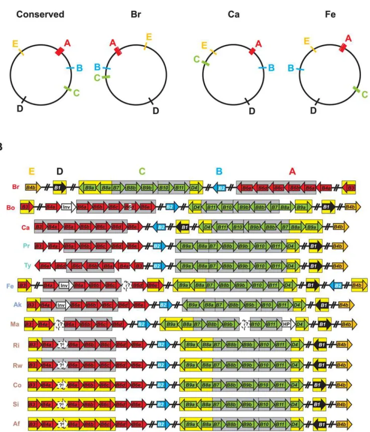
Rickettsia T4SS: an evolutionarily conserved archipelago Whether encoded on plasmids (common) or chromosomes (rare), the components of T4SSs are typically arranged in one operon or several adjacent operons [101,142]. Regarding T4SSs closely related to the vir system, genes encoding T4CPs (when present) are commonly not arrayed with VirB and VirB-like genes, but located in nearby operons that often include additional genes related to substrate processing [142]. Collectively, the 18 Vir genes within Rickettsia genomes are scattered throughout the circular chromosomes, with the T4CP gene (virD4) arrayedwithother Vir
components (Figure 9A). Given the conserved synteny inRickettsia genomes, especially in regards to the three derived groups [81], the various Vir components, now expanded to five genomic regions in most cases, can be organized into five ‘‘islets’’ (A–E) comprising an archipelago that spans the entire genome of each taxon. Ten of the 13 analyzed genomes have the five islets in similar locations, with islets C and D inR. canadensisand islet B inR. feliswithin regions of rearrangement unique to both genomes. The large rearrangement of most of theR. belliistr. RML369-C genome relative to all other Rickettsiagenomes positions its T4SS in a mirror image relative to the conserved arrangement.
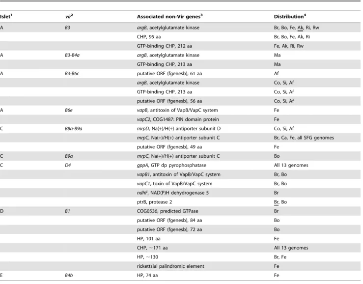
Operon prediction [143] within these islets was variable across the 13 genomes, and several islets were predicted to contain additional genes outside of those encoding the T4SS scaffold (Figure 9B, Table 2). Given the propensity for T4SS effectors and regulators to be encoded within close proximity to the scaffold genes [144], these genes will be of interest to future studies that aim to identify protein substrates (if any) specific to this transporter, as well as factors that regulate its expression. Two genes in particular are interesting in part due to their presence in all 13 genomes.gppAencodes an enzyme similar to RelA/SpoT homologs that functions as a mediator of the stringent response, hence coordinating a range of cellular activities in reaction to
Figure 8. Comparative analysis of VirB7 and VirB7-like lipoproteins with emphasis on the similarities betweenRickettsiaVirB7 and ComB7 proteins.In both panels, coordinates for each sequence are shown to the right, with numbers in parentheses depicting flanking residues of the alignment not shown. Green Cys = predicted lipoprotein cleavage site [197] and orange Cys = residue in disulphide bridge with VirB9/VirB9-like proteins. The ‘‘[KI]KSP’’ motif shared byRickettsiaspp. VirB7 proteins and ComB7 proteins fromHelicobacter pyloriandCampylobacter jejuniis shaded gray. See text for alignment details.(A)Multiple sequence alignment of nine VirB7 and VirB7-like sequences. Gray bar depicts region of the NMR structure for the VirB9 (TraOCT)/VirB7 (TraN) interaction inEscherichia coli(IncN plasmid R46), with the conserved ‘‘P[ILV]NK’’ motif in blue [141]. Taxon
abbreviations and associated NCBI accession numbers are as follows: Ec =E. coli TraN of IncN plasmid R46, NP_511194; At =Agrobacterium tumefaciensVirB7, NP_536291; Rt =R. typhihypothetical protein, YP_067241; Bs =Brucella suisVirB7, AAN33275; Bp =Bordetella pertussisTraI protein of plasmid pSB102, NP_361043; Bp2 =B. pertussisputative bacterial secretion system protein, NP_882291; Aa =Aggregatibacter actinomycetemco-mitans lipoprotein, NP_067577; Ec =E. coli TrwH protein, FAA00034; Bartonella henselaeTrwH-like protein, AAM82208. (B) Multiple sequence alignment of VirB7Ti,R. typhiputative VirB7 and the ComB proteins ofH. pylori(CAA10654) andC. jejuni(NP_863349). Invariant residues are denoted
changes in nutritional abundance [145]. Given that expression of the T4SS ofBrucellaspp. has been demonstrated to be gppA-like dependent [146], this protein is a potential candidate for regulation of the RickettsiaT4SS. The second conserved non-Vir gene encodes a conserved hypothetical protein within the predicted VirB1 operon, an ideal location for an effector protein given the role of VirB1 in murein degradation. blastp results using this protein as a query showed little similarity to other proteins (data not shown).In silicocharacteristics, such as lack of predicted signal peptides and a skew in positively charged residues in the C-terminal region, a common attribute of some T4SS effectors [2], make this curious protein a candidate for type IV secretion. Interestingly, the protein from R. sibirica bound VirB8 in a
bacterial two-hybrid screen [74]. Finally, several of the remaining non-Vir genes within predictedviroperons are attractive and open avenues for exploring lineage specific virulence factors associated with theRickettsiaT4SS.
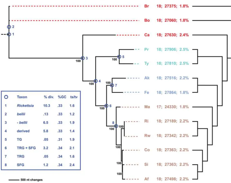
Combined phylogeny estimation from all 18 components of the RickettsiaT4SS corroborated the species tree generated from an analysis of over 700 coreRickettsiagenes (Figure 10, Figure S7). The inclusion of additional genomes released subsequent to our prior analyses, namelyR. massiliae, R. africae and R. rickettsii str. Iowa, does not overturn our tree-based classification ofRickettsia into four major groups: ancestral group (AG), typhus group (TG), transitional group (TRG), and spotted fever group (SFG) rickettsiae [81,147]. All but one of the phylogeny estimations of non-Vir genes (NB: seeTable 2for non-Vir genes associated withinviroperons). Genes predicted by fgenesb but not PATRIC are within dashed white boxes and annotated as ‘‘?’’. Genome codes are as follows: Br =R. belliistr. RML369-C, Bo =R. belliistr. OSU 85 389, Ca =R. canadensisstr. McKiel, Pr =R. prowazekiistr. Madrid E, Ty =R. typhistr. Wilmington, Fe =R. felisstr. URRWXCal2, Ak =R. akaristr. Hartford, Ma =R. massilaestr. MTU5, Ri =R. rickettsii str. Sheila Smith CWPP, Rw =R. rickettsiistr. Iowa, Co =R. conoriistr. Malish 7, Si =R. sibiricastr. 246, andR. africaestr. ESF-5. The genome codes are colored to reflect classification [81,147]: red = ancestral group rickettsiae (AG), turquoise = typhus group rickettsiae (TG), blue = transitional group rickettsiae (TRG), and brown = spotted fever group rickettsiae (SFG).
doi:10.1371/journal.pone.0004833.g009
Table 2.Non-Vir Genes Within Predicted Operons Encoding theRickettsiaT4SS.
Islet1 vir2
Associated non-Vir genes3 Distribution4
A B3 argB, acetylglutamate kinase Br, Bo, Fe, Ak, Ri, Rw
CHP, 95 aa Br, Bo, Fe, Ak, Ri
GTP-binding CHP, 212 aa Fe, Ak, Ri, Rw
A B3-B4a argB, acetylglutamate kinase Ma
GTP-binding CHP, 213 aa Ma
A B3-B6c putative ORF (fgenesb), 61 aa Af
argB, acetylglutamate kinase Co, Si, Af
GTP-binding CHP, 213 aa Co, Si, Af
putative ORF (fgenesb), 56 aa Co, Si, Af
A B6e vapB, antitoxin of VapB/VapC system Fe
vapC2, COG1487: PIN domain protein Fe
C B8a-B9a mrpD, Na(+)/H(+) antiporter subunit D Co, Si, Af
mrpC, Na(+)/H(+) antiporter subunit C Br, Ca, Fe, all SFG genomes
putative ORF (fgenesb), 49 aa Fe
C B9a mrpC, Na(+)/H(+) antiporter subunit C Bo
C D4 gppA, GTP dp pyrophosphatase All 13 genomes
vapB1, antitoxin of VapB/VapC system Br, Bo
vapC1, toxin of VapB/VapC system Br, Bo ndhF, NAD(P)H dehydrogenase 5 Br
ptrB, protease 2 Br, Bo
D B1 COG0536, predicted GTPase Br
putative ORF (fgenesb), 84 aa Bo
putative ORF (fgenesb), 72 aa Bo
HP, 101 aa Fe
CHP,,171 aa All 13 genomes
HP,,130 Br, Fe
rickettsial palindromic element Fe
E B4b HP, 74 aa Fe
1Corresponding to islets A (
virB3-virB6e), C (virB9a-virD4), D (virB1), E (virB4b) (SeeFigure 9).
2According to operon prediction by fgenesb [143].
3Consensus annotation from PATRIC [80] or fgenesb prediction. HP = hypothetical protein, CHP = conserved hypothetical protein. 4Taxa included within operon prediction. Underlined taxa depict split ORFs.
the independent Vir components failed to recover the species tree topology (Table 3; Figure S8), however deviations were minor and can be explained by the properties of the data (too few informative sites or homoplasy). In these latter cases, either the independent analyses failed to resolve the highly similar derived members of the SFG rickettsiae (R. rickettsii,R. conorii,R. sibirica,R. africae), for which the assignment of species names has received recent criticism [82], or failed to placeR. canadensisas basal to the TG, TRG and SFG rickettsiae. We previously reported that the fluctuating position of R. canadensis was dependant upon the analyzed gene(s) and phylogenetic utility of the said gene(s) [81], and our current analysis, as well as a very recent study [148], further supports this phenomenon. Altogether, phylogeny estima-tion supports a single inheritance of the 18 Vir components from theRickettsiaancestor, with one gene loss (virB6einR. massiliae), one split gene (virB6dinR. belliistr. OSU 85 389), three major gene rearrangements (Figure 9A), and several minor switches of coding strand (Figure 9B) the defining diversifying factors of this conserved system. Our analysis corroborates a recent study that analyzed a subset of Vir components (VirB3, VirB4, VirB8, VirB8,
VirB11) from 31 taxa of Alphaproteobacteria and demonstrated vertical inheritance of Vir genes across the entire Rickettsiales [149].
Lateral acquisition of Rickettsiales T4SS
Our synteny and phylogenetic analyses of the Rickettsia T4SS yield results consistent with previous studies on the T4SS operon structure of the closely related rickettsiaeAnaplasma,Ehrlichiaand Wolbachia, as these genomes have at least two virclusters (virB3 -virB4-virB6and virB8-virB9-virB10-virB11-virD4) in well-separated regions of their genomes, often with duplications of the VirB4, VirB6, VirB8, and VirB9 components [149,150,151,152]. Inter-estingly, the genome sequence of the sister taxon to Rickettsia, Orientia tsutsugamushi, revealed an unprecedented degree of Vir-like gene duplication (mostly comprising components of theE. coli tra operon), with 359 ORFs putatively coding for various components of a T4SS throughout 79 sites in the genome [153]. Thus a remarkable array of diverse T4SSs exists across species in the Rickettsiales, yet intrageneric conservation of these systems is apparently high. Interestingly, archipelagos ofvirislets seem to be
Figure 10. Phylogeny estimation of 18 putativevirgenes of theRickettsiaT4SS.Single most parsimonious tree of 12716 steps (6858 parsimonious characters of 28554 total characters). Branch support is from 1 million bootstrap replications. Taxon codes and coloring scheme as described in theFigure 9legend. Statistics to the right of taxon codes: number of T4SS genes; number of nts encoding T4SS; percentage of genome encoding T4SS. Cladogram on right depicts phylogeny of core orthologous groups (proteins) [80]. Inset shows statistics computed for eight nodes of the tree, following our classification scheme [81].
characteristic of all genomes in the Rickettsiales, and it has been shown in one system (Ehrlichia chaffeensis) that the same protein, EcxR, regulates all of its vir islets [150]. A search for a protein orthologous to EcxR in all of theRickettsiagenomes proved futile, although the small size of this protein (108 aa) is likely confounding blastp searches. It is expected that similar T4SS regulators will be identified in other Rickettsiales genomes, given the need to tightly regulate all of the scaffold components for efficient transporter function.
Collectively, conserved features across rickettsial T4SSs hint at a single inheritance that possibly fostered the route to obligate intracellular symbiosis (mitochondria, symbiotic rickettsiae) and subsequent pathogenesis after the divergence of the rickettsial ancestor from its free-living marine relativePelagibacter ubique[86]. Given that phylogeny estimation [18] and protein homology network-based clustering [29] of T4SSs imply the Rickettsialesvir system is closely related to the T4SSs from certain c- (Legionella spp. and Photobacterium profundum) and e-proteobacteria (H. pylori, Wolinella succinogenes and C. jejuni), it is likely ancestors of these distantly related organisms resided in a common environment and acquired similar T4SS genes from one or several progenitors. Several lines of evidence support this. First, Rickettsia bellii str. RML369-C [78],Legionella pneumophila[154] andH. pylori[155] are all capable of growth in various species of amoeba. Given the role of protozoa as reservoirs for amoeba-resistant organisms [156,157,158], it is likely amoeba provided a breeding ground
for rickettsial species and distantly related microbes [78]. Second, some members of the third class of Rickettsiales, the Holospor-aceae, are also found in amoeba [159,160,161]. This suggests at least two lineages branching from the Rickettsiales ancestor were capable of endosymbiosis within nucleated single-celled organisms (the mitochondrial endosymbiosis [162] being the other). Third, a gene encoding a Sec7-domain-containing protein, RalF, is known in prokaryotes only from Rickettsia and Legionella spp. [163]. In Legionella, RalF is a T4SS (Note: of the dot/icm type B T4SS) effector that functions as a guanine nucleotide exchange factor that recruits ADP-ribosylation factor to occupied phagosomes, permit-tingLegionellato replicate free from the host immune system and subvert vesicular trafficking [7]. While the NTD and associated central Sec-7-capping-domain are conserved in all Rickettsia genomes aside from SFG rickettsiae [81], a function forRickettsia RalF or association with the T4SS has still yet to be determined. Finally, the abovementioned similarities betweenRickettsiaVirB7/ VirB9a proteins and the ComB7/ComB9 proteins ofH. pyloriand C. jejunimay hint at a shared OM anchoring system for the mating channel common to VirB7Ti/VirB9T, although a similar role in
competence (versus protein translocation) inRickettsia cannot be ruled out.
Vir gene evolution via duplication and intrachromosomal recombination
It is not uncommon for bacteria to harbor multiple unrelated T4SSs; e.g., thecagandcomBsystems ofHelicobacter[164,165], the dot/icmandlvhsystems ofLegionella[27,166,167], and thevbh,virB and trw systems of Bartonella [168,169]. While many T4SSs are mainstays that define bacterial lifestyles, other T4SSs may be strain specific and highly plastic throughout bacterial populations, especially if encoded on plasmids [170]. InLegionella, components from thelvhsystem have been shown to complement analogs of the dot/icm system [27], suggesting functional resilience despite sequence plasticity. Growing studies utilizing cross-species heter-ologous complementation attest to this functional resilience [171,172]. Despite this, gene duplications within individual T4SSs are not the norm (Seubert et al., 2003; Alsmark et al., 2004), and the functional redundancy is likely best explained on a case-by-case basis. For example, a recent study of thetrwT4SS across sevenBartonellataxa revealed 7–8 copies oftrwL(virB3) and 2–5 copies of the trwJ-H operon (virB5-7) [173]. Phylogenetic analysis of the TrwL duplicates revealed probable neofunctiona-lization within a subset of the duplicates, while the flanking TrwJ and TrwH duplicates of the trwJ-H operon appeared to be products of LGT speculated to have arisen due to the coevolution of these OM proteins with erythrocyte surface structures.
Our recent generation ofRickettsiaOGs across 10 [81] and 12 [80] genomes unambiguously delineated the duplicate VirB4, VirB8 and VirB9 and quadruplicate VirB6 genes into discrete OGs. Sequence divergence and other summary statistics (Table 3) as well as phylogenetic analysis (Figure S4, Figure 11) strongly suggest all of these genes were vertically acquired (i.e., the duplications were ancestral). Analysis of the three duplicate components (VirB4, VirB8 and VirB9) suggests pseudogenization or neofunctionalization has occurred in one member from each family, as the suspects (virB4b,virB8aandvirB9b) comprise the only three T4SS genes that deviate from the average genomic base composition and drop below 30% GC (Table 3). The characteristics of these divergent duplicates (discussed above) are mapped onto individual phylogeny estimations (Figure 11). Given that avirB4active site mutant stabilized VirB3 and VirB8 and promoted T-pilus formation in A. tumefaciens [135], it is possible VirB4b still can function as an IM gate that stabilizes a
Table 3.Summary Statistics for the Vir Genes Encoding the RickettsiaT4SS.
Vir aa seqs nt seqs
div1 PI2 Tree3 div1 PI2 Tree3 GC4 ts/tv5 dN/dS6
D4 4.2 54 N 8.1 329 N 0.35 1.9 0.06
B4a 3.7 72 N 6.7 380 N 0.34 2.5 0.05
B4b 9.9 195 N 9.2 570 N 0.28 1.9 0.13
B11 4.8 33 N 9.7 186 N 0.37 1.9 0.06
B6a 14.7 374 N 12.8 1054 N 0.36 1.5 0.23
B6b 13.2 196 N 11.2 565 N 0.34 1.9 0.2
B6c 15.2 285 N 13.2 848 N 0.35 1.2 0.27
B6d 12.9 263 N 10.3 682 N 0.32 1.9 0.18
B6e 12.4 308 N 11.1 871 N 0.33 1.6 0.17
B8a 13.4 63 Y 10 163 N 0.26 1.7 0.26
B8b 9.4 55 N 9 161 N 0.31 1.3 0.13
B9a 4.7 27 N 7.6 130 N 0.35 1.8 0.06
B9b 10.8 43 N 8.2 98 N 0.26 2.5 0.14
B10 10.5 108 N 11 356 N 0.38 1.4 0.14
B3 4 8 N 6.2 43 N 0.31 1.4 0.1
B1 16.3 93 N 13.9 271 N 0.32 1.3 0.6
B2 11.6 35 N 10.5 104 N 0.32 1.9 0.26
B7 15.3 23 N 11 47 N 0.32 1.4 0.21
1Avg. pairwise differences across all sequences divided by avg. sequence
length.
2Number of parsimony informative characters.
3Tree corroborates (Y) or disagrees with (N) species tree. All trees are shown in Figure S4.
4%GC of all codon positions. 5Ratio of transitions to transversions.
6Ratio of non-synonymous to synonymous substitutions.
T4SS channel, facilitating (but not powering) the uptake or release of substrates. A full length VirB8 protein unable to dimerize or properly interact with other Vir components may still function in formation of a separate membrane-spanning channel. Also, a chaperone-assisted process may help VirB8b overcome the critical mutations in the linker between helixa4 and strandb4. However, given the asymmetrical position of the VirB8 and VirB9 duplicates around the putative VirB7 ORF (Figure 9), it is probable that these genes have experienced an intrachromosomal recombination event early in their evolution that deleted the CTD of VirB9b. In Rickettsia, intrachromosomal recombination has been implicated in the split of the rrs and rrl loci [174] and has likely shaped the atypical clustering of the Tuf and Fus genes [175]. Thus it is a common phenomenon inRickettsiagenomes. While neofunctiona-lization is difficult to suggest for VirB9b, given the deletion of the entire domain known to interact with VirB7, potential alternative functions for VirB8a and VirB4b are not unrealistic. However, in-frame pseudogenes that are still expressed may be degraded post-transcriptionally given the unusually high amount of genes involved in RNA degradation inRickettsiagenomes [71].
None of the proliferated VirB6 genes deviate from the average genomic base composition (Table 3), and given the conservation of the essential Trp residue in all sequences, as well as the constraints on hydrophobicity throughout large portions of proteins from all five duplicates (Figure 3), it is not possible to
rule out any VirB6 ortholog as a functional component of the RickettsiaT4SS. However, whilevirB6a-virB6dgenes have arrayed orthologs inO. tsutsugamushiand most of the Anaplasmataceae, a virB6eortholog inO. tsutsugamushiis not arrayed withvirB6a-virB6d, and no orthologs are present in the Anaplasmataceae genomes (data not shown). This correlates withvirB6ebeing the only deleted T4SS scaffold component across 13Rickettsiagenomes (R. massiliae str. MTU5). Nevertheless, selection for VirB6 duplications in Rickettsiales is evident given the trend for genome reduction [176], and the existence of at least four orthologs hints at a unique and constrained function. VirB6 proteins are similar to ComEC proteins [101], DNA channels involved in the uptake of environmental DNA [177]. Perhaps theRickettsiaVirB6 proteins are involved in DNA import, seeding multiple diverse uptake channels to maximize the potential for LGT, particularly in environments (protozoa, macrophage) with a high rate of congener contact. In support of this hypothesis, recent studies identified the Rickettsia accessory genome to be predominantly comprised of products and facilitators of the bacterial mobile gene pool [81,178]. The recent identification of plasmids [79,179,180] in Rickettsiahas shed light on the role of LGT in the sculpting of rickettsial genomes; however, a model for plasmid transfer has not been identified. Our identification of a Rickettsia T4SS that is highly similar to theLegionella lvhT4SS, which is primarily involved in conjugation [170], suggests a role in conjugation/DNA uptake
Figure 11. Phylogeny estimation of duplicate VirB4, VirB8 and VirB9 proteins.All trees estimated under parsimony (see text for details). Branch support is from 1,000 bootstrap replications. Taxon codes and coloring scheme as described in the Figure 9legend. Asterisks depict shortened branches that are shown in their full length below the tree. Skull and crossbones depict lineages leading to probable pseudogenization or neofunctionalization. Characteristics of these divergent sequences are listed.
needs consideration, particularly in light of the persistence of multiple channel proteins that defy the dynamics of rickettsial reductive evolution.
Lack ofvirB5correlates with rickettsial lifestyle
VirB5 and related proteins (e.g., TraC, TrwJ, LvhB5) are the minor components of the T-pilus [127,181,182,183], as they cofractionate with VirB2 and VirB7 [128,132] and bind both proteins in yeast two-hybrid screens [134]. virB6 expression stabilizes cellular levels of VirB5 as well as VirB3 [184], and a VirB5/VirB3 interaction via a yeast two-hybrid screen as well as a pull-down assay further supports the OM localization of VirB5 [134]. Signal sequences are predicted for most of the 157 VirB5 and VirB5-like proteins from diverse bacteria available on GenBank (data not shown). Like VirB2Ti, VirB5Tiinteracts with
VirB1* [119], and immuno-electron microscopy revealed that the protein localizes to the cell-bound region of the pilus as well as the distal regions of the pilus that contact other cells [185]. The crystal structure of TraC (a VirB5 analog) from the IncN plasmid pKM101 revealed a single domain consisting of three bundled helices and a globular appendage [186]. Further site-directed mutagenesis and functional complementation identified 14 highly conserved surface-exposed residues, as well as additional residues involved in TraC dimerization [186]. Despite important knowl-edge from these studies, the exact role VirB5 and VirB5-like proteins play in T4SS biogenesis and function remains unknown. blastp searches, as well as other tools designed to detect the strongly conserved residues located at the surface of TraC, failed to identify a VirB5 gene in any of the 13Rickettsiagenomes. As evidence is leaning more toward a role of VirB5 in host-cell recognition [185,186,187], the lack of avirB5inRickettsia is not surprising. A class of surface exposed proteins, Scas, have previously been implicated in host cell recognition and may trigger endocytosis [188]. Also, aside from recent evidence [78,79], pili have not been observed inRickettsia, and are not evident from all species. The structures reported forR. felis[79] andR. belliistr. RML 369-C [78] better resemble the flexible and tube-like Gram-negative bacterial conjugative pili, which are quite larger and easier to visualize than the T pilus and related structures [101]. Indeed, a diversity of extracellular pili and pili-like appendages associated with T4SSs exists, including needle like appendages [126,129], large sheathed structures [189,190], and fibrous meshes [191]. However, there is little evidence that the T pilus serves as a conduit for substrate transfer [2,140], and inA. tumefaciensT-DNA transfer occurs in the absence of T pilus production [6,42,131]. TheB. pertussisPtl T4SS also lacks a VirB5 gene, and it has been suggested that such a gene inB. pertussiswould be redundant, given the absence of cell-cell contact in the secretion of pertussis toxin [24]. Given the strictly intracellular lifestyle ofRickettsia, a VirB5 protein (and a T pilus) would also be redundant, as substrates would be secreted and imported directly from the host environment.
Conclusion
Previous studies on Gram negative bacterial T4SSs have hinted at a reduced scaffold inRickettsiaspp. relative toA. tumefaciensand other well characterized systems [27] (observations based largely on the R. prowazekii genome annotation), and indeed genomic studies on Rickettsia as well as other Rickettsiales genera have supported this assessment. However, the composition of the RickettsiaT4SS became more complex in recent years, with our past study determining that only four components (VirB1, VirB2, VirB5, VirB7) are missing compared to the vir T4SS of A. tumefaciens [81]. Reduced T4SSs, wherein only some genes are
present within genomes (relative to theA. tumefaciens virarchetype), have been suggested to have evolved different functions in certain bacteria [18]. However, trouble lurks within reliance on paradigms (and algorithms). For instance, two T4SSs of H. pylori, which independently function in pathogenicity (cag) and competence (comB), have recently been found to contain more components than automated annotation methods have predicted [164,165]. Regarding thecagsystem, strong evidence was found for additional putative T4SS components that are not comparable in aa sequence to any of the vir proteins [165]. Additionally, diverse components of the divergent dot/icmand lvhT4SSs of Legionella have been demonstrated to complement one another, underscor-ing the functional resilience of divergent T4SS proteins [27]. Thus caution must be undertaken when depending on automated genome annotation methods forin silicocharacterization of multi-component systems, such as the T4SS. The recent large-scale informatics analysis of 62 bacterial T4SSs revealed a highly conserved core set of T4SS components (VirB6, VirB8-VirB11, VirD4) coupled with a depauperate complex of the remaining Vir components, of which the latter were suggested to have been acquired in various independent events [29]. Our identification of three additional Rickettsia T4SS components (VirB1, VirB2, VirB7), all of which comprise much shorter sequences than the more conserved components (Table 4), implies that short (under 250 aa) or hyper-variable components are not easily revealed by automated methods. Finally, identifying all components of any secretion system may be hampered when selection does not favor the clustering of all genes within one operon, an apparent characteristic of some obligate intracellular bacteria [150,192].
Prior seminal reviews of T4SSs have focused largely on pathogenic bacteria, and it has been assumed that the Rickettsia vir-like T4SS plays a role in pathogenesis. Our study reveals a highly conserved T4SS across 13 genomes, some of which are not known to be pathogenic to invertebrate and vertebrate hosts. Despite an unknown function of this transporter, laboratory evidence is mounting for at least the expression, regulation and potential secretion of some of its components (Table 4). The lack of any identified effector proteins of theRickettsiaT4SS, coupled with an as-yet unknown mechanism for the high occurrence of LGT in these genomes, suggests that a function in DNA uptake and translocation, or some modified form of conjugation, cannot be overlooked. It is also possible that the Rickettsia T4SS is an amalgam of an effector translocation system and a DNA competence system, especially considering the redundancy of several of its components. These redundant components may serve as environment-specific factors facilitating the persistence of Rickettsia in its arthropod and vertebrate hosts, and probable protozoan reservoirs. The affinity to the Legionella lvh T4SS, which has recently been shown to serve as a virulence factor in the environmental spreading of Legionnaires’ disease [193], suggests the more appropriate annotation ofrvh(Rickettsialesvirhomolog) for this evolutionarily intriguing and enigmatic T4SS. Thus, Legionellaprovides a suitable genetic system for future character-ization of Rvh scaffold components, as well as for testing the translocation of predicted effector molecules.
Materials and Methods
Gene and Protein Sequences