The Arginine Methyltransferase PRMT6
Cooperates with Polycomb Proteins in
Regulating
HOXA
Gene Expression
Claudia Stein☯, René Reiner Nötzold☯, Stefanie Riedl☯, Caroline Bouchard,
Uta-Maria Bauer*
Institute for Molecular Biology and Tumor Research (IMT), Philipps-University of Marburg, Marburg, Germany
☯These authors contributed equally to this work. *[email protected]
Abstract
Protein arginine methyltransferase 6 (PRMT6) catalyses asymmetric dimethylation of tone H3 at arginine 2 (H3R2me2a), which has been shown to impede the deposition of his-tone H3 lysine 4 trimethylation (H3K4me3) by blocking the binding and activity of the MLL1 complex. Importantly, the genomic occurrence of H3R2me2a has been found to coincide with histone H3 lysine 27 trimethylation (H3K27me3), a repressive histone mark generated by the Polycomb repressive complex 2 (PRC2). Therefore, we investigate here a putative crosstalk between PRMT6- and PRC-mediated repression in a cellular model of neuronal differentiation. We show that PRMT6 and subunits of PRC2 as well as PRC1 are bound to the same regulatory regions of rostralHOXAgenes and that they control the
differentiation-associated activation of these genes. Furthermore, we find that PRMT6 interacts with sub-units of PRC1 and PRC2 and that depletion of PRMT6 results in diminished PRC1/PRC2 and H3K27me3 occupancy and in increased H3K4me3 levels at these target genes. Taken together, our data uncover a novel, additional mechanism of how PRMT6 contributes to gene repression by cooperating with Polycomb proteins.
Introduction
In mammals nine Protein Arginine Methyltransferases (PRMT1-9) have been identified, which transfer a methyl group from the ubiquitous cofactor S-adenosyl-L-methionine to the terminal guanidino nitrogens of arginine residues in target proteins. Subsequent to mono-methyl-arginine, type I PRMTs form asymmetric (ω-NG,ω-NG) dimethyl-arginine, whereas
type II enzymes give rise to symmetric (ω-NG,ω-N‘G) dimethyl-arginine [1]. Methylation of an
arginine does not alter the positive charge of the guanidinium side chain, but changes its struc-ture and the affinity between the substrate and its binding partners resulting in promotion or inhibition of interactions [2]. A multitude of nuclear and cytoplasmic proteins are substrates of arginine methylation, by which means PRMTs regulate a wide range of essential cellular func-tions [2].
OPEN ACCESS
Citation:Stein C, Nötzold RR, Riedl S, Bouchard C, Bauer U-M (2016) The Arginine Methyltransferase PRMT6 Cooperates with Polycomb Proteins in RegulatingHOXAGene Expression. PLoS ONE 11 (2): e0148892. doi:10.1371/journal.pone.0148892
Editor:Michael M. Nevels, University of St Andrews, UNITED KINGDOM
Received:December 22, 2015
Accepted:January 25, 2016
Published:February 5, 2016
Copyright:© 2016 Stein et al. This is an open access article distributed under the terms of the Creative Commons Attribution License, which permits unrestricted use, distribution, and reproduction in any medium, provided the original author and source are credited.
Data Availability Statement:All relevant data are within the paper and its Supporting Information files.
Funding:This work was supported by German Research Foundation/ Deutsche
Forschungsgemeinschaft (DFG) (http://www.dfg.de), grant no. TRR81 (project 3), BA2292/1-3. The funders had no role in study design, data collection and analysis, decision to publish, or preparation of the manuscript.
PRMT6 was identified in 2002 by using the high sequence conservation of the PRMT cata-lytic domain for homology search in the human genome [3]. The enzyme belongs to the type I enzymes and prefers mono-methylated arginine as substrate [4,5]. In agreement with its pri-mary nuclear localisation, PRMT6 methylates chromatin proteins. Similar to other PRMTs, PRMT6 typically targets glycin-arginine rich (GAR) motifs in substrates [6]. Accordingly, PRMT6 has been shown to modify HMGA1 and fibrillarin suggesting its involvement in chro-matin architecture, splicing and rRNA processing [3,7,8]. However, PRMT6 methylates also non-GAR motifs, such as in the Tat protein of HIV1 leading to restriction of viral transcription and replication [9]. Automethylation of PRMT6 in a non-GAR motif results in increased pro-tein stability and therefore activity [10]. Interestingly, PRMT6 was also reported to modify the DNA polymeraseβthereby enhancing base excision repair [11] and the androgen receptor contributing to neurodegeneration [12]. Furthermore, PRMT6 acts as a histone methyltrans-ferase on all four core histones, such as H3 at R2 [5,13], H2A at R29 [14] and H3 at R42 [15], thus leading to either transcriptional repression or activation. Due to transcriptional repression of tumor suppressor genes, PRMT6 promotes cell proliferation and blocks senescence [16–19].
The mechanism of transcriptional repression via asymmetric dimethylation of H3R2 (H3R2me2a) has intensively been studied. The H3R2me2a mark is a negative regulator of H3K4 trimethylation (H3K4me3), as it inhibits the activity of the H3K4 methyltransferase MLL1 and blocks recruitment of the MLL-complex subunit WDR5 to histone H3 [5,13,20]. Crystal structure analysis of WDR5 supports this antagonism between the two marks, as hydrogen bonding between the WD40 domain of WDR5 and the guanidino group of R2 is dis-turbed by asymmetric dimethylation [21,22]. Accordingly, overexpression of PRMT6 leads to global reduction of H3K4me3 levels and ChIP (chromatin immunoprecipitation) analysis showed a countercorrelation between the genomic occurrence of H3K4me3 and H3R2me2a [5,13]. Given that H3K4me3 generally marks the TSS (transcriptional start site) of actively transcribed as well as paused genes, reduction of the H3K4me3 levels by PRMT6-mediated H3R2me2a causes transcriptional repression, which has been demonstrated for a selection of PRMT6 target genes, such asHOXgenes, MYC-target genes andTSP-1[5,13,20,23]. In agree-ment with this, H3R2me2a is not enriched at promoters of active genes and occurs at silent genomic regions, such as pericentromeric chromatin [24,25]. Apart from interfering with the deposition of H3K4me3, H3R2me2a might also impair the recognition of H3K4me3, as some readers of H3K4me3 are blocked by the presence of an adjacent H3R2me2a mark, for example the TFIID subunit TAF3 and the NURF subunit BPTF [20,26,27]. However, it is not clear to which extent H3K4me3 and H3R2me2a might coexist on the same histone tail.
for the enzymatic activity of PRC1 by mono-ubiquitylating H2A at K119. CBX-containing PRC1 complexes recognise the H3K27me3 mark deposited by PRC2 via the chromodomain of the CBX subunit. Together, PRC1 and PRC2 achieve the stable transcriptional repression of PRC target genes.
Given that both H3K27me3 and H3R2me2a are repressive histone marks and co-occur at a subset of gene promoters, we asked here whether PRMT6 influences PRC-mediated repression of differentiation-associated target genes. We find that in NT2/D1 cells, a model for neuronal differentiation, PRMT6 and subunits of PRC1 as well as PRC2 are bound to the same regula-tory regions of rostralHOXAgenes and diminish their transcriptional activation. Further, we show that PRMT6 interacts with subunits of the PRC1 and PRC2 complexes. Interestingly, depletion of PRMT6 results on the one hand in declined PRC1/PRC2 recruitment as well as H3K27me3 levels and on the other hand in increased H3K4me3 levels at the rostralHOXA
genes. Taken together, our data uncover a novel, additional mechanism of how PRMT6 con-tributes to gene repression by cooperating with Polycomb proteins.
Materials and Methods
Cell lines, antibodies and plasmids
NT2/D1 and HEK293 cells were maintained in DMEM supplemented with 10% fetal calf serum (FCS, Gibco/BRL) at 37°C and 5% CO2. To induce neuronal differentiation of NT2/D1
cells with 0.1μM ATRA (all-transretinoic acid from Sigma) we followed the protocol
described elsewhere [36].
The following antibodies were employed: rabbit affinity-purified anti-PRMT6 was produced using His-tagged proteins corresponding to amino acids 60–375 of human PRMT6; anti-H3 (05–499) from Upstate; anti-H3R2me2a (04–808), anti-H3K27me3 (07–449) and anti-H3K4me3 (07–473) from Millipore; anti-Myc (9E11), anti-GFP (G6539), anti-Flag (M2) (F3165), mouse IgG (I5381) and rabbit IgG (I5006) from Sigma; anti-CDK2 (sc-163) from Santa Cruz; anti-CBX8 (A300-882A) from Bethyl Laboratories; anti-HPH1 [37], anti-HPH2 [38], anti-BMI1 [39], anti-CBX8 [40], anti-EZH2 [41], anti-RING1A [42], anti-RING1B [37] were kindly provided. The expression construct for HPH1 (Rae28) is described in [43]. The expression construct of Myc-HPH2 was a gift from H. Koseki (RIKEN Yokahma Institute). The expression constructs for CBX2, CBX4 and CBX8 were published by [40]. Flag-EZH2 was a gift from D. Reinberg.
Plasmid and siRNA transfection
Short interfering RNA (siRNA) oligonucleotide duplexes were obtained from Eurogentec or Dharmacon for targeting PRMT6, CBX8 and EZH2, respectively. The target sequences of siR-NAs are listed in the Supporting Information (S1 Text). Transfections of siRNAs in NT2/D1 cells were performed with the aid of Lipofectamine RNAiMAX (Invitrogen) according to the manufacturer’s instructions. Plasmids were transfected using the PEI or calcium phosphate methods.
Reverse transcription quantitative PCR (RT-qPCR) and chromatin
immunoprecipitation quantitative PCR (ChIP-qPCR)
Total RNA was isolated using Seqlab RNA-Mini-Kit according to the protocol and applied (0.5–1μg) to reverse transcription (RT) by incubation with oligo(dT)17primer and M-MLV
qPCR analysis gene-specific primers listed in the Supporting Information (S1 Text). Quanti-tative PCR was performed using Absolute QPCR SYBR Green Mix (Thermo Scientific) and the Mx3000P real-time detection system (Agilent). For RT-qPCRUBIQUITINgene tran-scription was used for normalisation. ChIP-qPCR results were calculated as % input. All experiments were performed at least three times and each experiment in triplicate. Repro-ducible and representative data sets are shown. Error bars represent mean +/- S.D. of tripli-cate reactions.
Gel filtration analysis
To prepare whole-cell extracts, cells were lysed in IPH-buffer (150 mM NaCl, 5 mM EDTA, 0.5% (v/v) NP-40, 50 mM Tris, pH 8.0) including protease inhibitors and incubated 30 minutes at 4°C under rotation. DNA was digested using Benzonase for at least one hour at 4°C. Subse-quently, proteins (3–5 mg) were separated on a Superose 6 HR 10/30 using the ÄktaTMPurifier 10 System (GE Healthcare) at a flow rate of 0.7 ml/minute and a pressure of 0.3–0.4 MPa. Six ml fractions were collected and either analysed by Western blot or used in co-immunoprecipi-tation followed by Western blot.
Results
PRMT6 and the PRC complexes occupy the same regulatory regions of
HOXA
genes during neuronal differentiation
The human embryonal carcinoma (EC) cell line NT2/D1 represents a well-characterised model system to study the regulation of theHOXAgene cluster (Fig 1Afor illustration of the
HOXAcluster) [36,44–46]. Upon all-transretinoic acid (ATRA) treatment NT2/D1 cells dif-ferentiate along the neuronal lineage, which is accompanied by transcriptional upregulation of rostral (3`)HOXAgenesHOXA1-5, whereas no or only subtle changes in expression of caudal (5`)HOXAgenesHOXA9-13have been observed [47,48]. These ATRA-induced tran-scriptional changes have been reported to be accompanied by a loss of PRC1 and PRC2 recruitment and a reduction of H3K27me3 levels at the rostralHOXAgenes [36]. Impor-tantly, theHOXA2gene is also a well-established target gene of PRMT6-mediated repression [5,20]. The finding that H3R2me2a and H3K27me3 coincide at human promoter regions [28] suggests a potential positive crosstalk between PRMT6- and Polycomb-mediated tran-scriptional repression. To further investigate PRMT6-occupied sites atHOXAgenes and how these relate to the binding mode of PRC1 and PRC2 at the gene cluster, we employed ChIP (chromatin immunoprecipitation) analysis of undifferentiated, 2 and 6 days ATRA-differen-tiated NT2/D1 cells. We determined the occurrence of PRMT6, its histone modification H3R2me2a, CBX8 as a representative for PRC1, EZH2 as a representative for PRC2 and H3K27me3 at regulatory regions and promoters of various rostralHOXAgenes, such as
HOXA1,A2andA5, as well as the caudalHOXAgenes,HOXA9andA10. We found that PRMT6 and H3R2me2a are present at the regulatory regions of all investigatedHOXAgenes in undifferentiated cells and that their occurrence remained unchanged at theHOXA9and
A10gene promoters throughout the time-course of differentiation (Fig 1B). In contrast, at the rostral genes,HOXA1,A2andA5, their occurrence was successively lost during differen-tiation (Fig 1B). This recruitment pattern of PRMT6 and H3R2me2a at the differentHOXA
PRMT6 and PRC complexes repress the differentiation-associated
transcriptional activation of rostral
HOXA
genes
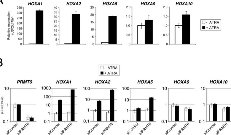
The genomic co-occurrence of PRMT6 and Polycomb proteins during neuronal differentiation of NT2/D1 cells suggests a potential positive crosstalk between the two repressive pathways in the transcriptional regulation ofHOXAgenes. We therefore aimed to investigate whether the presence of PRMT6 represses transcription ofHOXAgenes in NT2/D1 cells and how PRC complexes influence transcription of these genes. To this end, we initially confirmed by RT-qPCR the ATRA-driven transcriptional effects on rostral and caudalHOXAgene expression in NT2/D1 cells. As expected, upon ATRA treatmentHOXA1,A2andA5genes were transcrip-tionally activated, whereasHOXA9andA10transcript levels remained unchanged (Fig 2A). Next, we established siRNA-mediated depletion of PRMT6 in these cells by transfection of
Fig 1. PRMT6 and subunits of PRC1/PRC2 co-occupy the HOXA gene cluster.(A) Schematic representation of theHOXAgene cluster consisting of 11 genes (HOXA1-13) and located on chromosome 7. According to their expression pattern, the 3’-located genesHOXA1-5belong to the rostral genes and the 5’-locatedHOXA9-13to the caudal genes. (B) NT2/D1 cells were left untreated (0 d) or treated for 2 days (2 d) and 6 days (6 d) with 0.1μM ATRA.
Subsequently, the cells were subjected to ChIP analysis using PRMT6-, CBX8- as well as EZH2-specific antibodies and antibodies recognising the H3R2me2a and H3K27me3 marks. Isotype-specific IgG served as control antibody. Immunoprecipitated DNA was analysed in triplicates by qPCR with primers for theHOXA1,A2,A5,A9andA10genes. Mean values were calculated as percentage input (% Input) and error bars represent mean +/- S.D. of the triplicates.
doi:10.1371/journal.pone.0148892.g001
Fig 2. PRMT6 represses the ATRA-mediated transcriptional activation of rostralHOXAgenes, but has no influence on the transcription of caudal HOXAgenes.(A) NT2/D1 cells were left untreated (-) or treated for 2 days (+) with 0.1μM ATRA. Subsequently, total RNA was prepared and analysed in triplicates by RT-qPCR for transcript levels ofHOXA1,A2,A5,A9andA10normalised toUBIQUITINtranscription. Error bars represent mean +/- S.D. of the triplicates. Transcript levels of untreated cells were set to 1. (B) NT2/D1 cells were transfected with control siRNAs (siControl) or siRNA directed against PRMT6 (siPRMT6 = siPRMT6_1). Forty-eight hours post transfection cells were left untreated (-) or treated for 2 days (+) with 0.1μM ATRA. Subsequently,
total RNA was prepared and analysed in triplicates by RT-qPCR for transcript levels ofPRMT6,HOXA1,A2,A5,A9andA10normalised toUBIQUITIN
transcription. Error bars represent mean +/- S.D. of the triplicates. Transcript levels of untreated and siControl-transfected NT2/D1 cells were set to 1.
siRNA targeting PRMT6 (siPRMT6) or control siRNA (siControl). Subsequently, RT-qPCR analysis was performed to measure transcript levels ofPRMT6and the variousHOXAgenes in untreated as well as 2 days ATRA-treated NT2/D1 cells. Thereby, an efficient knockdown of
PRMT6was observed and we further showed that transcript levels ofPRMT6remain unchanged upon ATRA-induced differentiation in control cells (Fig 2B). Consistent with our observations in wild type NT2/D1 cells (Fig 2A),HOXA1andA2genes were transcriptionally induced (HOXA5weakly), whileHOXA9andA10transcript levels remained constant upon ATRA treatment in siControl transfected cells (Fig 2B). Interestingly, depletion of PRMT6 did not significantly influence the transcriptional activity of the rostral as well as caudalHOXA
genes in undifferentiated cells. However, it resulted in increased transcriptional activation of
HOXA1,A2andA5genes upon ATRA treatment compared to control transfected cells, whereasHOXA9andA10transcript levels were not altered (Fig 2B). Furthermore, the observa-tions for the rostralHOXAgenes were confirmed using alternative siRNAs directed against PRMT6 (S1 Fig). Taken together, these data suggest that PRMT6 acts as a transcriptional repressor of rostralHOXAgenes by fine-tuning the transcriptional activation of these genes in response to ATRA-induced neuronal differentiation of NT2/D1 cells.
We next asked whether PRC complexes, similar to PRMT6, are important for modulating the expression ofHOXAgenes during differentiation. Therefore, we additionally established siRNA-mediated depletion of CBX8 and EZH2 in NT2/D1 cells. An efficient knockdown was detected by Western blot analysis for all three proteins (Fig 3A). Similar to the effects upon PRMT6 depletion, RT-qPCR analysis revealed that depletion of CBX8 and EZH2 resulted in an increased transcriptional activation ofHOXA1,A2andA5genes in response to ATRA com-pared to control transfected cells, while the basal transcript levels of the rostralHOXAgenes remained unaltered in the absence of ATRA (Fig 3B). As observed upon PRMT6 depletion, the transcription of caudalHOXAgenes,HOXA9andA10, was not significantly affected by deple-tion of CBX8 and EZH2, neither in undifferentiated nor in differentiated cells (data not shown). These results indicate that PRMT6, PRC1 (represented by the CBX8 subunit) and PRC2 (represented by the EZH2 subunit) function as direct transcriptional repressors of rostral
HOXAgenes to control their differentiation-associated activation in NT2/D1 cells. Although transcriptional activation of these target genes is accompanied by reduced PRMT6, PRC1 and PRC2 occupancy as well as H3R2me2a and H3K27me3 levels at the corresponding regulatory regions (Fig 1B), PRMT6- and Polycomb-dependent repression still seem to be responsible for constraining rostralHOXAgene activation. Our observations suggest that PRMT6 and PRC1/ PRC2 are not required for maintaining the transcriptional repression ofHOXAgenes in the undifferentiated NT2/D1 cells, but rather confine their ATRA-induced gene activation.
PRMT6 interacts with PRC1 and PRC2 subunits
homologues on endogenous protein level (Fig 4C). In addition, CoIP-assays for the PSC homo-logues BMI1 and MEL18 indicated that endogenous BMI1 associates with PRMT6 (Fig 4C), whereas MEL18 showed no or only very weak interaction with the arginine methyltransferase (S2 Fig). The same results were obtained after addition of ethidium bromide in the IPs indicat-ing that these interactions are not DNA-mediated (data not shown). To answer the question of whether the interaction with PRC1 components is a general characteristic of arginine methyl-transferases, we performed similar analyses for PRMT1 and PRMT4. Thereby we demon-strated that these two PRMTs do not interact with BMI1 or HPH2 (S3 Fig) suggesting that the association with PRC1 subunits is a specific feature of PRMT6.
As several PRC1 complexes with alternate subunit composition have been identified in mammals [49,50], we next performed Superose 6 gel filtration analysis to determine whether PRMT6 associates with such PRC1-type complexes. We found that endogenous PRMT6 elutes in molecular weight fractions (maximum in fraction no. 12) of approximately 440 kD (Fig 4D,
S4 Fig), which is a molecular weight higher than expected from its monomeric (42 kD) or dimeric size. Notably, PRMT6 co-eluted with endogenous (Fig 4D) and exogenous (S4 Fig) PRC1 components, such as HPH2, CBX8, BMI1, RING1A and RING1B. CoIP assays of the PRMT6-containing gel filtration fractions indicated that PRMT6 interacts with CBX8 and HPH2 in these high-molecular weight fractions (S5 Fig) suggesting that PRMT6 stably associ-ates with the CBX-containing PRC1-type complex.
To examine whether PRMT6 interacts with PRC2 subunits, in particular EZH2, we tran-siently overexpressed Flag-tagged EZH2. As shown inFig 4E, endogenous PRMT6 co-immu-noprecipitated with exogenous as well as endogenous EZH2 (Fig 4E). Furthermore, gel filtration analysis showed that Flag-tagged EZH2 mainly co-elutes with PRMT6 in fraction no. 12, although EZH2 was also present in higher molecular weight fractions (no. 9 and 10) not containing PRMT6 (Fig 4F). We found that PRMT6 co-immunoprecipitates with EZH2 in such gel filtration fractions, in which both proteins co-eluted from the column (S6 Fig). Together these findings indicate that PRMT6, even though it does not seem to be an integral component of the core PRC complexes, transiently associates with PRC subunits as well as PRC complexes and thereby might influence the repressive capacity of these complexes.
PRMT6 influences PRC-mediated gene silencing at the rostral
HOXA
genes locus
As PRMT6 binding sites overlap with those of the PRC subunits CBX8 and EZH2 at regulatory regions within theHOXAgene locus and PRMT6 physically interacts with both proteins, we examined whether PRMT6 impacts Polycomb-dependent repression of the rostralHOXA
genes. Therefore, we performed ChIP analysis of PRC subunits CBX8 and EZH2 as well as H3K27me3 at rostralHOXAgenes in siPRMT6- or siControl-transfected NT2/D1 cells, which were either undifferentiated or ATRA-differentiated. The occupancy of PRMT6 was reduced at theHOXA2andA5gene promoters in the PRMT6-depleted conditions (Fig 5A). Interestingly, binding of CBX8 and EZH2 to theHOXA2andA5gene promoters was diminished upon depletion of PRMT6 both in the absence and presence of ATRA (Fig 5A). These events coin-cided with a decrease in H3K27me3 levels (Fig 5B) suggesting that PRMT6 positively
Subsequently, cells were harvested and 30μg total protein of each sample were analysed by Western blot
with the indicated antibodies. The asterisk indicates the specific signals for the CBX8 protein. (B) NT2/D1 cells were transfected as in (A). Forty-eight hours post transfection cells were left untreated (-) or treated for 2 days (+) with 0.1μM ATRA. Total RNA was then prepared and analysed in triplicates by RT-qPCR for
transcript levels ofHOXA1,A2andA5normalised toUBIQUITINtranscription. Error bars represent mean +/-S.D. of the triplicates. Transcript levels of untreated and siControl-transfected NT2/D1cells were set to 1.
Fig 4. PRMT6 interacts with subunits of PRC1 and PRC2.(A) HEK293 cells were transfected with untagged HPH1 or Myc-tagged HPH2 constructs (OE = overexpressed) and harvested 48 hours after transfection. Protein extracts were subjected to immunoprecipitation using antibodies against PRMT6 (α
-PRMT6) or isotype-specific IgG as control. Inputs (2%) and precipitates were subjected to Western blot analysis using antibodies against HPH1 (α-HPH1),
Myc-tag (α-Myc for HPH2 detection) and PRMT6 (α-PRMT6). The asterisk indicates an unspecific signal in theα-PRMT6 staining. (B) HEK293 cells were
transfected with GFP-tagged CBX2, CBX4 and CBX8 constructs (OE = overexpressed) and harvested 48 hours after transfection. Protein extracts were subjected to immunoprecipitation using antibodies against PRMT6 (α-PRMT6) or as control isotype-specific IgG. Input (2%) and precipitates were subjected
to Western blot analysis using antibodies against GFP (α-GFP for CBX detection) and PRMT6 (α-PRMT6). (C) For the detection of endogenous interactions,
HEK293 protein extracts were subjected to immunoprecipitation using antibodies against PRMT6 (α-PRMT6) or isotype-specific IgG as control. Input (2%)
and precipitates were subjected to Western blot analysis using antibodies against HPH1 (α-HPH1), HPH2 (α-HPH2), BMI1 (α-BMI1) and PRMT6 (α
-PRMT6). (D) For size fractionation by gel filtration chromatography, whole-cell protein extracts were prepared from HEK293 cells. Protein extracts were applied to a Superose 6 column and 6 ml fractions were collected. Five % of each fraction (no. 6–17) were analysed by Western blot using the indicated antibodies to detect PRMT6, HPH2, CBX8, BMI1, RING1A and RING1B, endogenously. The column was calibrated using standard protein markers. Accordingly, the molecular weight included in the fractions and the void volume (V0) are indicated. (E) For the detection of endogenous and exogenous
influences the deposition of this mark, possibly via its association with the PRC1 complex and EZH2. To test the hypothesis that PRMT6-mediated deposition of H3R2me2a might directly promote the catalytic activity of EZH2, we performedin vitromethyltransferase assays using recombinant PRC2 complex in the presence of unmodified and premodified H3 peptides. To first validate the assay we determined the methylation activity of EZH2 towards unmodified and H3K27me3-premodified peptides. While a methylation product was detected by autoradi-ography for the unmodified H3 peptide, no signal was obtained when the H3K27me3-premo-dified peptide was employed as a substrate confirming that H3K27 is the major methylation site of recombinant PRC2 complex in our assay (S7 Fig). Premodification at R2 either by mono-methylation or asymmetric dimethylation did not enhance the activity of EZH2 arguing against a positive crosstalk at the level of these histone modifications (S7 Fig). In agreement with previous observations [5], we found that depletion of PRMT6 results in increased levels of H3K4me3 at theHOXA2andA5gene promoters in NT2/D1 cells (Fig 5C). As H3K4me3 has been reported to inhibit recognition of the H3 N-terminus by PRC2 and thereby the PRC2 activity [51], PRMT6 might promote the occurrence of H3K27me3 and Polycomb-mediated repression by constraining the levels of H3K4me3.
Together, these data uncover a novel and additional mechanism of how PRMT6 contributes to gene repression during cellular differentiation. Apart from counteracting the occurrence of H3K4me3, PRMT6 interacts with components of PRC1/PRC2 and facilitates chromatin bind-ing of these subunits and the deposition of H3K27me3. Our observations suggest that PRMT6-and PRC1/PRC2-mediated repression mechanisms cooperate in fine-tuning PRMT6-and controlling appropriate timing and magnitude of ATRA-induced transcriptional activation of rostral
HOXAgenes.
Discussion
The PRMT family member PRMT6 catalyses asymmetric dimethylation of histone H3 at argi-nine 2 and thereby causes transcriptional repression of PRMT6-bound genes by antagonising the deposition of the active histone mark H3K4me3 by the MLL1 complex [5,13,20,23]. This mechanism of transcriptional repression has been shown to be relevant during differentiation processes, such as PRMT6 acts as a co-repressor of the transcription factor RUNX1 thereby inhibiting megakaryocytic genes in hematopoietic progenitor cells and later the erythroid-spe-cific gene expression program during megakaryocytic differentiation [29,30]. Furthermore, PRMT6 influences the expression of pluripotency and differentiation markers in embryonic stem cells [52]. Our own observations revealed that PRMT6 associates with differentiation-associated genes in neural progenitor cells [5], in line with high expression levels of PRMT6 in the developing mouse brain [53]. Therefore, we studied here the repressive role of PRMT6 and its functional relationship with neighbouring histone marks during neural differentiation using the EC model of NT2/D1 cells, which predominantly differentiate along the neuronal lineage upon exposure to ATRA.
H3R2me2a co-occurs at the genomic level with H3K27me3, a well-established repressive histone mark [28–30]. In addition, the presence of H3K27me3 and its writer as well as reader complexes (PRC2 and PRC1) have been described in detail at theHOXAgene cluster in NT2/ D1 cells [36,48,54]. In undifferentiated cells, theHOXAgene locus is marked by high levels of H3K27me3 and bound by EZH2 (PRC2) as well as CBX8 (PRC1). Upon ATRA-induced
(2%) and precipitates were analysed by Western blot using the indicated antibodies. (F) Whole-cell protein extracts were generated from HEK293 cells transfected with Flag-tagged EZH2 construct. Size fractionation by gel filtration chromatography was performed as described in (D) and fractions 6–16 were subjected to Western blot analysis using the indicated antibodies.
neuronal differentiation, H3K27me3 and PRCs are specifically displaced from the rostral
HOXAgenes (HOXA1-5) in agreement with their transcriptional activation, whereas the cau-dalHOXAgenes (HOXA9-13) keep high amounts of this repressive mark and the PRCs and unchanged expression levels [36,48,55]. Interestingly, PRMT6 and its histone mark H3R2me2a show a binding pattern at theHOXAcluster in undifferentiated and differentiating NT2/D1 cells similar to the one of H3K27me3 and the PRCs indicating that this locus is appropriate for studying a putative crosstalk between the PRMT6 and Polycomb proteins in target gene repression.
To elucidate the potential impact of PRMT6 as well as the PRCs on the transcriptional activ-ity of theHOXAlocus, we depleted the relevant enzymes and representative subunits of the complexes. We found that PRMT6 and PRC1/2 did not influence the expression of caudal
HOXAgenes, neither in undifferentiated nor in differentiating NT2/D1 cells. This is an agree-ment with previous observations showing that PRC1/2 binding does not strictly correlate with transcriptional silencing, but also occurs at transcriptionally active genes, which seems to hold true also for PRMT6 [36]. Furthermore, not all target genes of PRC1 and PRC2 are derepressed upon PRC depletion, but are thought to have acquired an additional, more permanent silenc-ing, such as DNA methylation or higher order chromatin structure, which makes the stable presence of PRC1/2 dispensable [31,36]. However, we surprisingly observed that PRMT6 and PRC1/2 specifically restrict the transcriptional activation of rostralHOXAgenes in differentiat-ing NT2/D1 cells, although the occupancy of PRMT6, PRC1/2 as well as their histone marks was reduced at these gene promoters upon differentiation. This further supports the view that binding of PRMT6 and PRCs is diminished at differentiation-specific genes to enable their transcriptional induction, while they still seem to be required for constraining and fine-tuning the transcriptional response.
Given that PRMT6 and subunits of PRC1 and PRC2 coincide in their binding pattern at ros-tralHOXAgenes and have a similar repressive influence on the ATRA-induced transcriptional activation of these genes, we investigated whether PRMT6 would directly regulate PRC-depen-dent repression. Depletion of PRMT6 resulted in reduced occupancy of CBX8 and EZH2 at
HOXA2andA5gene promoters and in decreased levels of H3K27me3, independently of the differentiation status of the cells. In differentiating NT2/D1 cells these observations are in agreement with an enhanced ATRA-mediated transcriptional activation ofHOXA2andA5, whereas the basal activity of these genes was not derepressed in undifferentiated cells. These results imply that PRMT6- and PRC-mediated repression cooperate in the transcriptional reg-ulation of cell-fate genes. Although we found that PRMT6 is able to physically interact with several subunits of PRC1 and with EZH2, we hypothesise that PRMT6 is not responsible for the chromatin recruitment of PRCs via protein-protein interactions. We postulate that PRMT6 associates with PRC components at the chromatin and stimulates a histone modification land-scape, such as increased H3R2me2a levels and decreased H3K4me3 levels, in the vicinity of PRC binding sites, which stabilises PRC binding and enhances the catalytic activity of EZH2. In agreement with this model, our data indicate that PRMT6 is not a stable component of core
Fig 5. PRMT6 influences PRC-mediated gene silencing at theHoxAgene locus.(A) NT2/D1 cells were transfected with control siRNAs (siControl) or siRNAs directed against PRMT6 (siPRMT6 = equal mixture of siPRMT6_1 and siPRMT6_2). Forty-eight hours post transfection cells were left untreated (-) or treated for 2 days (+) with 0.1μM ATRA. Subsequently, cells were subjected to ChIP analysis using PRMT6-, CBX8- as well as EZH2-specific antibodies.
IgG served as control antibody for these three antibodies and is displayed together with the PRMT6 ChIP in the upper graph. Immunopreciptiated DNA was analysed in triplicates by qPCR with primers for theHOXA2andA5genes. Mean values are calculated as percentage input (% input) and error bars represent mean +/- S.D. of the triplicates. (B, C) NT2/D1 cells were transfected and treated as described in (A). Cells were then subjected to ChIP analysis using antibodies recognising histone H3 and the histone marks H3K27me3 (B) and H3K4me3 (C). Immunoprecipitated DNA was analysed in triplicates by qPCR with primers for theHOXA2andA5genes. Mean values are calculated as enrichment relative to histone H3 and error bars represent mean +/- S.D. of the triplicates.
PRC complexes, but rather support that PRMT6 transiently associates with PRC subunits and complexes to regulate their repressive capacity.
Given the complex interplay between different histone modifications and the fact that a pre-existing modification can promote or inhibit the subsequent occurrence of another histone mark, such as H3R2me2a counteracting the deposition of H3K4me3 (5, 13), we asked whether the positive crosstalk between PRMT6 and PRCs is mediated via the H3R2me2a mark. There-fore, we assayed the catalytic activity of EZH2 on unmodified as well as H3R2me2a-premodi-fied H3 peptides. Monomethylation or asymmetric dimethylation at R2 did not enhance the activity of EZH2 indicating that PRMT6 does not directly influence the EZH2 activity via depositioning the H3R2me2a mark. However, H3K4me3 levels were increased at theHOXA2
as well asA5gene promoter in PRMT6-depleted NT2/D1 cells, in particular upon ATRA treat-ment. As H3K4me3 has been reported to inhibit the recognition of histone H3 N-terminus by PRC2 and thereby the PRC2 activity [51], we hypothesise that PRMT6 promotes the occur-rence of H3K27me3 and consequently the recruitment of CBX8 at rostralHOXAgenes in an indirect manner via associating with PRC complexes and thereby restricting the occurrence and magnitude of H3K4me3.
In summary, we postulate a model, in which neither PRMT6 nor PRCs are necessary for maintaining the transcriptional status (low basal activity) of theHOXAgene cluster in undif-ferentiated NT2/D1 cells despite their presence at the chromatin. This situation also applies to the caudalHOXAgenes in differentiating cells suggesting that here additional, more perma-nent silencing mechanisms have been acquired. However, upon ATRA treatment rostral
HOXAgenes are transcriptionally activated, which is facilitated by an increase in H3K4me3 levels via MLL1 complex recruitment with the concomitant reduction of PRMT6 and PRC1/2 recruitment and their corresponding histone marks. Even during this transcriptional activation a balance of activating and silencing mechanisms seem to be necessary to enable an adequate transcriptional response, since PRMT6 and PRCs are important for constraining the ATRA-mediated transcriptional response of rostralHOXAgenes. Altogether, our results confirm the previously reported countercorrelation between PRMT6 and MLL1 activities and uncover a novel cooperation between PRMT6- and PRC-mediated repression.
Supporting Information
S1 Fig. Alternative siRNAs targeting PRMT6 confirm the function of PRMT6 in repressing the ATRA-mediated transcriptional activation of rostralHOXAgenes.NT2/D1 cells were
transfected with control siRNAs (siControl) or 8 alternative siRNAs directed against PRMT6 (including the mixture of siPRMT6_1 and siPRMT6_2, which was used inFig 3andFig 5). Forty-eight hours post transfection cells were left untreated (-) or treated for 2 days (+) with 0.1μM ATRA. Subsequently, total RNA was prepared and analysed in triplicates by RT-qPCR
for transcript levels ofPRMT6,HOXA1,A2andA5normalised forUBIQUITINtranscription. Error bars represent mean +/- S.D. of the triplicates. Transcript levels of untreated and siCon-trol-transfected NT2/D1 cells were set to 1.
(TIF)
S2 Fig. PRMT6 does not or only weakly interact with MEL18.HEK293 cells were transfected with GFP-tagged MEL18 construct and harvested 48 hours after transfection. Protein extracts were subjected to immunoprecipitation using antibodies for PRMT6 (α-PRMT6) or as controls beads alone as well as isotype-specific IgG. Input (2%) and precipitates were subjected to West-ern blot analysis using antibodies against GFP (α-GFP for MEL18 detection) and PRMT6 (α -PRMT6).
S3 Fig. PRMT1 and PRMT4 do not interact with subunits of the PRC1 complex, such as BMI1 and HPH2.HEK293 protein extracts were subjected to immunoprecipitation using anti-bodies for PRMT1 (α-PRMT1), PRMT4 (α-PRMT4) or as controls beads alone as well as iso-type-specific IgG. Input (2%) and precipitates were subjected to Western blot analysis using antibodies against BMI1 (α-BMI1), HPH2 (α-HPH2), PRMT1 (α-PRMT1) and PRMT4 (α -PRMT4). The asterisk indicates the specific signals for the PRMT4 protein.
(TIF)
S4 Fig. PRMT6 co-elutes with the PRC1 subunits HPH2 and CBX8 in the same high molec-ular weight fractions.For size fractionation by gel filtration chromatography, HEK293 cells were transfected with GFP-tagged CBX8 and Myc-tagged HPH2 constructs. Subsequently whole-cell protein extracts were applied to a Superose 6 column and 6 ml fractions were col-lected. Five % of each fraction (no. 6–15) were analysed by Western blot using the indicated antibodies to detect GFP (α-GFP for CBX8 detection), Myc-tag (α-Myc for HPH2 detection) and PRMT6 (α-PRMT6). The column was calibrated using standard protein markers. Accord-ingly, the molecular weight included in the fractions and the void volume (V0) are indicated.
(TIF)
S5 Fig. PRMT6 stably interacts with CBX8 and HPH2 in co-elution gel filtration fractions.
HEK293 cells were transfected with GFP-tagged CBX8 and Myc-tagged HPH2 constructs. Whole-cell protein extracts (WCE) and the corresponding Superose 6 gel filtration fractions (no. 6–15) were subjected to immunoprecipitation using antibodies against PRMT6 (α-P6,α -PRMT6) or as control isotype-specific IgG (only for WCE). Input (2%) of WCE and precipi-tates were analysed by Western blot using antibodies against GFP (α-GFP for CBX8 detection), Myc-tag (α-Myc for HPH2 detection) and PRMT6 (α-PRMT6).
(TIF)
S6 Fig. PRMT6 stably interacts with EZH2 in co-elution gel filtration fractions.HEK293 cells were transfected with Flag-tagged EZH2 construct. Whole-cell protein extract (WCE) and the corresponding Superose 6 gel filtration fractions (no. 6–16) were subjected to IP using anti-bodies against PRMT6 (α-P6,α-PRMT6) or as control isotype-specific IgG (only for WCE). Input (2%) of WCE and precipitates were subjected to Western blot analysis using antibodies against Flag-tag (α-Flag for EZH2 detection) and PRMT6 (α-PRMT6).
(TIF)
S7 Fig. The catalytic activity of recombinant PRC2 is not enhanced by R2me2a-premodifi-cation of H3.Recombinant PRC2 complex (400 ng) and 4μg of either unmodified, R2me1,
R2me2a or K27me3 premodified H3 peptides (aa 1–30) were incubated in the presence or absence of [14C-methyl]-SAM overnight at 30°C. Subsequently, the methyltransferase reactions were analysed by SDS-PAGE, blotting and autoradiography. Ponceau S staining of the blot was used as loading control for the different H3 peptides.
(TIF)
S1 Text. Supporting Information Materials and Methods.
(DOCX)
Acknowledgments
laboratory for their suggestions and help. This work was supported by the Deutsche For-schungsgemeinschaft (DFG) through project grant numbers TRR81/A3 and BA2292/1-3 to U. M.B.
Author Contributions
Conceived and designed the experiments: CS RRN SR CB UMB. Performed the experiments: CS RRN SR. Analyzed the data: CS RRN SR CB UMB. Contributed reagents/materials/analysis tools: CB. Wrote the paper: UMB.
References
1. Di Lorenzo A, Bedford MT. Histone arginine methylation. FEBS Lett. 2011; 585: 2024–2031. Available: http://www.ncbi.nlm.nih.gov/entrez/query.fcgi?cmd=Retrieve&db=PubMed&dopt=Citation&list_uids= 21074527doi:10.1016/j.febslet.2010.11.010PMID:21074527
2. Gayatri S, Bedford MT. Readers of histone methylarginine marks. Biochim Biophys Acta—Gene Regul Mech. Elsevier B.V.; 2014; 1839: 702–710. doi:10.1016/j.bbagrm.2014.02.015
3. Frankel A, Yadav N, Lee J, Branscombe TL, Clarke S, Bedford MT. The novel human protein arginine N-methyltransferase PRMT6 is a nuclear enzyme displaying unique substrate specificity. J Biol Chem. 2002; 277: 3537–3543. Available:http://www.ncbi.nlm.nih.gov/entrez/query.fcgi?cmd=Retrieve&db= PubMed&dopt=Citation&list_uids=11724789PMID:11724789
4. Lakowski TM, Frankel A. A kinetic study of human protein arginine N-methyltransferase 6 reveals a dis-tributive mechanism. J Biol Chem. 2008; 283: 10015–10025. Available:http://www.ncbi.nlm.nih.gov/ entrez/query.fcgi?cmd=Retrieve&db=PubMed&dopt=Citation&list_uids=18263580doi:10.1074/jbc. M710176200PMID:18263580
5. Hyllus D, Stein C, Schnabel K, Schiltz E, Imhof A, Dou Y, et al. PRMT6-mediated methylation of R2 in histone H3 antagonizes H3 K4 trimethylation. Genes Dev. 2007; 21: 3369–3380. PMID:18079182
6. Thandapani P, O’Connor TR, Bailey TL, Richard S. Defining the RGG/RG motif. Mol Cell. 2013; 50: 613–623. Available:http://www.ncbi.nlm.nih.gov/entrez/query.fcgi?cmd=Retrieve&db=PubMed&dopt= Citation&list_uids=23746349doi:10.1016/j.molcel.2013.05.021PMID:23746349
7. Miranda TB, Webb KJ, Edberg DD, Reeves R, Clarke S. Protein arginine methyltransferase 6 specifi-cally methylates the nonhistone chromatin protein HMGA1a. Biochem Biophys Res Commun. 2005; 336: 831–835. Available:http://www.ncbi.nlm.nih.gov/entrez/query.fcgi?cmd=Retrieve&db= PubMed&dopt=Citation&list_uids=16157300PMID:16157300
8. Sgarra R, Lee J, Tessari MA, Altamura S, Spolaore B, Giancotti V, et al. The AT-hook of the chromatin architectural transcription factor high mobility group A1a is arginine-methylated by protein arginine methyltransferase 6. J Biol Chem. 2006; 281: 3764–3772. Available:http://www.ncbi.nlm.nih.gov/ entrez/query.fcgi?cmd=Retrieve&db=PubMed&dopt=Citation&list_uids=16293633PMID:16293633
9. Boulanger MC, Liang C, Russell RS, Lin R, Bedford MT, Wainberg MA, et al. Methylation of Tat by PRMT6 regulates human immunodeficiency virus type 1 gene expression. J Virol. 2005; 79: 124–131. Available:http://www.ncbi.nlm.nih.gov/entrez/query.fcgi?cmd=Retrieve&db=PubMed&dopt= Citation&list_uids=15596808PMID:15596808
10. Singhroy DN, Mesplede T, Sabbah A, Quashie PK, Falgueyret JP, Wainberg MA. Automethylation of protein arginine methyltransferase 6 (PRMT6) regulates its stability and its anti-HIV-1 activity. Retrovi-rology. 2013; 10: 73. Available:http://www.ncbi.nlm.nih.gov/entrez/query.fcgi?cmd=Retrieve&db= PubMed&dopt=Citation&list_uids=23866860doi:10.1186/1742-4690-10-73PMID:23866860
11. El-Andaloussi N, Valovka T, Toueille M, Steinacher R, Focke F, Gehrig P, et al. Arginine methylation regulates DNA polymerase beta. Mol Cell. 2006; 22: 51–62. Available:http://www.ncbi.nlm.nih.gov/ entrez/query.fcgi?cmd=Retrieve&db=PubMed&dopt=Citation&list_uids=16600869PMID:16600869
12. Scaramuzzino C, Casci I, Parodi S, Lievens PMJ, Polanco MJ, Milioto C, et al. Protein Arginine Methyl-transferase 6 Enhances Polyglutamine-Expanded Androgen Receptor Function and Toxicity in Spinal and Bulbar Muscular Atrophy. Neuron. The Authors; 2015; 85: 88–100. doi:10.1016/j.neuron.2014.12. 031PMID:25569348
13. Guccione E, Bassi C, Casadio F, Martinato F, Cesaroni M, Schuchlautz H, et al. Methylation of histone H3R2 by PRMT6 and H3K4 by an MLL complex are mutually exclusive. Nature. 2007; 449: 933–937. Available:http://www.ncbi.nlm.nih.gov/entrez/query.fcgi?cmd=Retrieve&db=PubMed&dopt= Citation&list_uids=17898714PMID:17898714
nih.gov/entrez/query.fcgi?cmd=Retrieve&db=PubMed&dopt=Citation&list_uids=21774791doi:10. 1186/1756-8935-4-11PMID:21774791
15. Casadio F, Lu X, Pollock SB, LeRoy G, Garcia BA, Muir TW, et al. H3R42me2a is a histone modifica-tion with positive transcripmodifica-tional effects. Proc Natl Acad Sci U S A. 2013; 110: 14894–14899. Available: http://www.ncbi.nlm.nih.gov/entrez/query.fcgi?cmd=Retrieve&db=PubMed&dopt=Citation&list_uids= 23980157doi:10.1073/pnas.1312925110PMID:23980157
16. Stein C, Riedl S, Ruthnick D, Notzold RR, Bauer UM. The arginine methyltransferase PRMT6 regulates cell proliferation and senescence through transcriptional repression of tumor suppressor genes. Nucleic Acids Res. 2012; 40: 9522–9533. Available:http://www.ncbi.nlm.nih.gov/entrez/query.fcgi? cmd=Retrieve&db=PubMed&dopt=Citation&list_uids=22904088doi:10.1093/nar/gks767PMID: 22904088
17. Phalke S, Mzoughi S, Bezzi M, Jennifer N, Mok WC, Low DH, et al. p53-Independent regulation of p21Waf1/Cip1 expression and senescence by PRMT6. Nucleic Acids Res. 2012; 40: 9534–9542. Available:http://www.ncbi.nlm.nih.gov/entrez/query.fcgi?cmd=Retrieve&db=PubMed&dopt= Citation&list_uids=22987071doi:10.1093/nar/gks858PMID:22987071
18. Neault M, Mallette FA, Vogel G, Michaud-Levesque J, Richard S. Ablation of PRMT6 reveals a role as a negative transcriptional regulator of the p53 tumor suppressor. Nucleic Acids Res. 2012; 40: 9513– 9521. Available:http://www.ncbi.nlm.nih.gov/entrez/query.fcgi?cmd=Retrieve&db=PubMed&dopt= Citation&list_uids=22904064doi:10.1093/nar/gks764PMID:22904064
19. Kleinschmidt MA, de Graaf P, van Teeffelen HA, Timmers HT. Cell cycle regulation by the PRMT6 argi-nine methyltransferase through repression of cyclin-dependent kinase inhibitors. PLoS One. 2012; 7: e41446. Available:http://www.ncbi.nlm.nih.gov/entrez/query.fcgi?cmd=Retrieve&db=PubMed&dopt= Citation&list_uids=22916108doi:10.1371/journal.pone.0041446PMID:22916108
20. Iberg AN, Espejo A, Cheng D, Kim D, Michaud-Levesque J, Richard S, et al. Arginine methylation of the histone H3 tail impedes effector binding. J Biol Chem. 2008; 283: 3006–3010. Available:http:// www.ncbi.nlm.nih.gov/entrez/query.fcgi?cmd=Retrieve&db=PubMed&dopt=Citation&list_uids= 18077460PMID:18077460
21. Couture JF, Collazo E, Trievel RC. Molecular recognition of histone H3 by the WD40 protein WDR5. Nat Struct Mol Biol. 2006; 13: 698–703. Available:http://www.ncbi.nlm.nih.gov/entrez/query.fcgi?cmd= Retrieve&db=PubMed&dopt=Citation&list_uids=16829960PMID:16829960
22. Ruthenburg AJ, Wang W, Graybosch DM, Li H, Allis CD, Patel DJ, et al. Histone H3 recognition and presentation by the WDR5 module of the MLL1 complex. Nat Struct Mol Biol. 2006; 13: 704–712. Avail-able:http://www.ncbi.nlm.nih.gov/entrez/query.fcgi?cmd=Retrieve&db=PubMed&dopt=Citation&list_ uids=16829959PMID:16829959
23. Michaud-Levesque J, Richard S. Thrombospondin-1 is a transcriptional repression target of PRMT6. J Biol Chem. 2009; 284: 21338–21346. Available:http://www.ncbi.nlm.nih.gov/entrez/query.fcgi?cmd= Retrieve&db=PubMed&dopt=Citation&list_uids=19509293doi:10.1074/jbc.M109.005322PMID: 19509293
24. Barski A, Cuddapah S, Cui K, Roh TY, Schones DE, Wang Z, et al. High-resolution profiling of histone methylations in the human genome. Cell. 2007; 129: 823–837. Available:http://www.ncbi.nlm.nih.gov/ entrez/query.fcgi?cmd=Retrieve&db=PubMed&dopt=Citation&list_uids=17512414PMID:17512414
25. Rosenfeld J a, Wang Z, Schones DE, Zhao K, DeSalle R, Zhang MQ. Determination of enriched histone modifications in non-genic portions of the human genome. BMC Genomics. 2009; 10: 143. doi:10. 1186/1471-2164-10-143PMID:19335899
26. van Ingen H, van Schaik FM, Wienk H, Ballering J, Rehmann H, Dechesne AC, et al. Structural insight into the recognition of the H3K4me3 mark by the TFIID subunit TAF3. Structure. 2008; 16: 1245–1256. Available:http://www.ncbi.nlm.nih.gov/entrez/query.fcgi?cmd=Retrieve&db=PubMed&dopt= Citation&list_uids=18682226doi:10.1016/j.str.2008.04.015PMID:18682226
27. Vermeulen M, Mulder KW, Denissov S, Pijnappel WW, van Schaik FM, Varier RA, et al. Selective anchoring of TFIID to nucleosomes by trimethylation of histone H3 lysine 4. Cell. 2007; 131: 58–69. Available:http://www.ncbi.nlm.nih.gov/entrez/query.fcgi?cmd=Retrieve&db=PubMed&dopt= Citation&list_uids=17884155PMID:17884155
28. Guccione E, Martinato F, Finocchiaro G, Luzi L, Tizzoni L, Dall’Olio V, et al. Myc-binding-site recogni-tion in the human genome is determined by chromatin context. Nat Cell Biol. 2006; 8: 764–770. Avail-able:http://www.ncbi.nlm.nih.gov/entrez/query.fcgi?cmd=Retrieve&db=PubMed&dopt=Citation&list_ uids=16767079PMID:16767079
30. Herglotz J, Kuvardina ON, Kolodziej S, Kumar A, Hussong H, Grez M, et al. Histone arginine methyla-tion keeps RUNX1 target genes in an intermediate state. Oncogene. 2013; 32: 2565–2575. Available: http://www.ncbi.nlm.nih.gov/entrez/query.fcgi?cmd=Retrieve&db=PubMed&dopt=Citation&list_uids= 22777353doi:10.1038/onc.2012.274PMID:22777353
31. Margueron R, Reinberg D. The Polycomb complex PRC2 and its mark in life. Nature. 2011; 469: 343– 349. doi:10.1038/nature09784PMID:21248841
32. Cao R, Wang L, Wang H, Xia L, Erdjument-Bromage H, Tempst P, et al. Role of histone H3 lysine 27 methylation in Polycomb-group silencing. Science (80-). 2002; 298: 1039–1043. Available:http://www. ncbi.nlm.nih.gov/entrez/query.fcgi?cmd=Retrieve&db=PubMed&dopt=Citation&list_uids=12351676 PMID:12351676
33. Kuzmichev A, Nishioka K, Erdjument-Bromage H, Tempst P, Reinberg D. Histone methyltransferase activity associated with a human multiprotein complex containing the Enhancer of Zeste protein. Genes Dev. 2002; 16: 2893–2905. Available:http://www.ncbi.nlm.nih.gov/entrez/query.fcgi?cmd=
Retrieve&db=PubMed&dopt=Citation&list_uids=12435631PMID:12435631
34. Müller J, Hart CM, Francis NJ, Vargas ML, Sengupta A, Wild B, et al. Histone methyltransferase activity of a Drosophila Polycomb group repressor complex. Cell. 2002; 111: 197–208. doi: 10.1016/S0092-8674(02)00976-5PMID:12408864
35. Levine SS, Weiss A, Erdjument-Bromage H, Shao Z, Tempst P, Kingston RE. The core of the polycomb repressive complex is compositionally and functionally conserved in flies and humans. Mol Cell Biol. 2002; 22: 6070–6078. Available:http://www.ncbi.nlm.nih.gov/entrez/query.fcgi?cmd=Retrieve&db= PubMed&dopt=Citation&list_uids=12167701PMID:12167701
36. Bracken AP, Dietrich N, Pasini D, Hansen KH, Helin K. Genome-wide mapping of Polycomb target genes unravels their roles in cell fate transitions. Genes Dev. 2006; 20: 1123–1136. Available:http:// www.ncbi.nlm.nih.gov/entrez/query.fcgi?cmd=Retrieve&db=PubMed&dopt=Citation&list_uids= 16618801PMID:16618801
37. Suzuki M, Mizutani-Koseki Y, Fujimura Y, Miyagishima H, Kaneko T, Takada Y, et al. Involvement of the Polycomb-group gene Ring1B in the specification of the anterior-posterior axis in mice. Develop-ment. 2002; 129: 4171–4183. Available:http://www.ncbi.nlm.nih.gov/entrez/query.fcgi?cmd= Retrieve&db=PubMed&dopt=Citation&list_uids=12183370PMID:12183370
38. Miyagishima H, Isono K, Fujimura Y, Iyo M, Takihara Y, Masumoto H, et al. Dissociation of mammalian Polycomb-group proteins, Ring1B and Rae28/Ph1, from the chromatin correlates with configuration changes of the chromatin in mitotic and meiotic prophase. Histochem Cell Biol. 2003; 120: 111–119. Available:http://www.ncbi.nlm.nih.gov/entrez/query.fcgi?cmd=Retrieve&db=PubMed&dopt= Citation&list_uids=12883906PMID:12883906
39. Bracken AP, Kleine-Kohlbrecher D, Dietrich N, Pasini D, Gargiulo G, Beekman C, et al. The Polycomb group proteins bind throughout the INK4A-ARF locus and are disassociated in senescent cells. Genes Dev. 2007; 21: 525–530. Available:http://www.ncbi.nlm.nih.gov/entrez/query.fcgi?cmd=Retrieve&db= PubMed&dopt=Citation&list_uids=17344414PMID:17344414
40. Ren X, Vincenz C, Kerppola TK. Changes in the distributions and dynamics of polycomb repressive complexes during embryonic stem cell differentiation. Mol Cell Biol. 2008; 28: 2884–2895. Available: http://www.ncbi.nlm.nih.gov/entrez/query.fcgi?cmd=Retrieve&db=PubMed&dopt=Citation&list_uids= 18316406doi:10.1128/MCB.00949-07PMID:18316406
41. Pasini D, Bracken AP, Jensen MR, Lazzerini Denchi E, Helin K. Suz12 is essential for mouse develop-ment and for EZH2 histone methyltransferase activity. Embo J. 2004; 23: 4061–4071. Available:http:// www.ncbi.nlm.nih.gov/entrez/query.fcgi?cmd=Retrieve&db=PubMed&dopt=Citation&list_uids= 15385962PMID:15385962
42. Schoorlemmer J, Marcos-Gutierrez C, Were F, Martinez R, Garcia E, Satijn DP, et al. Ring1A is a tran-scriptional repressor that interacts with the Polycomb-M33 protein and is expressed at rhombomere boundaries in the mouse hindbrain. Embo J. 1997; 16: 5930–5942. Available:http://www.ncbi.nlm.nih. gov/entrez/query.fcgi?cmd=Retrieve&db=PubMed&dopt=Citation&list_uids=9312051PMID:9312051
43. Nomura M, Takihara Y, Shimada K. Isolation and characterization of retinoic acid-inducible cDNA clones in F9 cells: one of the early inducible clones encodes a novel protein sharing several highly homologous regions with a Drosophila polyhomeotic protein. Differentiation. 1994; 57: 39–50. Avail-able:http://www.ncbi.nlm.nih.gov/entrez/query.fcgi?cmd=Retrieve&db=PubMed&dopt=Citation&list_ uids=8070621PMID:8070621
44. Boyer LA, Plath K, Zeitlinger J, Brambrink T, Medeiros LA, Lee TI, et al. Polycomb complexes repress developmental regulators in murine embryonic stem cells. Nature. 2006; 441: 349–353. Available: http://www.ncbi.nlm.nih.gov/entrez/query.fcgi?cmd=Retrieve&db=PubMed&dopt=Citation&list_uids= 16625203PMID:16625203
www.ncbi.nlm.nih.gov/entrez/query.fcgi?cmd=Retrieve&db=PubMed&dopt=Citation&list_uids= 16630818PMID:16630818
46. Boncinelli E, Simeone a, Acampora D, Mavilio F. HOX gene activation by retinoic acid. Trends Genet. 1991; 7: 329–34. Available:http://www.ncbi.nlm.nih.gov/pubmed/1685814PMID:1685814
47. Andrews PW. Retinoic acid induces neuronal differentiation of a cloned human embryonal carcinoma cell line in vitro. Dev Biol. 1984; 103: 285–293. Available:http://www.ncbi.nlm.nih.gov/entrez/query. fcgi?cmd=Retrieve&db=PubMed&dopt=Citation&list_uids=6144603PMID:6144603
48. Houldsworth J, Heath SC, Bosl GJ, Studer L, Chaganti RSK. Expression profiling of lineage differentia-tion in pluripotential human embryonal carcinoma cells. Cell Growth Differ. 2002; 13: 257–264. PMID: 12114215
49. Tavares L, Dimitrova E, Oxley D, Webster J, Poot R, Demmers J, et al. RYBP-PRC1 Complexes Medi-ate H2A Ubiquitylation at Polycomb Target Sites Independently of PRC2 and H3K27me3. Cell. 2012; 148: 664–678. doi:10.1016/j.cell.2011.12.029PMID:22325148
50. Gao Z, Zhang J, Bonasio R, Strino F, Sawai A, Parisi F, et al. PCGF Homologs, CBX Proteins, and RYBP Define Functionally Distinct PRC1 Family Complexes. Mol Cell. Elsevier Inc.; 2012; 45: 344– 356. doi:10.1016/j.molcel.2012.01.002PMID:22325352
51. Schmitges FW, Prusty AB, Faty M, Stutzer A, Lingaraju GM, Aiwazian J, et al. Histone methylation by PRC2 is inhibited by active chromatin marks. Mol Cell. 2011; 42: 330–341. Available:http://www.ncbi. nlm.nih.gov/entrez/query.fcgi?cmd=Retrieve&db=PubMed&dopt=Citation&list_uids=21549310doi: 10.1016/j.molcel.2011.03.025PMID:21549310
52. Lee YH, Ma H, Tan TZ, Ng SS, Soong R, Mori S, et al. Protein arginine methyltransferase 6 regulates embryonic stem cell identity. Stem Cells Dev. 2012; 21: 2613–2622. Available:http://www.ncbi.nlm.nih. gov/entrez/query.fcgi?cmd=Retrieve&db=PubMed&dopt=Citation&list_uids=22455726doi:10.1089/ scd.2011.0330PMID:22455726
53. McKee AE, Minet E, Stern C, Riahi S, Stiles CD, Silver PA. A genome-wide in situ hybridization map of RNA-binding proteins reveals anatomically restricted expression in the developing mouse brain. BMC Dev Biol. 2005; 5: 14. Available:http://www.ncbi.nlm.nih.gov/entrez/query.fcgi?cmd=Retrieve&db= PubMed&dopt=Citation&list_uids=16033648PMID:16033648
54. Xu M, Zhao G-N, Lv X, Liu G, Wang LY, Hao D-L, et al. CTCF Controls HOXA Cluster Silencing and Mediates PRC2-Repressive Higher-Order Chromatin Structure in NT2/D1 Cells. Mol Cell Biol. 2014; 34: 3867–3879. doi:10.1128/MCB.00567-14PMID:25135475