Blockade of Hedgehog Signaling Synergistically
Increases Sensitivity to Epidermal Growth
Factor Receptor Tyrosine Kinase Inhibitors in
Non-Small-Cell Lung Cancer Cell Lines
Xiao-Yan Bai1, Xu-Chao Zhang1, Su-Qing Yang1, She-Juan An1, Zhi-Hong Chen1, Jian Su1, Zhi Xie1, Lan-Ying Gou1,2, Yi-Long Wu1*
1Department of Pulmonary Oncology, Guangdong Lung Cancer Institute, Guangdong General Hospital & Guangdong Academy of Medical Science, Guangzhou 510080, China,2Southern Medical University, Guangzhou 510515, China
Abstract
Aberrant activation of the hedgehog (Hh) signaling pathway has been implicated in the epi-thelial-to-mesenchymal transition (EMT) and cancer stem-like cell (CSC) maintenance; both processes can result in tumor progression and treatment resistance in several types of human cancer. Hh cooperates with the epidermal growth factor receptor (EGFR) signaling pathway in embryogenesis. We found that the Hh signaling pathway was silenced in EGFR-TKI-sensitive non-small-cell lung cancer (NSCLC) cells, while it was inappropriately acti-vated in EGFR-TKI-resistant NSCLC cells, accompanied by EMT induction and ABCG2 overexpression. Upregulation of Hh signaling through extrinsic SHH exposure downregu-lated E-cadherin expression and elevated Snail and ABCG2 expression, resulting in gefiti-nib tolerance (P<0.001) in EGFR-TKI-sensitive cells. Blockade of the Hh signaling
pathway using the SMO antagonist SANT-1 restored E-cadherin expression and downregu-late Snail and ABCG2 in EGFR-TKI-resistant cells. A combination of SANT-1 and gefitinib markedly inhibited tumorigenesis and proliferation in EGFR-TKI-resistant cells (P<0.001). These findings indicate that hyperactivity of Hh signaling resulted in EGFR-TKI resistance, by EMT introduction and ABCG2 upregulation, and blockade of Hh signaling synergistically increased sensitivity to EGFR-TKIs in primary and secondary resistant NSCLC cells. E-cad-herin expression may be a potential biomarker of the suitability of the combined application of an Hh inhibitor and EGFR-TKIs in EGFR-TKI-resistant NSCLCs.
Introduction
Lung cancer is a leading cause of cancer death worldwide, and poses a significant risk to human health. Median survival of patients with advanced non-small-cell lung cancer (NSCLC) who receive standard chemotherapy is only 9–12 months[1]. The advent of molecularly tar-geted therapies has shed much light on lung cancer treatment. Epidermal growth factor
OPEN ACCESS
Citation:Bai X-Y, Zhang X-C, Yang S-Q, An S-J, Chen Z-H, Su J, et al. (2016) Blockade of Hedgehog Signaling Synergistically Increases Sensitivity to Epidermal Growth Factor Receptor Tyrosine Kinase Inhibitors in Non-Small-Cell Lung Cancer Cell Lines. PLoS ONE 11(3): e0149370. doi:10.1371/journal. pone.0149370
Editor:Jingwu Xie, Indiana University School of Medicine, UNITED STATES
Received:December 28, 2014
Accepted:January 31, 2016
Published:March 4, 2016
Copyright:© 2016 Bai et al. This is an open access article distributed under the terms of theCreative Commons Attribution License, which permits unrestricted use, distribution, and reproduction in any medium, provided the original author and source are credited.
Data Availability Statement:All relevant data are within the paper and its Supporting Information files.
Funding:The authors have no support or funding to report.
receptor tyrosine kinase inhibitors (EGFR-TKIs) have become the standard first-line treatment for advanced NSCLC with sensitive EGFR mutations[2,3]. However, almost all patients inevita-bly develop resistance within 6–12 months after the initiation of EGFR-TKI treatment[4]. Although several mechanisms, including the T790M secondary mutation[5,6], MET amplifica-tion[7,8], hepatocyte growth factor (HGF) overexpression[9], and the KRAS mutation[10,11], have been reported, overcoming EGFR-TKI resistance remains a challenge in clinical practice.
Emerging evidence suggests that induction of the epithelial-to-mesenchymal transition (EMT) in malignancy results in tumor progression and drug resistance[12,13]. Cancer stem-like cells (CSCs) are another reason for resistance to conventional tumor therapy[14]. Recent reports indicated that EMT processes could generate CSCs[15]. The hedgehog (Hh) signaling pathway, a major regulator of many fundamental processes, closely regulates cell proliferation, differentiation, EMT, and stem cell maintenance in vertebrate embryonic development, and aberrant Hh activation in adult tissues has been implicated in tumorigenesis, self-renewal, and drug resistance in several types of human cancer, including lung cancer[16–18]. Three Hh homologs have been identified in humans: Sonic hedgehog (SHH), Indian hedgehog (IHH), and Desert hedgehog (DHH)[19,20]. The Hh signaling pathway is initiated by the Hh ligand binding to a 12-transmembrane protein,“patched”(PTCH). In the absence of the Hh ligand, PTCH represses the activity of Smoothened (SMO), a G-like protein-coupled receptor, by pre-venting its localization to the primary cilium. Once Hh binds to PTCH, the inhibition of SMO is relieved, allowing SMO to translocate to the primary cilium and to transduce the Hh signal to the GLI family of zinc-finger transcription factors (GLI1, GLI2, GLI3)[20,21]. The GLIs then translocate into the nucleus to regulate the activation of Hh target genes involved in vari-ous processes, such as feedback regulation, proliferation, EMT, and self-renewal[22,23].
During embryogenesis, the Hh signaling pathway cooperates with the EGFR signaling path-way in the process of neocortical stem cell proliferation[23]. Accumulating evidence indicates that cooperative interactions between the Hh and EGFR pathways result in synergistic regulation of GLI target gene expression and contribute to the malignant transformation of cancer cells,in vitroandin vivo[24,25]. Taken together, we supposed that stimulation of the Hh signaling path-way may bypass or attenuate the therapeutic efficacy of EGFR-TKIs in NSCLC. Thus, we used EGFR-TKI-sensitive and -resistant NSCLC cell models to first demonstrate that upregulation of Hh signaling contributed to EGFR-TKI-resistance. We found that the combination of an Hh sig-naling inhibitor and EGRF-TKIs had a marked synergistic effect in NSCLC cells.
Materials and Methods
Antibodies and reagents
Gefitinib was provided by AstraZeneca (Cheshire, UK). The Hh inhibitor SANT-1 was purchased from TOCRIS Bioscience. Recombinant human Shh N-terminus was purchased from R&D Sys-tems. Antibodies against Snail (#9585), E-cadherin (#3195), and ABCG2 (#4477) were obtained from Cell Signaling Technology. Antibodies against GLI1 (ab92611) were purchased from Abcam.
Cell lines
due to the EGFR L858R and T790M mutations. PC9 cells harbor the EGFR exon 19 frame dele-tion and are highly sensitive to EGFR-TKIs. NSCLC cell lines were maintained in RPMI 1640 medium supplemented with 10% fetal bovine serum, penicillin (100 UI/mL), and streptomycin (100μg/mL). All cells were cultured at 37°C in a humidified atmosphere with 5% CO2.
Patients
NSCLC tumor specimens containing the EGFR exon 19 deletion or L858R sensitive mutation, EGFR T790M secondary mutation, and KRAS mutation were obtained from Guangdong Gen-eral Hospital (Guangzhou, China) under institutional review board approval. This study was approved by the Ethics Committee of Guangdong General Hospital (YUE medical ethics no. 2013185 (R2)). All patients provided written informed consent. The presence of EGFR and KRAS mutations in each specimen was determined by PCR-based direct sequencing.
Immunocytochemistry and immunohistochemistry
NSCLC cells were fixed and permeabilized with cold acetone; frozen sections (5μm) were fixed
in cold methanol for 10 min and air-dried. These slides were immersed in 3% H2O2for 10 min to block endogenous peroxidases, then incubated with 10% goat serum albumin for 10 min at room temperature to block non-specific antibody binding. Subsequently, cells were stained with GLI1, E-cadherin, Snail, and ABCG2 primary antibodies (1:100) overnight at 4°C. After washing with PBS, cells were incubated with Envision+/HRP against rabbit (DAKO GK400305, Glostrup, Denmark) for 30 min, followed by 3,3’-diaminobenzidine (DAB) detection. Slides were counterstained with hematoxylin and after dehydration were mounted permanently. Neg-ative controls were performed in all cases by omitting the primary antibodies.
All slides were evaluated independently by two pathologists who were blinded to the case information. Cases with staining in>10% of cells were considered positive. Immunohisto-chemical reactivity was graded on a scale of 0 to 3 according to staining intensity and the per-centage of immunopositive cells, as follows: 0, no staining,<10% positive cells, 1, weak staining in>10% of tumor cells or moderate staining in 10–40% of tumor cells, 2, moderate staining in>40% of tumor cells or strong staining in 10–40% of tumor cells, or 3, strong stain-ing in>40% of tumor cells[26].
RNA isolation and quantitative real-time PCR analysis
Total RNA was extracted using TRIZOL reagent (Invitrogen, Carlsbad, CA, USA) from the cell lines according to the manufacturer’s instructions. Total RNA was quantified using the ultravio-let spectrophotometer (Nanodrop ND-1000). RNA integrity was assessed using 1% denaturing agarose gel electrophoresis. The cDNA synthesis was performed using high capacity cDNA reverse transcription kit (ABI, USA) according to the manufacturer’s instructions. Q-PCR reac-tions were performed using TaqMan gene expression master mix (ABI, USA),β-ACTIN was used as an endogenous control to normalize the data. All qPCR reactions were performed in trip-licate. Primer sequences used in this study were the following: 5’-AGCGTGAGCCTGAATCTG TG-3’(forward) and 5’-CAGCATGTACTGGGCTTTGAA-3’(reverse) for GLI1, 5’-AAAGACC TGTACGCCAACAC-3’(forward) and 5’-GTCATACTCCTGCTTGCTGAT-3’(reverse) for
β-ACTIN. The relative RNA expression level of GLI1 was calculated with 2-Δctmethods.
Western blot analysis
assay (‘RIPA’) buffer, supplemented with protease inhibitors PMSF. The protein concentration of each lysate was determined using the bicinchoninic acid assay. Proteins(30ug/well) were sep-arated by SDS-PAGE and transferred onto PVDF membranes. The proteins were blocked by 5% Proteins non-fat milk for 1h at room temperature. Proteins were detected by incubation of the membranes in the presence of the following primary antibodies: GAPDH (1:1000), ABCG2 (1:500), Snail (1:500), E-cadherin (1:500) and Gli1 (1:500) and then incubated with horseradish peroxidase-conjugated secondary antibody at room temperature for 1h. Antibody binding was detected by an enhanced chemiluminescence kit (Thermo, Rockford, IL, USA).
Films were scanned and analyzed with image J software for protein quantification. The rela-tive protein levels were counted using a comparison to untreated control.
Cell proliferation assay
Cells were seeded in 96-well plates at a density of 2,000 /well for PC9, 3,000 /well for H1975, and 1,500 /well for A549. Following overnight attachment, cells were treated, with five repli-cates, with SHH-N 1μg/mL (R&D Systems) for 24 h, followed by various concentrations of
gefitinib for 72 h, or treated directly with various concentrations of inhibitors for 72 h. The number of viable cells was assessed in the five replicate wells per assay condition using the MTT assay (Sigma) according to the manufacturer’s instructions. Each experiment was repeated at least three times independently.
Clonogenic assay
Cells were seeded in six-well plates at a density of 1×10²/well. Following overnight attachment, cells were treated, in triplicate, with the drug on days 0, 3, and 6. After incubation for 10 days at 37°C in a humidified atmosphere with 5% CO2, cells were fixed with methanol and acetic acid (3:1, v/v). Next, colonies were stained with 0.5% crystal violet and quantified by directly count-ing colonies. The experiment was repeated at least three times independently.
Statistical analyses
Statistical analyses were performed using the SPSS software (ver. 13.0; SPSS Inc., Chicago, IL). Results are presented as means ± SE of at least three experiments. First, data were tested for a normal distribution and homoscedasticity. Differences in Q-PCR, western blot gray scale val-ues, and cell clone formation, between groups were assessed by one-way ANOVA, differences in cell proliferation were tested by a factorial analysis, and intragroup differences were evalu-ated using the LSD test if the data were normally distributed and showed homoscedasticity. Otherwise, a Welch test was used, and intragroup differences were evaluated using Dunnett’s T3 test. Differences in ranked data among groups were assessed using a Kruskal-Wallis H-test. Correlations of ranked data were analyzed using Spearman’s rank correlation test.P
values<0.05 were considered to indicate statistical significance in all cases.
Results
Differences in Hh signaling pathway activity between
EGFR-TKI-sensitive and -resistant NSCLC cells
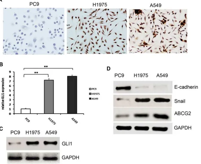
expression was negative in the EGFR-TKI-sensitive cell line PC9. We confirmed this result by Q-PCR and Western blot analysis. As shown inFig 1B and 1C, GLI1 was expressed at a very low level in PC9 compared with H1975 and A549 cells(P>0.001andP = 0.005respectively). Previous studies indicated that Hh signaling regulates EMT via upregulation of the transcrip-tion factor Snail and downregulatranscrip-tion of E-cadherin[27,28]. The stem cell marker ABCG2 is also a direct target of the Hh signaling pathway[29]. To further clarify the Hh pathway differ-ences between EGFR-TKI-sensitive and -resistant cells, these three important downstream tar-get genes were examined by Western blotting. We found that Snail expression was
considerably weaker in the sensitive PC9 cell line compared with the EGFR-TKI-resistant cell lines H1975 and A549 (P= 0.001). E-cadherin expression in PC9 cells was quite high, while its expression was very weak in the EGFR-TKI-resistant cell lines H1975 and A549 (P<0.001;Fig 1D). This result was consistent with previous reports that Snail expression was inversely correlated with that of E-cadherin[30,31]. ABCG2 expression was also quite high in
Fig 1. Hh pathway activity in EGFR-TKI-sensitive and -resistant cells.(A). Differences in GLI1 expression in EGFR-TKI-sensitive and -resistant NSCLC cells determined with immunocytochemistry. GLI1 was expressed in the nuclei of EGFR-TKI-resistant cell lines A549 and H1975, but GLI1 expression was negative in the EGFR-TKI-sensitive cell line PC9. (B). Relative GLI1 mRNA expression in EGFR-TKI-sensitive and -resistant NSCLC cells determined by Q-PCR; The expression of PC9 was significantly lower than that of H1975 and A549;**(P<0.01). (C). Differences in GLI1 expression in EGFR-TKI-sensitive and -resistant NSCLC cells determined by western blot. (D) Differences in E-cadherin, Snail, and ABCG2 expression in EGFR-TKI-sensitive and -resistant NSCLC cells determined by western blot.
the EGFR-TKI-resistant cell lines H1975 and A549 compare with its expression in the
EGFR-TKI-sensitive PC9 cell line (P<0.001;Fig 1D). Together, these results indicated a signifi-cant difference in the activity of the Hh signaling pathway between EGFR-TKI-sensitive and -resistant cells. The Hh signaling pathway was aberrantly activated in EGFR-TKI-resistant cells, while it was silenced in EGFR-TKI-sensitive cells.
Upregulation of Hh signaling pathway activity by exposure to SHH lead
to gefitinib tolerance accompanied by EMT induction and ABCG2
upregulation in EGFR-TKI-sensitive NSCLC cells
Based on the results above, we hypothesized that aberrant activation of the Hh signaling path-way may contribute to the EGFR-TKI resistance in NSCLC by affecting EMT and CSC mainte-nance. To examine this possibility, we tested the role of Shh signaling in NSCLC cells. First, EGFR-TKI-sensitive PC9 cells were treated with N-Shh (0.5μg/mL). Immunocytochemistry
result showed that GLI1 expression in PC9 cells was negative without exposure to N-Shh, but it was expressed in the nuclei after N-Shh treatment for 24 and 48 h (Fig 2A). Western blotting and Q-PCR results showed that GLI1 expression was obviously elevated after exposure to N-Shh for 24 h and 48 h (P= 0.008 andP<0.001 respectively;Fig 2B and 2C). These results demonstrated that Hh signaling was activated by extrinsic N-Shh in PC9 cells.
Next, EGFR-TKI-sensitive cells PC9 were treated with increasing concentrations of gefitinib with or without exposure to extrinsic N-Shh (0.5μg/mL), then their viability was assessed. Our
results showed that, compared with the gefitinib single-agent group, exposure to extrinsic N-Shh significantly increased gefitinib tolerance in PC9 cells (P= 0.001;Fig 2DandS1andS2
Tables). These findings show that aberrant activation of the Shh signaling pathway leads to EGFR-TKI resistance in NSCLC cells.
To examine the molecular mechanisms underlying the contribution of Shh signaling to EGFR-TKI resistance in NSCLC cells, we examined Snail, E-cadherin, and ABCG2 expression at 0, 24, and 48 h after treatment of PC9 cells with N-Shh (0.5μg/mL) by Western blotting. As
shown inFig 2E, after exposure to N-Shh for 24 h, the expression of Snail was elevated (P<0.001), the expression of E-cadherin was obviously attenuated (P= 0.003), and ABCG2 expression was markedly upregulated in PC9 cells (P= 0.008). These effects were sustained for 48 h following N-Shh stimulation. These results confirmed that hyperactivation of Hh signal-ing contributed to EGFR-TKI resistance in NSCLC cells through activation of the EMT transi-tion and the ABCG2 upregulatransi-tion.
Hh inhibition reversed EMT induction and decreased ABCG2 expression
in EGFR-TKI-resistant NSCLC cells
Next, to further assess the molecular mechanisms of Hh signaling in EGFR-TKI-resistant NSCLC cells, we examined GLI1, Snail, E-cadherin, and ABCG2 expression at 0, 24, and 48 h after treatment of the EGFR-TKI-resistant cell lines H1975 and A549 with SANT-1 (40μM).
The combination of gefitinib and SANT-1 synergistically inhibited
tumorigenesis and proliferation in EGFR-TKI-resistant NSCLC cell lines
To further demonstrate the role of the Hh signaling pathway in NSCLC cells, we blocked Hh signaling using the SMO inhibitor SANT-1. The IC50of SANT-1 in most NSCLC cell lines is ~40μM[18], and the IC50of gefitinib in EGFR-TKI-sensitive cells is ~20 nM[32]. Thus, the
Fig 2. Upregulation of Hh signaling pathway activity by exposure to SHH lead to gefitinib tolerance accompanied by EMT induction and ABCG2 upregulation in EGFR-TKI-sensitive NSCLC cells.(A) Immunocytochemical analysis of GLI1 in PC9 cells after N-Shh (0.5μg/mL) treatment for 0, 24, and
48h. GLI1 expression in PC9 cells was negative without exposure to N-Shh. GLI1 was expressed in the nuclei after N-Shh treatment for 24 and 48 h. (B) Western blot analysis of GLI1 in PC9 cells after N-Shh (0.5μg/mL) treatment for 0, 24, and 48h. (C) Q-PCR analysis of GLI1 mRNA relative expression in
PC9 cells after N-Shh (0.5μg/mL) treatment for 0, 24, and 48h; GLI1 expression was obviously elevated after exposure to N-Shh for 24 h and 48 h;**
(P<0.01). (D) Proliferation of PC9 cells was assessed by MTT assay after treatment with the indicated concentrations of gefitinib with or without pre-exposure to extrinsic N-Shh (0.5μg/mL) for 24h; compared with the gefitinib single-agent group, exposure to extrinsic N-Shh significantly increased gefitinib tolerance
EGFR-TKI-resistant cell lines A549 and H1975were treated with 40μM SANT-1 alone, 20 nM
gefitinib alone, or a combination of SANT-1 and gefitinib. As shown inFig 3C and 3D, SANT-1 and gefitinib alone did not inhibit clonogenic growth (P= 0.252 andP= 0.187 respectively). However, colonies very barely formed in the group subjected to combined SANT-1 and gefiti-nib treatment (P<0.001). These results indicate that SANT-1 and gefitinib may have a syner-gistic effect in resistant NSCLC cells. To confirm this, we treated the EGFR-TKI-resistant NSCLC cell lines A549 and H1975 with increasing concentrations of SANT-1 alone, gefitinib alone, and combinations of SANT-1 and gefitinib, and then assessed their prolifera-tion. The results showed that A549 cells were resistant not only to gefitinib but also to SANT-1 (P= 0.503;Fig 3EandS3andS4Tables). This result is consistent with a previous report that A549 cells showed hyperactivation of Hh signaling, but were resistant to Hh-signaling inhibi-tors [18]. However, the combination of gefitinib and SANT-1 inhibited the proliferation of A549 cells (P<0.001;Fig 3EandS3andS4Tables). Although, compared with gefitinib, SANT-1 was more effective in H1975 cells (P= 0.002), H1975 cells showed the‘best’response to the combination of gefitinib + SANT1 (P<0.001;Fig 3FandS5andS6Tables). Taken together, these results confirmed that the combination of an Hh signaling inhibitor and EGFR-TKIs had marked synergistic effects on EGFR-TKI-resistant NSCLC cells.
Fig 3. Hh inhibition reversed EMT and decreased the ABCG2 expression in EGFR-TKI-resistant NSCLC cells and the combination of gefitinib and SANT1 synergistically inhibited tumorigenesis and proliferation in EGFR-TKI-resistant NSCLC cell lines.(A) and (B), Western blot analysis of the Hh signaling pathway activity and its target genes E-Cadherin, Snail and ABCG2 in EGFR-resistant cells A549 and H1975 after SANT-1 treatment for 0, 24, and 48 h. (C) and (D), colony formation quantitative analysis in A549 and H1975 cells; gefitinib alone or SANT1 alone did not inhibit clonogenic growth effectively. However, colonies very barely formed in the group subjected to combined SANT-1 and gefitinib treatment in A549 and H1975 cells;*(P<0.05),**(P<0.01). (E) and (F), Proliferation of A549 and H1975 cells was assessed by MTT assay after treatment with the indicated concentrations of gefitinib alone, SANT-1 alone, or the combination of gefitinib and SANT-1;*(P<0.05),**(P<0.01).
Activity of the Hh signaling pathway in EGFR-TKI-sensitive and
-resistant NSCLC tissues
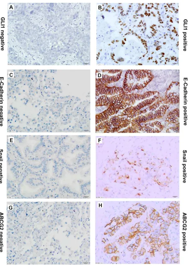
To further understand the difference in Hh signaling pathway activity between EGFR-TKI-sen-sitive and -resistant NSCLCs, expression of GLI1, ABCG2, Snail, and E-cadherin was assessed by IHC in four NSCLC tissues that contained the EGFR exon 19 deletion or the L858R sensitive mutation, four tissues that contained the EGFR T790M secondary mutation, and four tissues that contained the KRAS mutation (Fig 4). The results indicated that GLI1 was expressed in both EGFR-TKI-sensitive and -resistant tissues. Because of the small size of the samples, GLI1 expression showed no statistically significant difference among the three groups (P= 0.108). However, the mean rank of EGFR-TKI-sensitive tissues was 5.25, that of secondary resistant mutation tissues was 4.88, and the mean rank of KRAS mutation tissues was 9.38. Thus, a trend towards higher GLI1 expression was found in tissues that contained the KRAS mutation (Tables1and2). KRAS mutation-harboring NSCLCs may have higher Hh signaling pathway activity compared with those with EGFR-TKI-sensitive mutations and secondary resistant mutations.
ABCG2 expression did not differ significantly among EGFR-TKI-sensitive, secondary resis-tant, and primary resistant NSCLC tissues (P= 0.340). E-cadherin was moderately or strongly expressed in most NSCLC tissues (9/12,P= 0.555). Conversely, Snail was negative or weakly expressed in most tissues (11/12,P= 0.658) (Tables1and2).
Correlation analysis showed that E-cadherin expression was significantly negatively corre-lated with Snail expression (r= 0.582,P= 0.047). This result was consistent with the cell exper-iments. Additionally, GLI1 expression was negatively correlated with E-cadherin expression (r= 0.408,P= 0.188), and positively correlated with Snail (r= 0.372,P= 0.300) and ABCG2 (r= 0.386,P= 0.215) expression. Although the correlations lacked statistical significance because of the small sample size, the tendency was consistent with the cell experiments.
Discussion
The Hh signaling pathway is an effective therapeutic target in the treatment and prevention of many types of human cancer. Inappropriate activation of the Hh signaling pathway has been reported in NSCLC[17,18,33,34]. Previous studies have shown that EMT induction is associ-ated with sensitivity to EGFR-TKI in lung cancer[35–37]. The hedgehog signaling pathway tightly regulates EMT induction through its downstream target genes, such as Snail, ZEB1, and TWIST2[22]. Downregulation or loss of E-cadherin expression is a hallmark of EMT in embry-onic development and cancer progression. Snail expression is inversely correlated with E-cad-herin in epithelial tumor cell lines[30,31]. ABCG2 is one of the major multidrug resistance (MDR) pumps; it is also a stem cell marker and is closely associated with resistance to several drugs[38,39]. The Hh signaling pathway directly regulates the activity of ABCG2[29]. Hh antagonists can inhibit the activity of ABCG2 and resensitize NSCLC cells to mitoxantrone and topotecanin vitro[40]. Hh and EGFR signaling cooperatively regulate stem cell prolifera-tion in the postnatal and adult brain[23,41]. Simultaneous blockade of Hh and EGFR signaling inhibited proliferation and induced apoptosis, and improved the cytotoxic effects of docetaxel in metastatic prostate cancer cells[42]. Given this background, we hypothesized that the Hh signaling pathway might contribute to EGFR-TKI resistance in NSCLC by disregulated the EMT and ABCG2 activity.
Fig 4. Representative immunostaining of GLI1, ABCG2, E-Cadherin and Snail in NSCLC tissues.(A) and (B), negative control and positive GLI1 expression in NSCLC tissues. (C) and (D), negative control and positive E-Cadherin expression in NSCLC tissues. (E) and (F), negative control and positive Snail expression in NSCLC tissues.(G) and (H), negative and positive ABCG2 expression in NSCLC tissues.
the Hh pathway was silenced in EGFR-TKI-sensitive cells. The difference in Hh signaling activ-ity between EGFR-TKI-sensitive and -resistant cells suggested that aberrant activation of the Hh signaling pathway may contributes to EGFR-TKI resistance by inducing an EMT pheno-type and ABCG2 upregulation. To confirm these findings, Hh signaling was upregulated using extrinsic SHH in the EGFR-TKI-sensitive PC9 cell line. Upregulation of Hh activity resulted in induction of EMT and elevation of ABCG2 expression. Hh signaling hyperactivity also resulted in EGFR-TKI tolerance in otherwise EGFR-TKI-sensitive cells. These results confirmed that aberrant activation of Hh signaling resulted in the development of EGFR-TKI resistance in NSCLC cells through induction of EMT and ABCG2 overexpression. Inhibition of Hh signal-ing by the Hh inhibitor SANT-1 enhanced E-cadherin expression and downregulated Snail and ABCG2 expression. This result demonstrated that blockade of Hh signaling can reverse the EMT phenotype and maybe reduce CSC abundance. A previous study indicated that restor-ing E-cadherin expression increased sensitivity to EGFR-TKIsin vitroandin vivo[38]. Based on these results, we had reason to believe that targeting the Hh signaling pathway could affect EGFR-TKI resistance in NSCLC cells. Thus, we next treated EGFR-TKI-resistant cells with gefitinib or SANT-1 alone or gefitinib plus SANT-1. The results indicated that compared with either gefitinib or SANT-1 alone, the combination of gefitinib plus SANT-1 significantly
Table 1. Expression of GLI1, ABCG2, E-cadherin and Snail in NSCLC tissues tested by immunohistochemistry.
ID Gender Age Gene statues EGFR-TKI GLI1 ABCG2 E-cadherin Snail
L1756 F 64 DEL Sensitive 0 0 3 0
L2365 M 59 DEL Sensitive 1 1 3 0
L2186 F 63 DEL Sensitive 2 0 1 1
L2160 F 50 L858R Sensitive 0 0 3 0
L1433 M 45 T790M+del Resistance 2 1 2 1
L1439 M 56 T790M Resistance 1 0 0 1
L0353 M 43 T790M+del Resistance 1 2 3 0
L1423 F 53 T790M+L858R Resistance 0 0 2 0
L1301 M 66 KRAS Resistance 2 2 2 2
L1677 M 61 KRAS Resistance 3 0 0 0
L1834 M 63 KRAS Resistance 2 2 3 0
L0357 F 58 KRAS Resistance 2 2 2 1
doi:10.1371/journal.pone.0149370.t001
Table 2. Expression differences of GLI1, ABCG2, E-cadherin and Snail in EGFR-TKIs sensitive and resistant tissues.
Protein EGFR-TKI Mean rank χ2-value P-value
GLI1 Sensitive 5.25 4.460 0.108
Secondary resistance 4.88
Primary resistance 9.38
ABCG2 Sensitive 5.13 2.155 0.340
Secondary resistance 6.00
Primary resistance 8.38
E-cadherin Sensitive 8.00 1.179 0.555
Secondary resistance 5.75
Primary resistance 5.75
Snail Sensitive 5.38 0.838 0.658
Secondary resistance 6.75
Primary resistance 7.38
inhibited tumorigenesis and cell viability. Taken together, our findings suggest associations among Hh signaling, EMT, ABCG2 overexpression, and EGFR-TKI resistance in NSCLC cells for the first time.
Deregulation of Hh signaling contributes to tumorigenesis or accelerates tumor growth in an Hh ligand-independent or -dependent manner[43]. In most basal cell carcinomas (BCCs) and medulloblastomas, loss-of-function mutations in PTCH and gain-of-function mutations in SMO both lead to ligand-independent, mutation-driven activation of the Hh pathway[44–
46]. This type of tumor can benefit from treatment with a single Hh inhibitor. In the develop-ment of several other types of cancer—such as prostate, breast, pancreatic, breast, and lung cancer—over- or ectopic expression of Hh ligands, which activate signaling in an autocrine or paracrine manner, lead to ligand-dependent Hh pathway activation[34,47–51]. In this mecha-nism, the Hh signaling pathway plays a secondary role in tumor maintenance and growth. Thus, targeting the Hh pathway alone in these tumors is less likely to be successful unless com-bined with other appropriate chemotherapy or targeted therapy, or as a maintenance therapy [52]. Our data indicated that although SANT-1 reversed the EMT phenotype and decreased ABCG2 expression in EGFR-TKI-resistant cells, it had a minor inhibitory effect, especially in the A549 cell line. In contrast, the combination of SANT-1 with gefitinib effectively inhibited cellular proliferation and tumorigenesis. Patients with NSCLC may not benefit from single-agent Hh inhibitors. Combinations of Hh inhibitors and EGFR-TKIs may be an effective thera-peutic strategy for primary and secondary EGFR-TKI-resistant NSCLCs. Compared with cyto-toxic chemotherapy, targeted drugs possess the features of superior responses and fewer adverse effects, and thus have a broader treatment spectrum. Patients with ECOG performance status scores>2 lose the opportunity to receive‘traditional’cytotoxic therapy. However, these patients can receive targeted therapy. Determining individual therapeutic schedules according to their molecular profiles is an increasing trend in cancer treatment. Our research indicated that an Hh inhibitor increased the sensitivity to EGFR-TKIs in EGFR-TKI- resistant cells that have an EMT phenotype and ABCG2 overexpression. NSCLCs harboring KRAS mutations may have even higher Hh signaling pathway activity. E-cadherin was strongly expressed in EGFR-TKI-sensitive NSCLC tissues. E-cadherin expression was negatively related to GLI1 expression. Previous studies have also reported that EMT is a determinant of EGFR-TKIs in NSCLCsin vitroandin vivo[35,36]. Thus, E-cadherin expression may be a potential bio-marker of suitability for combined application of Hh inhibitors and EGFR-TKIs in EGFR-TKI-resistant NSCLCs.
In conclusion, our study suggests that Hh signaling hyperactivity resulted in EGFR-TKI resistance due to EMT induction and ABCG2 upregulation. The combination of an Hh inhibi-tor and EGFR-TKIs may be an effective therapy for EGFR-TKI-resistant NSCLCs that harbor the EMT phenotype and ABCG2 overexpression. E-cadherin may be a useful biomarker to determine the suitability of application of Hh inhibitors in EGFR-TKI-resistant NSCLCs.
Supporting Information
S1 Table. The raw date of the proliferation of PC9 cells after treatment with indicated con-centrations of Gefitinib with or without the pre exposure of extrinsic N-Shh (0.5ug/ml) for 24 hours analyzed by factorial analysis.
(DOCX)
S2 Table. The proliferation of PC9 cells after treatment with indicated concentrations of Gefitinib with or without the pre exposure of extrinsic N-Shh (0.5ug/ml) for 24 hours.
S3 Table. The raw date of the proliferation effects after treatment with different concentra-tion of Gefitinib single agent, SANT-1 single agent or the combinaconcentra-tion of Gefitinib and SANT-1 on A549 cells analyzed by factorial analysis.
(DOCX)
S4 Table. The effects of proliferation after treatment with different concentration of Gefiti-nib single agent, SANT-1 single agent or the combination of GefitiGefiti-nib and SANT-1 on A549 cells.
(DOCX)
S5 Table. The raw date of the proliferation effects after treatment with different concentra-tion of Gefitinib single agent, SANT-1 single agent or the combinaconcentra-tion of Gefitinib and SANT-1 on H1975 cells analyzed by factorial analysis.
(DOCX)
S6 Table. The effects of proliferation after treatment with different concentration of Gefiti-nib single agent, SANT-1 single agent or the combination of GefitiGefiti-nib and SANT-1 on H1975 cells.
(DOCX)
Acknowledgments
The authors thank Dr. Tony Mork (Chinese University of Hong Kong) for kind of providing H1975and PC9 cell lines. The English in this document has been checked by at least two pro-fessional editors, both native speakers of English. For a certificate, please see:http://www. textcheck.com/certificate/6rs3Qt.
Author Contributions
Conceived and designed the experiments: XYB XCZ SJA YLW. Performed the experiments: XYB SQY ZX ZHC JS LYG. Analyzed the data: XYB XCZ SJA YLW. Contributed reagents/ materials/analysis tools: XYB ZHC YLW. Wrote the paper: XYB.
References
1. Ciuleanu T, Brodowicz T, Zielinski C, Kim JH, Krzakowski M, Laack E, et al. (2009) Maintenance peme-trexed plus best supportive care versus placebo plus best supportive care for non-small-cell lung can-cer: a randomised, double-blind, phase 3 study. Lancet 374: 1432–1440. doi:10.1016/S0140-6736
(09)61497-5PMID:19767093
2. Zhou C, Wu YL, Chen G, Feng J, Liu XQ, Wang C, et al. (2011) Erlotinib versus chemotherapy as first-line treatment for patients with advanced EGFR mutation-positive non-small-cell lung cancer (OPTI-MAL, CTONG-0802): a multicentre, open-label, randomised, phase 3 study. Lancet Oncol 12: 735– 742. doi:10.1016/S1470-2045(11)70184-XPMID:21783417
3. Rosell R, Carcereny E, Gervais R, Vergnenegre A, Massuti B, Felip E, et al. (2012) Erlotinib versus standard chemotherapy as first-line treatment for European patients with advanced EGFR mutation-positive non-small-cell lung cancer (EURTAC): a multicentre, open-label, randomised phase 3 trial. Lancet Oncol 13: 239–246. doi:10.1016/S1470-2045(11)70393-XPMID:22285168
4. Costa DB, Kobayashi S, Tenen DG, Huberman MS (2007) Pooled analysis of the prospective trials of gefitinib monotherapy for EGFR-mutant non-small cell lung cancers. Lung Cancer 58: 95–103. PMID:
17610986
5. Balak MN, Gong Y, Riely GJ, Somwar R, Li AR, Zakowski MF, et al. (2006) Novel D761Y and common secondary T790M mutations in epidermal growth factor receptor-mutant lung adenocarcinomas with acquired resistance to kinase inhibitors. Clin Cancer Res 12: 6494–6501. PMID:17085664 6. Pao W, Miller VA, Politi KA, Riely GJ, Somwar R, Zakowski MF, et al. (2005) Acquired resistance of
7. Bean J, Brennan C, Shih JY, Riely G, Viale A, Wang L, et al. (2007) MET amplification occurs with or without T790M mutations in EGFR mutant lung tumors with acquired resistance to gefitinib or erlotinib. Proc Natl Acad Sci U S A 104: 20932–20937. PMID:18093943
8. Turke AB, Zejnullahu K, Wu YL, Song Y, Dias-Santagata D, Lifshits E, et al. (2010) Preexistence and clonal selection of MET amplification in EGFR mutant NSCLC. Cancer Cell 17: 77–88. doi:10.1016/j.
ccr.2009.11.022PMID:20129249
9. Yamada T, Matsumoto K, Wang W, Li Q, Nishioka Y, Sekido Y, et al. (2010) Hepatocyte growth factor reduces susceptibility to an irreversible epidermal growth factor receptor inhibitor in EGFR-T790M mutant lung cancer. Clin Cancer Res 16: 174–183. doi:10.1158/1078-0432.CCR-09-1204PMID:
20008840
10. Metro G, Chiari R, Duranti S, Siggillino A, Fischer MJ, Giannarelli D, et al. (2012) Impact of specific mutant KRAS on clinical outcome of EGFR-TKI-treated advanced non-small cell lung cancer patients with an EGFR wild type genotype. Lung Cancer 78: 81–86. doi:10.1016/j.lungcan.2012.06.005PMID:
22770374
11. Ludovini V, Bianconi F, Pistola L, Chiari R, Minotti V, Colella R, et al. (2011) Phosphoinositide-3-kinase catalytic alpha and KRAS mutations are important predictors of resistance to therapy with epidermal growth factor receptor tyrosine kinase inhibitors in patients with advanced non-small cell lung cancer. J Thorac Oncol 6: 707–715. doi:10.1097/JTO.0b013e31820a3a6bPMID:21258250
12. Sabbah M, Emami S, Redeuilh G, Julien S, Prevost G, Zimber A, et al. (2008) Molecular signature and therapeutic perspective of the epithelial-to-mesenchymal transitions in epithelial cancers. Drug Resist Updat 11: 123–151. doi:10.1016/j.drup.2008.07.001PMID:18718806
13. Guarino M, Rubino B, Ballabio G (2007) The role of epithelial-mesenchymal transition in cancer pathol-ogy. Pathology 39: 305–318. PMID:17558857
14. Garvalov BK, Acker T (2011) Cancer stem cells: a new framework for the design of tumor therapies. J Mol Med (Berl) 89: 95–107.
15. Morel AP, Lievre M, Thomas C, Hinkal G, Ansieau S, Puisieux A. (2008) Generation of breast cancer stem cells through epithelial-mesenchymal transition. PLoS One 3: e2888. doi:10.1371/journal.pone. 0002888PMID:18682804
16. McMahon AP, Ingham PW, Tabin CJ (2003) Developmental roles and clinical significance of hedgehog signaling. Curr Top Dev Biol 53: 1–114. PMID:12509125
17. Watkins DN, Berman DM, Burkholder SG, Wang B, Beachy PA, Baylin SB. (2003) Hedgehog signalling within airway epithelial progenitors and in small-cell lung cancer. Nature 422: 313–317. PMID:
12629553
18. Yuan Z, Goetz JA, Singh S, Ogden SK, Petty WJ, Black CC, et al. (2007) Frequent requirement of hedgehog signaling in non-small cell lung carcinoma. Oncogene 26: 1046–1055. PMID:16909105 19. Lum L, Beachy PA (2004) The Hedgehog response network: sensors, switches, and routers. Science
304: 1755–1759. PMID:15205520
20. Kalderon D (2000) Transducing the hedgehog signal. Cell 103: 371–374. PMID:11081624 21. Hooper JE, Scott MP (2005) Communicating with Hedgehogs. Nat Rev Mol Cell Biol 6: 306–317.
PMID:15803137
22. Katoh Y, Katoh M (2009) Hedgehog target genes: mechanisms of carcinogenesis induced by aberrant hedgehog signaling activation. Curr Mol Med 9: 873–886. PMID:19860666
23. Palma V, Ruiz IAA (2004) Hedgehog-GLI signaling regulates the behavior of cells with stem cell proper-ties in the developing neocortex. Development 131: 337–345. PMID:14681189
24. Kasper M, Schnidar H, Neill GW, Hanneder M, Klingler S, Blaas L, et al. (2006) Selective modulation of Hedgehog/GLI target gene expression by epidermal growth factor signaling in human keratinocytes. Mol Cell Biol 26: 6283–6298. PMID:16880536
25. Bigelow RL, Jen EY, Delehedde M, Chari NS, McDonnell TJ (2005) Sonic hedgehog induces epidermal growth factor dependent matrix infiltration in HaCaT keratinocytes. J Invest Dermatol 124: 457–465. PMID:15675968
26. Taylor CR (2006) Quantifiable internal reference standards for immunohistochemistry: the measure-ment of quantity by weight. Appl Immunohistochem Mol Morphol 14: 253–259. PMID:16932014 27. Karhadkar SS, Bova GS, Abdallah N, Dhara S, Gardner D, Maitra A, et al. (2004) Hedgehog signalling
in prostate regeneration, neoplasia and metastasis. Nature 431: 707–712. PMID:15361885 28. Gupta S, Takebe N, Lorusso P (2010) Targeting the Hedgehog pathway in cancer. Ther Adv Med
29. Singh RR, Kunkalla K, Qu C, Schlette E, Neelapu SS, Samaniego F, et al. (2011) ABCG2 is a direct transcriptional target of hedgehog signaling and involved in stroma-induced drug tolerance in diffuse large B-cell lymphoma. Oncogene 30: 4874–4886. doi:10.1038/onc.2011.195PMID:21625222 30. Yokoyama K, Kamata N, Fujimoto R, Tsutsumi S, Tomonari M, Taki M, et al. (2003) Increased invasion
and matrix metalloproteinase-2 expression by Snail-induced mesenchymal transition in squamous cell carcinomas. Int J Oncol 22: 891–898. PMID:12632084
31. Yokoyama K, Kamata N, Hayashi E, Hoteiya T, Ueda N, Fujimoto R, et al. (2001) Reverse correlation of E-cadherin and snail expression in oral squamous cell carcinoma cells in vitro. Oral Oncol 37: 65– 71. PMID:11120485
32. Cheng H, An SJ, Zhang XC, Dong S, Zhang YF, Fujimoto R, et al. (2011) In vitro sequence-dependent synergism between paclitaxel and gefitinib in human lung cancer cell lines. Cancer Chemother Phar-macol 67: 637–646. doi:10.1007/s00280-010-1347-4PMID:20495920
33. Singh S, Wang Z, Liang FD, Black KE, Goetz JA, Tokhunts R, et al. (2011) Hedgehog-producing cancer cells respond to and require autocrine Hedgehog activity. Cancer Res 71: 4454–4463. doi:10.1158/
0008-5472.CAN-10-2313PMID:21565978
34. Gialmanidis IP, Bravou V, Amanetopoulou SG, Varakis J, Kourea H, Papadaki H. (2009) Overexpres-sion of hedgehog pathway molecules and FOXM1 in non-small cell lung carcinomas. Lung Cancer 66: 64–74. doi:10.1016/j.lungcan.2009.01.007PMID:19200615
35. Thomson S, Buck E, Petti F, Griffin G, Brown E, Ramnarine N, et al. (2005) Epithelial to mesenchymal transition is a determinant of sensitivity of non-small-cell lung carcinoma cell lines and xenografts to epi-dermal growth factor receptor inhibition. Cancer Res 65: 9455–9462. PMID:16230409
36. Yauch RL, Januario T, Eberhard DA, Cavet G, Zhu W, Fu L, et al. (2005) Epithelial versus mesenchy-mal phenotype determines in vitro sensitivity and predicts clinical activity of erlotinib in lung cancer patients. Clin Cancer Res 11: 8686–8698. PMID:16361555
37. Witta SE, Gemmill RM, Hirsch FR, Coldren CD, Hedman K, Ravdel L, et al. (2006) Restoring E-cad-herin expression increases sensitivity to epidermal growth factor receptor inhibitors in lung cancer cell lines. Cancer Res 66: 944–950. PMID:16424029
38. Krishnamurthy P, Ross DD, Nakanishi T, Bailey-Dell K, Zhou S, Mercer KE, et al. (2004) The stem cell marker Bcrp/ABCG2 enhances hypoxic cell survival through interactions with heme. J Biol Chem 279: 24218–24225. PMID:15044468
39. Sarkadi B, Ozvegy-Laczka C, Nemet K, Varadi A (2004) ABCG2—a transporter for all seasons. FEBS Lett 567: 116–120. PMID:15165903
40. Zhang Y, Laterra J, Pomper MG (2009) Hedgehog pathway inhibitor HhAntag691 is a potent inhibitor of ABCG2/BCRP and ABCB1/Pgp. Neoplasia 11: 96–101. PMID:19107236
41. Palma V, Lim DA, Dahmane N, Sanchez P, Brionne TC, Herzberg CD, et al. (2005) Sonic hedgehog controls stem cell behavior in the postnatal and adult brain. Development 132: 335–344. PMID:
15604099
42. Mimeault M, Johansson SL, Vankatraman G, Moore E, Henichart JP, Depreux P, et al. (2007) Com-bined targeting of epidermal growth factor receptor and hedgehog signaling by gefitinib and cyclopa-mine cooperatively improves the cytotoxic effects of docetaxel on metastatic prostate cancer cells. Mol Cancer Ther 6: 967–978. PMID:17363490
43. Gupta S, Takebe N, Lorusso P (2010) Targeting the Hedgehog pathway in cancer. Ther Adv Med Oncol 2: 237–250. doi:10.1177/1758834010366430PMID:21789137
44. Goodrich LV, Milenkovic L, Higgins KM, Scott MP (1997) Altered neural cell fates and medulloblastoma in mouse patched mutants. Science 277: 1109–1113. PMID:9262482
45. Xie J, Murone M, Luoh SM, Ryan A, Gu Q, Zhang C, et al. (1998) Activating Smoothened mutations in sporadic basal-cell carcinoma. Nature 391: 90–92. PMID:9422511
46. Raffel C, Jenkins RB, Frederick L, Hebrink D, Alderete B, Fults DW, et al. (1997) Sporadic medulloblas-tomas contain PTCH mutations. Cancer Res 57: 842–845. PMID:9041183
47. Ma X, Chen K, Huang S, Zhang X, Adegboyega PA, Evers BM, et al. (2005) Frequent activation of the hedgehog pathway in advanced gastric adenocarcinomas. Carcinogenesis 26: 1698–1705. PMID:
15905200
48. Thayer SP, di Magliano MP, Heiser PW, Nielsen CM, Roberts DJ, Lauwers GY, et al. (2003) Hedgehog is an early and late mediator of pancreatic cancer tumorigenesis. Nature 425: 851–856. PMID:
14520413
50. Azoulay S, Terry S, Chimingqi M, Sirab N, Faucon H, Gil Diez de Medina S, et al. (2008) Comparative expression of Hedgehog ligands at different stages of prostate carcinoma progression. J Pathol 216: 460–470. doi:10.1002/path.2427PMID:18825689
51. Rubin LL, de Sauvage FJ (2006) Targeting the Hedgehog pathway in cancer. Nat Rev Drug Discov 5: 1026–1033. PMID:17139287