Arq Bras Oftalmol. 2008;71(4):576-8
Abscesso orbitário secundário à dacriocistite aguda: relato de caso
Trabalho realizado no Departamento de Oftalmologia, Otorrinolaringologia e Cirurgia de Cabeça e Pescoço da Faculdade de Medicina de Ribeirão Preto - Universida-de Universida-de São Paulo.
1Médica Residente do Hospital das Clínicas da Universi-dade de São Paulo - USP - Ribeirão Preto (SP) - Brasil. 2Médico Residente do Hospital das Clínicas da USP
-Ribeirão Preto (SP) - Brasil.
3 Médica contratada do Setor de Oculoplástica. Hospital das Clínicas USP - Ribeirão Preto (SP) - Brasil. 4Professor Titular e Chefe do Setor de Oculoplástica do
Hospital das Clínicas da USP Ribeirão Preto (SP) -Brasil.
Endereço para correspondência: Antonio Augusto Ve-lasco e Cruz. Departamento de Oftalmologia, Otorrinola-ringologia e Cirurgia de Cabeça e Pescoço, Hospital das Clínicas - Campus. Av. Bandeirantes, 3900 - Ribeirão Preto (SP) CEP 14049-900
E-mail: [email protected] Recebido para publicação em 22.07.2007 Aprovação em 18.11.2007
Nota Editorial: Depois de concluída a análise do artigo sob sigilo editorial e com a anuência da Dra. Joelice dos Santos Araújo sobre a divulgação de seu nome como revisora, agradecemos sua participação neste processo. Marcia Clivati Martins1
José Reinaldo da Silva Ricardo2 Patrícia Mitiko Santello Akaishi3 Antonio Augusto Velasco e Cruz4
Orbital abscess secondary to acute dacryocystitis:
case report
Keywords: Lacrimal apparatus diseases; Dacryocystitis/complications; Orbital diseases/ etiology; Cellulitis; Abscess; Bacterial infections; Case reports [Publication type]
Acute dacryocystitis usually induces preseptal infection. In rare instances the infection that is confined to the lacrimal sac can extend to the orbital contents resulting in orbital cellulitis. We present a case of intraconal abscess secondary to acute dacryocystitis and review the literature of orbital cellulitis resulting from acute lacrimal sac infection.
ABSTRACT
RELATOS DE CASOS
INTRODUCTION
The immediate consequence of nasolacrimal duct obstruction is epipho-ra. In chronic cases conjunctivitis and mucoid discharge ensue. Acute da-cryocystitis may develop when significant bacterial growth occurs in the stagnant fluid of the lacrimal sac. The main complication of acute dacryo-cystitis is lacrimal sac abscess that often leads to draining fistulas to the skin. The infection can also spread to the lower eyelid tissues, becoming a preseptal cellulitis. Orbital extension rarely can occur(1-10) and may result in intraconal abscess formation(2-5,8). We present a case of intraconal abscess secondary to acute dacryocystitis and review the literature of orbital cellu-litis caused by acute dacryocystitis.
CASE REPORT
Arq Bras Oftalmol. 2008;71(4):576-8
577 Orbital abscess secondary to acute dacryocystitis: case report
anesthesia. The orbit was approached through a subciliary inci-sion. The abscess was located and drained, releasing a large amount of pus. After drainage of the abscess, irrigation of the lacrimal sac through the lower canaliculus clearly demonstrated that the sac was ruptured. The skin was closed and a small Penrose drain was left in the wound. On the third postoperative day the patient was afebrile, with a marked decrease in eyelid swelling and improved ocular motility of the left eye. Culture of the tissues grew strictly anaerobic bacteria (Peptostreptococcus
prevotii and Prevotella melaninogenica). After resolution of
the infection her final visual acuity was 20/20 in both eyes and an external dacryocystorhinostomy (DCR) was performed.
DISCUSSION
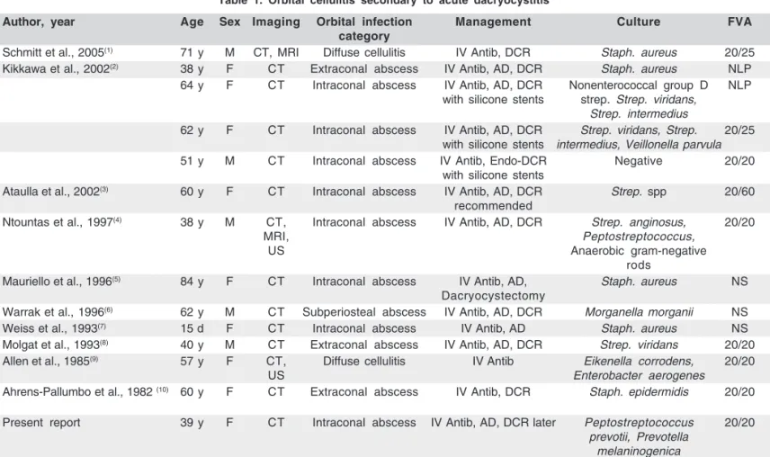
Dacryocystitis is a rare cause of orbital infection. A litera-ture search using the Medline data base showed only 10 reports of orbital cellulitis (13 cases) caused by acute dacryo-cystitis (Table 1). For 7 patients (53.8%) the infection reached the intraconal space forming an abscess. The rarity of orbital extension has been attributed to the fact that the lacrimal sac is a preseptal structure. In fact, the orbital septum inserts posteriorly at the lacrimal crest, acting as an anatomic barrier. In addition, other anatomical elements such as the lacrimal fascia, the posterior limb of the medial canthal ligament, and deep heads of the pretarsal and preseptal orbicularis muscles (Horner muscle) also act as a barrier to posterior extension.
Once the infection ruptures the lacrimal sac the posterior barriers may be overcome by the infection, leading to orbital cellulitis. Because of the anterior and inferior location of the lacrimal sac a channel of communication can be formed bet-ween the medial and inferior rectus muscles directly to the intraconal space, leading to an intraconal abscess(2-3,5).
Table 1 shows that the microorganisms found in dacryo-cystitis-induced orbital cellulitis do not differ from the germs found in uncomplicated dacryocystitis. In most cases (53.8%) single isolates of gram-positive bacteria are found. There were only four polymicrobial infections (30.8%), with strictly anae-robic bacteria in two of them.
Orbital abscess formation can occur even in patients using oral antibiotics and can rapidly lead to visual loss(2). Intrave-nous antibiotic therapy is necessary in all cases of orbital cellulitis; because of its bacteriology we consider the use of intravenous oxacillin, 2 g every 6 hours, and ceftriaxone, 2 g every 12 hours, as the first empirical option.
Although intravenous antibiotic therapy is necessary, sur-gical drainage of the abscess is mandatory for the resolution of the acute orbital picture. DCR can be delayed until complete resolution of the acute infectious disease.
The present report demonstrates that acute dacryocystitis can be added to the list of causes of orbital abscess and may be a cause of visual loss. In view of this type of complication, we conclude that patients with acute infection of the lacrimal sac should be carefully monitored.
RESUMO
A dacriocistite aguda comumente evolui para infecção pré-septal. Raramente a infecção localizada no saco lacrimal pode estender-se ao conteúdo orbitário resultando em celulite orbi-tária. Apresentamos um caso de abscesso orbitário intraconal secundário à dacriocistite aguda e uma revisão de literatura de celulite orbitária causada por infecção aguda do saco lacrimal.
Descritores: Doenças do aparelho lacrimal;
Dacriocistite/com-plicações; Doenças orbitárias/etiologia; Celulite; Abscesso; Infecções bacterianas; Relatos de casos [Tipo de publicação] Figure 1 - Clinical aspect at presentation. Note non axial proptosis and
chemosis.
Figure 2 - Coronal computed tomographic scan showing a left lacrimal sac abscess adjacent to an extra- and intraconal orbital abscess
578Orbital abscess secondary to acute dacryocystitis: case report
Arq Bras Oftalmol. 2008;71(4):576-8
Table 1. Orbital cellulitis secondary to acute dacryocystitis
Author, year Age Sex Imaging Orbital infection Management Culture FVA category
Schmitt et al., 2005(1) 71 y M CT, MRI Diffuse cellulitis IV Antib, DCR Staph. aureus 20/25 Kikkawa et al., 2002(2) 38 y F CT Extraconal abscess IV Antib, AD, DCR Staph. aureus NLP
64 y F CT Intraconal abscess IV Antib, AD, DCR Nonenterococcal group D NLP with silicone stents strep. Strep. viridans,
Strep. intermedius
62 y F CT Intraconal abscess IV Antib, AD, DCR Strep. viridans, Strep. 20/25 with silicone stents intermedius, Veillonella parvula
51 y M CT Intraconal abscess IV Antib, Endo-DCR Negative 20/20 with silicone stents
Ataulla et al., 2002(3) 60 y F CT Intraconal abscess IV Antib, AD, DCR Strep. spp 20/60 recommended
Ntountas et al., 1997(4) 38 y M CT, Intraconal abscess IV Antib, AD, DCR Strep. anginosus, 20/20
MRI, Peptostreptococcus,
US Anaerobic gram-negative
rods
Mauriello et al., 1996(5) 84 y F CT Intraconal abscess IV Antib, AD, Staph. aureus NS Dacryocystectomy
Warrak et al., 1996(6) 62 y M CT Subperiosteal abscess IV Antib, AD, DCR Morganella morganii NS Weiss et al., 1993(7) 15 d F CT Intraconal abscess IV Antib, AD Staph. aureus NS Molgat et al., 1993(8) 40 y M CT Extraconal abscess IV Antib, AD, DCR Strep. viridans 20/20 Allen et al., 1985(9) 57 y F CT, Diffuse cellulitis IV Antib Eikenella corrodens, 20/20
US Enterobacter aerogenes
Ahrens-Pallumbo et al., 1982 (10) 60 y F CT Extraconal abscess IV Antib, DCR Staph. epidermidis 20/20
Present report 39 y F CT Intraconal abscess IV Antib, AD, DCR later Peptostreptococcus 20/20
prevotii, Prevotella melaninogenica
FVA= final visual acuity; y= years; d= days; CT= computed tomography; MRI= magnetic resonance imaging; US= ultrasonography; IV Antib.= intravenous antibiotic
therapy; DCR= external dacryocystorhinostomy; AD= abscess drainage; Endo-DCR= endonasal dacryocystorhinostomy; Staph.= Staphylococcus; Strep.=
Streptococcus; NLP= no light perception; NS= not stated
REFERENCES
1. Schmitt NJ, Beatty RL, Kennerdell JS. Superior ophthalmic vein thrombosis in a patient with dacryocystitis-induced orbital cellulitis. Ophthal Plast Re-constr Surg. 2005;21(5):387-9.
2. Kikkawa DO, Heinz GW, Martin RT, Nunery WN, Eiseman AS. Orbital cellulitis and abscess secondary to dacryocystitis. Arch Ophthalmol. 2002;120(8):1096-9. 3. Ataullah S, Sloan B. Acute dacryocystitis presenting as an orbital abscess.
Clin Experiment Ophthalmol. 2002;30(1):44-6.
4. Ntountas I, Morschbacher R, Pratt D, Patel BC, Anderson RL, McCann JD. An orbital abscess secondary to acute dacryocystitis. Ophthalmic Surg Lasers. 1997;28(9):758-61.
5. Mauriello JA, Wasserman BA. Acute dacryocystitis: an unusual cause of life-threatening orbital abscess with frozen globe. Ophthal Plast Reconstr Surg. 1996;12(4):294-5.
6. Warrak E, Khoury P. Orbital abscess secondary to acute dacryocystitis. Can J Ophthalmol. 1996;31(4):201-2.
7. Weiss GH, Leib ML. Congenital dacryocystitis and retrobulbar abscess. J Pediatr Ophthalmol Strabismus. 1993;30(4):271-2.
8. Molgat YM, Hurwitz JJ. Orbital abscess due to acute dacryocystitis. Can J Ophthalmol. 1993;28(4):181-3.
9. Allen MV, Cohen KL, Grimson BS. Orbital cellulitis secondary to dacryo-cystitis following blepharoplasty. Ann Ophthalmol. 1985;17(8):498-9. 10. Ahrens-Palumbo MJ, Ballen PH. Primary dacryocystitis causing orbital