Original Article
1 4 6 Arq Bras Oftalmol. 2015;78(3):146-9 http://dx.doi.org/10.5935/0004-2749.20150038
INTRODUCTION
Preterm birth is a significant public health concern, as it is associa-ted with a high risk of infant mortality, various morbidities in both the neonatal period and later age, and significant socio-economic difficulties(1,2). Prematurely born children are disadvantaged in terms of perinatal mortality and long-term growth(3-5), and they have low birth weight and shorter eyes as compared with full-term children(6,7).
ABSTRACT
Purpose: To analyze ocular biometry parameters and evaluate their relationship with gestational age, birth weight, and postmenstrual age in prematurely born infants.
Methods: The right eyes of 361 premature infants born before the 36th gestational week were evaluated. Birth weight, gestational week, and gender were recorded. An A-scan Biometer was used for obtaining axial measurements, including anterior chamber depth, lens thickness, vitreous length, and total axial length. Results: Gestational age and birth weight values ranged from 23 to 36 weeks and from 560 to 2,670 g, respectively. The mean gestational age and birth weight were 30.8 ± 2.8 weeks and 1,497.9 ± 483.6 g, respectively. During the first examination (4-5 weeks of postnatal age), birth weight and gestational age of the infants cor-related significantly and positively with lens thickness, vitreous length, and axial length (r>0.5, p<0.001), but not with anterior chamber depth (r<0.5). Increased vitreous and axial lengths correlated significantly with increasing postmenstrual age of the infants (r=0.669, p<0.001; r=0.845, p<0.001, respectively).
Conclusions: Lens thickness, vitreous length, and axial length, but not anterior chamber depth, were significantly correlated with birth weight and gestational age. All four parameters increased with increasing postmenstrual age, with higher correlations for vitreous and axial lengths than for anterior chamber depth and lens thickness. It was concluded that axial elongation resulted primarily from increasing posterior chamber length.
Keywords: Anterior chamber; Axial length, eye; Biometry; Gestational age; Premature; Birth weight
RESUMO
Objetivo: Medir os comprimentos axiais dos componentes oculares e avaliar a re-lação com a idade gestacional, peso ao nascer e idade pós-menstrual em crianças nascidas prematuramente.
Método: O olho direito de 361 crianças prematuras, que nasceram com menos de 36 semanas de gestação, foram avaliados. O peso ao nascer, semanas de gestação e gênero foram registrados. Um biômetro A-scan foi utilizado para a obtenção das medidas axiais da profundidade da câmara anterior, espessura do cristalino, compri-mento vítreo e compricompri-mento axial total.
Resultados: A idade gestacional e os valores de peso ao nascimento variaram de 23 a 36 semanas e de 560 a 2.670 g, respectivamente. A idade gestacional e o peso ao nascer foram 30,8 ± 2,8 semanas e 1.497,9 ± 483,6 g. Ao primeiro exame (4 a 5 semanas de idade pós-natal), o peso ao nascimento e a idade gestacional dos recém-nascidos apresentaram correlação positiva, estatisticamente significativa, com a espessura do cristalino, comprimento vítreo e comprimento axial total (r>0,5 p<0,001), mas não com a profundidade da câmara anterior (r<0,5). O alongamento de comprimento vítreo e do comprimento axial total se correlacionaram significativamente com o aumento da idade pós-menstrual dos lactentes (r=0,669; p<0,001 e r=0,845; p<0,001, respectivamente).
Conclusões: A espessura do cristalino, o comprimento vítreo e o comprimento axial total, mas não profundidade da câmara anterior, foram significativamente corre-lacionados com o peso ao nascimento e com a idade gestacional. Todos os quatro componentes aumentaram com a idade pós-menstrual, apresentando correlações mais elevadas do comprimento vítreo e comprimento axial total do que da profundidade da câmara anterior e espessura do cristalino. Concluiu-se que o alongamento axial resultou principalmente do aumento do comprimento da câmara posterior.
Descritores: Camada anterior; Comprimento axial do olho; Biometria; Idade gesta-cional; Infant; Premature; Peso ao nascer
At present, ultrasound and optical biometry (partial coherence la-ser interferometry) are used to measure intraocular distances. Ho wever, eye measurements in newborns are only possible using ul tra sonic methods. Ultrasound biometry, commonly referred to as A-scan and B-scan, is utilized for diagnostic testing and biometric mea surements(8). A-scan ultrasonography provides a one-dimensional measurement of length in the axial plane. Additionally, it facilitates the monitoring
The relationship of birth weight, gestational age, and postmenstrual age with
ocular biometry parameters in premature infants
A relação entre o peso ao nascer, idade gestacional e idade pós-menstrual com a biometria ocular
em bebês prematuros
Ozdemir Ozdemir1, zuhal Ozen Tunay1, damla erginTurk acar1, muhammeT kazim erOl2, ender Sener3, ugur acar4
Submitted for publication: October 27, 2014 Accepted for publication: February 19, 2015
1 Ophthalmology Department, Zekai Tahir Burak Women’s Health Education and Research Hospital, Ankara, Turkey.
2 Ophthalmology Department, Antalya Education and Research Hospital, Antalya, Turkey. 3 Ophthalmology Department, Tokat Medical Park Hospital, Tokat, Turkey.
4 Ophthalmology Department, Kastamonu Faculty of Medicine, Hacettepe University, Ankara, Turkey.
Funding: No specific financial support was available for this study.
Disclosure of potential conflicts of interest: None of the authors have any potential conflict of interest to disclose.
Corresponding author: Ozdemir Ozdemir. Göz Hastalıkları Polikliniği, Zekai Tahir Burak Kadın
Sağlığı Eğitim ve Araştırma Hastanesi, Talatpaşa Bulvarı, Altındağ, Ankara, Turkey. E-mail: [email protected]
Ozdemir O, et al.
147 Arq Bras Oftalmol. 2015;78(3):146-9 eye growth during infancy(9). The ultrasound axial length of the eye is
measured using either contact or immersion techniques. The contact technique is used more frequently, while measuring the axial length of children’s eyes by pediatric cataract surgeons(10).
This study is aimed to measure ocular biometric parameters in pre-mature infants and to investigate their relationship with birth weight, gestational age, and postmenstrual age.
METHODS
I
NFANTSPrematureinfants enrolled in the retinopathy of prematurity (ROP) screening and who were born between September 1, 2013 and Janua-ry 1, 2014 at Zekai Tahir Burak Women’s Health Education and Research Hospital were selected for this cohort study. The inclusion criterion was birth at ≤36th gestation week. Infants with all types of congenital anomalies were excluded, even mild ones, and those with congenital eye abnormalities were also excluded from the study. Infants who had received previous treatment, such as laser photocoagulation and/or intravitreal injections, were also excluded.
Examinations for ROP and biometry measurement were initiated between postnatal weeks 4 and 5. Follow-up examinations were planned at approximately 1-3-week intervals depending on ROP results and retinal findings. The measurements were performed at appro ximately one-month intervals. Biometry measurements were performed on both eyes, but only data from the right eye were inclu-ded in the analysis.
This study was approved by the local ethics committee of Zekai Tahir Burak Women’s Health Education and Research Hospital and performed in accordance with the ethical standards stipulated in the Declaration of Helsinki. Parents or guardians of all infants gave infor-med consent prior to the examinations.
B
IRTHPARAMETERSData on birth weight (g) and gestational age (weeks) were obtai-ned from medical records, the hospital physician, or nurse records. The birth weight of a newborn was measured using an electronic weighing machine. Newborns were weighed without clothes within the first few hours of delivery. Gestational age was determined on the basis of the first day of the last normal menstrual period and the day of delivery, or on the basis of prenatal ultrasonography(11).
E
YEEXAMINATIONSANDMEASUREMENTSTopical phenylephrine hydrochloride 2.5% with topical tropica-mide 1% were administered two times at an interval of 10 min, and fundoscopy was performed only after a minimum of 30 min after the latter administration(12). The eyelids were retracted using a pediatric speculum following the administration of the topical anesthetic 0.5%-proparacaine hydrochloride. An indirect ophthalmoscope (Hei-ne Optotechnik, Herrsching, Germany) was used for fundoscopy with scleral indentation. ROP was graded according to The International Classification of Retinopathy of Prematurity(13).
Anterior chamber depth, lens thickness, vitreous length, and axial length were measured with an A-scan biometer (Compact Touch 3-in-1 Ultrasound system, B-scan, Biometry, Pachymetry; Cedex, France). The A-scan probe was placed gently on the center of the cornea, perpen-dicular to its axis. Researchers were careful to avoid the indentation of the cornea. The average value of at least five mea surements was recorded for each eye.
D
ATAANALYSISStatistical analysis was conducted using Statistical Package for the Social Sciences™ 16.0 (SPSS Inc. Chicago, IL). Results are reported as means ± standard deviation. The one-sample Kolmogorov-Smirnov test was used for determining normally distributed variables, and
one-way analysis of variance (ANOVA) test was used for evaluating the homogeneity of variance.
For comparison of gender with biometry parameters (anterior chamber depth, lens thickness, vitreous length, and axial length), the measurements during the first examination were evaluated, and a parametric test, t-test, for independent samples, was used. The Pear-son product-moment correlation coefficient was used to evalua te the relationship of biometry parameters with birth weight and gestational age during the first examination and with postmenstrual age at all examinations. Differences were considered significant at a probability (p) level <0.05, and correlation coefficients were considered to be significant at r>0.5.
RESULTS
The study population comprised 185 females and 176 males, adding up to a total of 361 infants. Gestational age and birth weight values ranged from 23 to 36 weeks and from 560 to 2,670 g, respecti-vely. The mean gestational age and birth weight were 30.8 ± 2.8 weeks and 1,497.9 ± 483.6 g, respectively. During the follow-up period, 159 (approximately 44.0%) infants developed ROP, whereas 202 (approxi-mately 56.0%) did not. Stage 1, stage 2 and stage 3 ROP were deve-loped by 95 (59.7%), 48 (30.1%), and 16 (10.0%) infants, respectively. The first examination took place at the 4th or 5th postnatal week. Data from at least one examination for each infant was used in the study. The mean number of examinations was 2.6 per infant, yielding a total of 939 examinations.
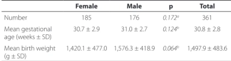
Mean birth weight and gestational age demonstrated no signi-ficant difference between girls and boys at the initial examination (p>0.05) (Table 1). In male infants, the mean anterior chamber depth was 0.10 mm longer, the vitreous length was 0.06 mm longer, and axial length was 0.09 mm longer, while the mean lens thickness was 0.05 mm shorter as compare with the female infants. However, female and male preterm infants did not differ significantly in any aspect of biometric parameters (p>0.05). The postmenstrual age of the infants ranged from 28 to 56 weeks during follow-up, and the mean postmenstrual age during the first examination was 35.2 ± 5.7 weeks (Table 2).
Table 1. Comparison of mean gestational age and mean birth weight among male and female infants
Female Male p Total
Number 185 176 0.172a 361
Mean gestational
age (weeks ± SD) 30.7 ± 2.9 31.0 ± 2.7
0.124b 30.8 ± 2.8
Mean birth weight (g ± SD)
1,420.1 ± 477.0 1,576.3 ± 418.9 0.064b 1,497.9 ± 483.6
a= one-sample Kolmogorov-Smirnov test; b= independent-sample t-test; SD= standard
deviation; n= number; g= gram.
Table 2. Comparison of mean biometry parameters during the irst examination among male and female infants by the t-test for indepen-dent samples
Mean ± SD Female (n=185) Male (n=176) p Total Postmenstrual age
(week) 34.90 ± 3.10 35.60 ± 3.40
0.124 35.20 ± 3.80
Anterior chamber depth (mm)
02.14 ± 0.28 02.24 ± 0.32 0.097 02.19 ± 0.36
Lens thickness (mm) 03.68 ± 0.64 03.61 ± 0.34 0.095 03.64 ± 0.53 Vitreous length (mm) 10.20 ± 1.52 10.26 ± 1.87 0.716 10.23 ± 1.04 Axial length (mm) 16.02 ± 1.05 16.11 ± 0.68 0.129 16.06 ± 0.73
The relationship of birth weight, gestational age, and postmenstrual age with ocular biometry parameters in premature infants
148 Arq Bras Oftalmol. 2015;78(3):146-9
The correlation between birth weight and gestational age with biometry parameters is shown in table 3. During the first examination, the mean postmenstrual age was 35.2 ± 3.8 weeks, and the birth weight and gestational age of the infants correlated significantly and positively with lens thickness, vitreous length, and axial length (r>0.5, p<0.001). However, no strong correlation was found between birth weight and gestational age with anterior chamber depth (r<0.5).
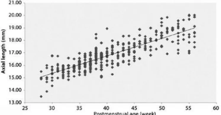
Increased vitreous and axial lengths correlated significantly with increasing postmenstrual age of the infants (r=0.669, p<0.001; r=0.845, p<0.001, respectively). Anterior chamber depth and lens thickness increased with increasing postmenstrual age. However, the correlation between anterior chamber depth and lens thickness with postmenstrual age was weak (r=0.432, p<0.001; r=0.412, p<0.001, respectively) (Figures 1-4).
DISCUSSION
The eye undergoes significant growth between the neonatal period and adulthood. Investigations of the globe in neonates and infants have demonstrated that the posterior segment of the globe is
relatively less developed than the anterior segment. These parameters change rapidly over the first 18 months of age(14-16). The A-mode of ultrasound (amplitude mode) is a type of ultrasound in which a single transducer scans a line through the body with echoes plotted on a screen as a function of depth. Generally, in children, A-scan biometry is used for measuring the anterior-posterior diameter of eye. Ultrasono-graphy is commonly used for clinical examinations of infants because of its safety and non-invasive character(17).
The mean axial length of the full-term newborn eye is 16.8 mm, while in adults it is 23.6 mm(18). However, the axial length of the eye in term infants varies according to the method of measurement. Lengths obtained by ultrasonographic biometry tend to be shorter than the lengths obtained by pathologic studies. It was reported that the newborn eye had a mean axial length between 17.1 and 17.5 mm. At term, the anterior chamber depth averages 2.05 mm, with a range of 1.8-2.4 mm(19). Isenberg et al.(20) demonstrated that the mean axial length was 16.2 mm, the anterior chamber depth was 2.0 mm, lens thickness was 3.8 mm, and vitreous chamber depth was 10.5 mm for term newborns. Globe size and axial length undergo dramatic changes during infancy. Although the anterior chamber depth of a newborn eye is approximately 75%-80% of that of adult eyes, their pos-terior segment at birth is less than half the size of that of the adult eye(14). Similarly, in the present study, we found that the axial length elonga-tion is mostly due to vitreous chamber elongaelonga-tion during the growth of postmenstrual age of the infants, i.e., at the age of 28th-56th week.
There are few reports in the literature on ocular biometric parame-ters in premature infants. Axial length continues to increase from birth. High refractive errors are common in the neonatal period following full-term and preterm birth and are related with poor emmetropiza-tion. It was argued that premature birth signals increased the risk of Table 3. Correlation between birth weight and gestational age with
biometry parameters during the irst examination
Anterior chamber depth
Lens thickness
Vitreous
length Axial length Birth weight r=0.037,
p=0.775
r=0.551, p<0.001
r=0.612, p<0.001
r=0.577, p<0.001 Gestational age r=0.125,
p=0.001
r=0.582, p<0.001
r=0.680, p<0.001
r=0.634, p<0.001
(r= Pearson correlation).
Figure 1. Relationship between anterior chamber depth and postmenstrual age (r=0.432, df=938, p<0.001). The regression line it to the data has a slope of 0.029.
Figure 2. Relationship between lens thickness and postmenstrual age (r=0.412, df= 938, p<0.001). The regression line it to the data has a slope of 0.039.
Figure 3. Relationship between vitreous length and postmenstrual age (r=0.669, df=938, p<0.001). The regression line it to the data has a slope of 0.115.
Ozdemir O, et al.
149 Arq Bras Oftalmol. 2015;78(3):146-9 abnormal refractive development(21-24).Kobayashi et al.(25) measured
anterior segments in 39 premature infants (at the gestational age of 25-39 weeks) using ultrasound biomicroscopy. They found that the mean anterior chamber depth was 1.3 mm at the 34.4 postcon-ceptional week. These values appear quite low compared with our findings; we detected an anterior chamber depth of 2.10 mm at the 34th postmenstrual week (Figure 1).
We found that the mean anterior chamber depth was 2.19 mm, lens thickness was 3.64 mm, vitreous length was 10.23 mm, and axial length was 16.06 mm at the 35th postmenstrual week. Our results are in agreement with the investigation of Cook et al.(26) with regard to the development of biometric parameters in premature infants with or without retinopathy of prematurity (Figures 1-4). These authors obtained a mean axial length between 16.37 and 16.66 mm, anterior chamber depth between 2.14 and 2.26, posterior segment length between 10.18 and 10.47, and lens thickness between 3.93 and 4.04 mm. Similarly, we have previously reported the mean anterior chamber depth as 2.1 ± 0.4 mm, lens thickness as 4.1 ± 0.7 mm, vitreous length as 10.3 ± 1.5 mm, and axial length as 16.4 ± 1.3 mm in 138 eyes of 69 premature infants with ROP(27).
In the present study, the anterior chamber depth, vitreous length, and axial length were slightly greater in boys than girls. On the other hand, the lens was slightly thicker in girls than boys; however, these differences were insignificant. In a similar manner, the mean axial length in the male gender for term neonates was reported to be 0.2 mm longer than that for the female neonates(19). Laws et al.(28) also found that male infants had longer axial lengths. This may be related to larger biparietal or occipitofrontal head diameter and to heavier weight of male infants. In another study, axial growth was measured at 3 and 9 months of age with similar findings(15).
Both birth weight and gestational age have an effect on ocular growth(26). Saw et al.(29) examined the association of birth parameters with biometry in children aged 7-9 years, and suggested that children who were born heavier or who were born more mature had longer axial lengths and deeper vitreous chambers. However, they found no significant association between birth weight and lens thickness or anterior chamber depth. We documented that the birth weight and gestational age had a significant effect on lens thickness and vitreous and axial lengths. However, their impact on anterior chamber depth was minimal. As mentioned, infants with high gestational age and birth weight have larger head circumference and longer ocular biometric parameters.
The relationship between the size of the eyeball and other factors, such as birth weight, gestational age, and postmenstrual age has re-cently been documented(9,28-30). Axial length grows linearly during the postnatal period in premature infants(28). Axer-Siegel et al.(30) reported that in preterm infants with maturation, the anterior chamber depth and the axial length are enlarged, whereas lens thickness remains sta-ble. In our study, both vitreous and axial lengths showed strongly po-sitive correlation with postmenstrual age. However, anterior chamber depth and lens thickness showed little positive change in correlation with the growth in postmenstrual age.
It is important to clarify the role of birth parameters on ocular biometric measures, such as anterior chamber depth, lens thickness, vitreous length, and axial length, in premature infants. While the an-terior chamber depth, vitreous length, and axial length were slightly longer in boys, the lens was slightly thicker in girls at the average 35th postmenstrual week. Birth weight and gestational age had a signifi-cant effect on lens thickness, vitreous length, and axial length but had little impact on anterior chamber depth in this study. Moreover, the vitreous length and axial length showed strongly positive correlation with postmenstrual age. However, anterior chamber depth and lens thickness showed little change in correlation with the growth in post-menstrual age.
In this study, there were several limitations, including a short study period, lack of a control group including full-term infants, and the fact that it is a single-center study. In conclusion, our study demonstrated that axial length elongation is mostly due to an increase of the
pos-terior chamber length during the growth of premature infants in the postmenstrual 28th-56th week.
REFERENCES
1. Ferguson KK, O’Neill MS, Meeker JD. Environmental contaminant exposures and pre-term birth: a comprehensive review. J Toxicol Environ Health B Crit Rev. 2013;16(2): 69-113.
2. Kochanek KD, Kirmeyer SE, Martin JA, Strobino DM, Guyer B. Annual summary of vital statistics: 2009. Pediatrics 2012;129(2):338-48.
3. Morley R, Cole TJ, Powell R, Lucas A. Growth and development in premature twins. Arch Dis Child. 1989;64(7):1042-5.
4. Sarikabadayi YU, Aydemir O, Ozen ZT, Aydemir C, Tok L, Oguz SS, et al. Screening for re tinopathy of prematurity in a large tertiary neonatal intensive care unit in Turkey: frequency and risk factors. Ophthalmic Epidemiol. 2011;18(6):269-74.
5. Aydemir O, Sarikabadayi YU, Aydemir C, Ozen ZT, Tok L, Erdeve O, et al. Adjusted poor weight gain for birth weight and gestational age as a predictor of severe ROP in VLBW infants. Eye (Lond). 2011;25(6):725-9.
6. Fledelius HC. Prematurity and the eye. Ophthalmic 10-year follow-up of children of low and normal birth weight. Acta Ophthalmol Suppl.1976;128:3-245.
7. Fledelius HC, Fledelius C. Eye size in threshold retinopathy of prematurity, based on a Danish preterm infant series: early axial eye growth, pre-and postnatal aspects. Invest Ophthalmol Vis Sci. 2012;53(7):4177-84.
8. Mundt GH Jr, Hughes Wf Jr. Ultrasonics in ocular diagnosis. Am J Ophthalmol. 1956; 41(3):488-98.
9. Modrzejewska M, Grzesiak W, Karczewicz D, Zaborski D. Refractive status and ocular axial length in preterm infants without retinopathy of prematurity with regard to birth weight and gestational age. J Perinat Med. 2010;38(3):327-31.
10. Trivedi RH, Wilson ME. Axial length measurements by contact and immersion techni-ques in pediatric eyes with cataract. Ophthalmology 2011;118(3):498-502. 11. Engle WA, American Academy of Pediatrics Committee on Fetus and Newborn. Age
terminology during the perinatal period. Pediatrics 2004;114(5):1362-4.
12. Cohen AM, Cook N, Harris MC, Ying GS, Binenbaum G. The pain response to mydriatic eyedrops in preterm infants. J Perinatol. 2013;33(6):462-5.
13. An International classification of retinopathy of prematurity. The Committee for the Classification of Retinopathy of Prematuriry. Arch Ophthalmol. 1984;102(8):1130-4. 14. Eustis HS, Guthrie ME. Postnatal development. In: Wright KW, Spiegel PH, editors.
Pediatric Ophthalmology and Strabismus. 2o ed. New York: Springer-Verlag; 2003. p.39-53. 15. Mutti DO, Mitchell GL, Jones LA, Friedman NE, Frane SL, Lin WK, et al. Axial growth and changes in lenticular and corneal power during emmetropization in infants. Invest Ophthalmol Vis Sci. 2005;46(9):3074-80.
16. Flitcroft D, Knight-Nanan D, Bowell R, Lanigan B, O’Keefe M. Intraocular lenses in chil-dren: changes in axial length, corneal curvature, and refraction. Br J Ophthalmol. 1999; 83(3):265-9.
17. Ramji FG, Slovis TL, Baker JD. Orbital sonography in children. Pediatr Radiol. 1996;26(4): 245-58.
18. Gordon RA, Donzis PB. Refractive development of the human eye. Arch Ophthalmol. 1985;103(6):785-9.
19. Gunton KB, Nelson LB, Olitsky SE. Neonatal ophthalmology: ocular development in childhood. In: Harley RD, Nelson LB, Olitsky SE, editors. Harley’s Pediatric Ophthalmo-logy, 4o ed. Philadelphia: Lippincott Williams & Wilkins; 2005. p.66-52.
20. Isenberg SJ, Neumann D, Cheong PY, Ling YL, McCall LC, Ziffer AJ. Growth of the inter-nal and exterinter-nal eye in term and preterm infants. Ophthalmology 1995;102(5):827-30. 21. Saunders KJ, McCulloch DL, Shepherd AJ, Wilkinson AG. Emmetropisation following
preterm birth. Br J Ophthalmol. 2002;86(9):1035-40.
22. Hebbandi SB, Bowen JR, Hipwell GC, Ma PJ, Leslie GI, Arnold JD. Ocular sequelae in extremely premature infants at 5 years of age. J Paediatr Child Health. 1997;33(4):339-42. 23. Kushner BJ. Strabismus and amblyopia associated with retinopathy of prematurity.
Arch Ophthalmol. 1982;100(2):256-61.
24. Pennefather PM, Tin W, Strong NP, Clarke MP, Dutton J, Cottrell DG. Refractive errors in children born before 32 weeks gestation. Eye (Lond). 1997;11(Pt 5):736-43. Comment in: Eye (Lond). 1997;11(Pt 5):580-1.
25. Kobayashi H, Kiryu J, Kobayashi K, Kondo T. Ultrasound biomicroscopic measurement of anterior chamber angle in premature infants. Br J Ophthalmol. 1997;81(6):460-4. 26. Cook A, White S, Batterbury M, Clark D. Ocular growth and refractive error
develop-ment in premature infants with or without retinopathy of prematurity. Invest Oph thalmol Vis Sci. 2008;49(12):5199-207.
27. Ozdemir O, Tunay ZÖ, Petriçli IS, Acar DE, Acar U, Erol MK. Analysis of the horizontal corneal diameter, central corneal thickness, and axial length in premature infants. Arq Bras Oftalmol. 2014;77(4):225-7.
28. Laws DE, Haslett R, Ashby D, O’Brien C, Clark D. Axial length biometry in infants with retinopathy of prematurity. Eye (Lond). 1994;8(Pt 4):427-30.
29. Saw SM, Tong L, Chia KS, Koh D, Lee YS, Katz J. The relation between birth size and the results of refractive error and biometry measurements in children. Br J Ophthalmol. 2004;88(4):538-42.