Gender Differences in Ventricular Volumes and Left Ventricle
Ejection Fraction Estimated by Myocardial Perfusion Imaging:
Comparison of Quantitative Gated SPECT (QGS) and Segami
Software Programs
Alice Tatsuko Yamada, Guilherme de Carvalho Campos Neto, José Soares Júnior, Maria Clementina P. Giorgi, Fernando
Araújo, José Claudio Meneghetti, Alfredo José Mansur
Instituto do Coração do Hospital das Clínicas – FMUSP - São Paulo, SP - Brazil
Summary
Objective: To test for gender differences in the measurements obtained by Segami and Quantitative Gated SPECT (QGS) software programs.
Methods: 181 asymptomatic individuals without heart disease were submitted to myocardial perfusion imaging. End-diastolic volumes (EDV), end-systolic volumes (ESV) and left ventricular ejection fraction (LVEF) were measured by QGS and Segami software programs to evaluate the influence of gender, age, weight, height, heart rate, systolic blood pressure, diastolic blood pressure, body mass index and body surface area.
Results: The means in the QGS method were: EDV (women= 68 ml; men= 95 ml; p<0.001), LVEF (women= 66.24%;
men= 58, 7%) and Segami: EDV (women= 137 ml; men= 174 ml), LVEF (women= 62.67%; men= 58, 52%). There were significant differences between men and women in the EDV (p<0.001), ESV (p<0.001) and LVEF (p=0.001) that persisted after adjusting for body surface area.
Conclusion: Ventricular volumes were significantly lower and LVEF was significantly higher in women, estimated by QGS or Segami software programs.
Key words: Tomography, emission-computed, single-photon; sex; stroke volume.
Mailing address: Alice Tatsuko Yamada •
Instituto do Coração do Hospital das Clínicas (INCOR) – Avenida Dr. Enéas de Carvalho Aguiar, 44 – 05403-000 – São Paulo, SP - Brazil
E-mail: [email protected]
Manuscript received April 5, 2006, revised manuscript received June 1, 2006; accepted on July 7, 2006.
Introduction
Ventricular volumes and left ventricle ejection fraction (LVEF) may be estimated in the myocardial perfusion single-photon emission computed tomography (SPECT) by different software programs.
The Cedars-Sinai Quantitative Gated SPECT software package (QGS) uses the edge-detection scheme for assessment of ventricular volumes and LVEF. These parameters were demonstrated to be influenced by gender, with lower ventricular volumes and higher LVEF values estimated for women1. However, the non-linear underestimation
of myocardial volumes using QGS may have resulted in over-reduction of ventricular volumes and major LVEF calculations in women when compared to men, because the underestimation increases at smaller volumes2.
The Segami software program developed by researchers of Stanford University defines the ventricular wall position by statistical parameters of count rate distribution. In computer simulations, methods that do not require detection of edges
for LVEF calculation were demonstrated to be more accurate when imaging patients with small hearts3. Gender influence
on the measurements of the left ventricle by Segami software program is less well known.
We performed this study to evaluate gender-related differences of left ventricular volumes and LVEF measured by myocardial perfusion imaging measured by the QGS and Segami software program in asymptomatic individuals without evidence of heart disease.
Methods
After written informed consent, participants were submitted to a treadmill electrocardiography exercise stress test, laboratory work-up, and myocardial perfusion gated SPECT.
Inclusion criteria - Individuals without cardiac symptoms, older than 18 years, presenting with normal physical examination, normal electrocardiogram and chest X-ray were included in the study.
Exclusion criteria - Individuals with a history of cardiovascular disease, systemic hypertension, Trypanosoma cruzi infection (Chagas’ disease microorganism), diabetes mellitus, thyroid-stimulating hormone lower than 0.05 or higher than 8 mg/dL, chronic obstructive pulmonary disease, asthma, renal failure, chronic inflammatory diseases, osteoarticular diseases, chronic anemia or neoplasia, abnormal 12-lead resting electrocardiogram, echocardiogram or treadmill electrocardiographic exercise stress test were excluded from the study.
Study sample - One hundred and eighty-one individuals were enrolled in the study, 85 (47%) men and 96 (53%) women. Their ages ranged between 20 and 80 (mean 43.8; standard deviation 12.2) years. Baseline characteristics of the studied population are presented in table 1.
Exercise test - The exercise tests were performed on the Fukuda Denshi Ml-8000 Star model according to the Ellestad exercise protocol4. The criteria for interruption of the exercise
were physical exhaustion or exceeded maximum heart rate for the age. The electrocardiogram response during stress was measured 0.08 s after the J point and was compared with baseline values. The response was considered ischemic if horizontal or downsloping ST-segment depression of > or = 1 mm or upsloping depression > or = a 1.5 mm.
Myocardial perfusion gated SPECT – The individuals were submitted to a same-day Tc-99m sestamibi (296 MBq) ungated rest/Tc-99m sestamibi (1110 MBq) stress protocol. Gated SPECT acquisition started within 30 minutes of exercise completion using a dual-headed gamma camera equipped with low-energy high-resolution collimators (ADAC Vertex Plus). Imaging was performed over 180º (64 projections) with a total imaging time of approximately 20 minutes. R-R interval was divided into 8 parts, correspondent to the different moments of the cardiac cycle. From the duration of a defined basic cycle in the beginning of the acquisition, a maximum variation of 20% for the subsequent cycles was established. Images were reconstructed with an iterative algorithm using
a low-pass filter with an order of 5.0 and a cut-off frequency of 0.7 cycles/cm. The resulting transaxial image sets were re-oriented into short axis sets to which both QGS and Segami algorithms were applied.
The determination of ventricular volumes and LVEF was described in detail and previously validated using QGS5 and
Segami6 software programs.
An observer verified the automatic left ventricle delimitation by the algorithms and carried out alterations when necessary. The Segami method requires some operator intervention to mask and threshold the gated images. Masking is done by manually fitting an ellipsoidal mask around the left ventricle to exclude activity coming from the right ventricle and non-cardiac structures. Thresholding is performed by subtracting from each pixel the average count density measured in a small region of interest drawn at the base of the left ventricular cavity on the end-diastolic bin. The myocardium is then sampled in the three-dimensional coordinates along radii originating in the center of the ventricular cavity. The left ventricular wall position is calculated as the first moment of the count rate distribution along each radius. Information is used to calculate left ventricular volume.
Studied variables - We studied left ventricle end-diastolic volume, left ventricle end-systolic volumes and LVEF measured by QGS in comparison to those obtained by the Segami software program in relation to gender, age, body weight, height, body mass index, heart rate, systolic and diastolic blood pressure.
Statistical analysis - Data are expressed as mean ± 1 standard error. Comparisons between men and women regarding basal characteristics and ventricular parameters were performed using Student’s t-test. Pearson’s correlation coefficient was used for statistical analysis of relationship between age and ventricular measurements. Analysis of Covariance (ANCOVA) was performed to compare the groups; the effects of other variables were taken into account. The comparison between QGS and Segami was analyzed by using Pearson´s correlation, intraclass correlation coefficient (ICC) and paired t- test. A value of p<0.05 was considered statistically significant. The above correction was made for clarity.
Ethics committee - The study was approved by the institutional Committee on Ethics and Human Research (File SDC 296/97/90; Cappesq 615/97), and all participants signed an informed consent before inclusion in the study.
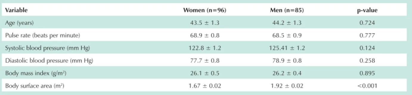
Table 1- Baseline characteristics of patients relative to gender
Variable Women (n=96) Men (n=85) p-value
Age (years) 43.5 ± 1.3 44.2 ± 1.3 0.724
Pulse rate (beats per minute) 68.9 ± 0.8 68.5 ± 0.9 0.777
Systolic blood pressure (mm Hg) 122.8 ± 1.2 125.41 ± 1.2 0.124
Diastolic blood pressure (mm Hg) 77.7 ± 0.8 78.9 ± 0.8 0.258
Body mass index (g/m2) 26.1 ± 0.5 26.2 ± 0.4 0.895
Body surface area (m2) 1.67 ± 0.02 1.92 ± 0.02 <0.001
Results
Myocardial perfusion imaging - All individuals had stress myocardial perfusion imaging without uptake defects.
Comparison between genders - Baseline characteristics comparison of the men and women showed that body surface area was significantly lower in women. There were no statistically significant differences in relation to age, heart rate, systolic and diastolic blood pressure between men and women (Table 1).
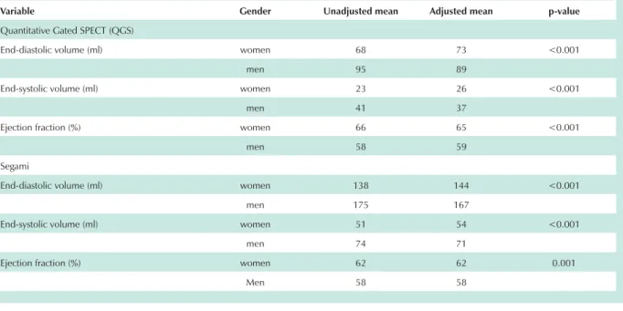
Ventricular volumes were significantly lower and LVEF were significantly higher in women, as estimated by QGS and Segami software programs (Table 2).
Left ventricular diastolic and systolic volumes were related to body surface area. In the QGS software program estimate, the correlation coefficients were r = 0.584 for end-diastolic volume and r=0.536 for end-systolic volume (p<0.001). In the Segami software program measurements, correlation coefficients were r = 0.502 for end-diastolic volume and r=0.459 for end-systolic volume (p<0.0001). LVEF demonstrated an inverse relationship with the body
surface area in both software programs, as demonstrated by r = - 0.412 for QGS and by r = - 0.193 for Segami software programs (p < 0.009).
An adjustment for the body surface area was done in the ventricular volumes and LVEF, and the differences between women and men persisted (Table 2).
Comparison of QGS and Segami software programs - Comparison of QGS and Segami software programs revealed that the Segami estimations demonstrated higher volumes systematically (Table2).
Pearson’s correlation results between volumes measurements obtained with Segami and QGS were around 0.7. However, the intraclass correlation coefficients showed poor agreement and the differences were significant (Table 3).
Bland-Altman analysis was performed for comparison of LVEF measurements obtained with Segami and QGS software programs in men (Graphic 1) and women (Graphic 2). The curves revealed a trend to lower values (negative differences) with Segami software compared to QGS and for a higher ejection fraction, especially in women.
Table 2 – Comparison of ventricular volumes and left ventricular ejection fraction between men and women with adjustment for the body surface area (ANCOVA)
Variable Gender Unadjusted mean Adjusted mean p-value
Quantitative Gated SPECT (QGS)
End-diastolic volume (ml) women 68 73 <0.001
men 95 89
End-systolic volume (ml) women 23 26 <0.001
men 41 37
Ejection fraction (%) women 66 65 <0.001
men 58 59
Segami
End-diastolic volume (ml) women 138 144 <0.001
men 175 167
End-systolic volume (ml) women 51 54 <0.001
men 74 71
Ejection fraction (%) women 62 62 0.001
Men 58 58
Table 3 – Results of comparisons between ventricular volumes and left ventricle ejection fraction measured by Quantitative Gated SPECT (QGS) and Segami software programs
Correlation coefficients Comparison between means
Variable Pearson ICC
QGS Mean ± SE
Segami
Mean ± SE Paired t-test
End-diastolic volume 0.784 0.190 80.8 ± 1.8 155.3 ± 2.8 <0.001
End-systolic volume 0.800 0.332 31.8 ± 1.2 62.4 ± 1.6 <0.001
Ejection fraction 0.554 0.526 62.4 ± 0.6 60.7 ± 0.5 0.002
Graphic 2 - Bland-Altman analysis of comparison between QGS and Segami measurements of LVEF in women.
Graphic 1 - Bland-Altman analysis of comparison between QGS and Segami measurements of LVEF in men.
Reference values for left ventricular ejection fraction and volume measurements - Based on the percentiles 5% and 95%, the inferior limit of normality for LVEF was in the QGS method: 44% for men and 51% for women and in the Segami method: 48,3% for men and 53% for women. Reference values for ventricular volumes and LVEF are shown in table 4.
Age related differences - Correlation between age and ventricular measurements estimated by the QGS software program were not found in our study. The Segami software program showed correlations between volumes and age with low coefficients (below 0.20). The values of the correlations are shown in table 5.
Discussion
Our study evaluated a sample of Brazilian population without heart disease in a wide range of age (between 20 and 80) and found some reference values for left ventricular volumes and LVEF for men and women, measured by QGS and Segami software programs.
Gender-related differences - Using the QGS software program, Rozanski et al7 and De Bondt et al1, found that the
ventricular volumes and LVEF presented significant differences
Table 4 – Reference values for ventricular volumes and left ventricular ejection fraction estimated by Quantitative Gated SPECT (QGS) and Segami software programs
Women Men
Percentile 5% Percentile 95% Percentile 5% Percentile 95%
QGS
End-diastolic volume (ml) 48 95 60 144
End-systolic volume (ml) 12 41 18 70
Ejection fraction (%) 51 78 44 70
Segami
End-diastolic volume (ml) 100 192 120 248
End-systolic volume (ml) 38 78 46 112
Ejection fraction (%) 53 70 48 67
Table 5 - Correlation between age and ventricular volumes and left ventricular ejection fraction estimated by Quantitative Gated SPECT
(QGS) and Segami software
Pearson correlation Sig. (2-tailed)
QGS
End-diastolic volume -0.136 0.068
End-systolic volume -0.125 0.093
Ejection fraction 0.090 0.226
Segami
End-diastolic volume -0.196 0.008
End-systolic volume -0.161 0.030
Ejection fraction 0.013 0.857
References
1. De Bondt P, Van de Wiele C, De Sutter J, De Winter F, De Backer G, Dierckx RA. Age- and gender specific-differences in left ventricular cardiac function and volumes determined by gated SPECT. Eur J Nucl Med. 2001; 28: 620-4.
2. Nakajima K, Taki J, Higuchi T, Kawano M, Taniguchi M, Maruhashi K, et al. Gated SPECT quantification of small hearts: mathematical simulation and clinical application. Eur J Nucl Med. 2000; 27: 1372-9.
3. Feng B, Sitek A, Gullberg GT. Calculation of the left ventricular ejection fraction without edge detection: application to small hearts. J Nucl Med. 2002; 43: 786-94.
4. Ellestad MH, Davis FA. Stress testing: principles and practice. Philadelphia: S.A. Davis; 1995.
5. Germano G, Kiat H, Kavanagh PB, Moriel M, Mazzanti M, Su H, et al. Automatic quantification of ejection fraction from gated myocardial perfusion SPECT. J Nucl Med. 1995; 36: 2138-47.
6. Everaert H, Franken PR, Flamen P, Goris ML, Momen A, Bossuyt A. Left ventricular ejection fraction from gated SPECT myocardial perfusion studies: a method based on the radial distribution of count rate density across the myocardial wall. Eur J Nucl Med. 1996; 23: 1628-33.
7. Rozanski A, Nichols K, Yao SS, Malholtra S, Cohen R, DePuey G. Development and application of normal limits for left ventricular ejection fraction and volume measurements from 99mTc-sestamibi myocardial perfusion gated SPECT. J Nucl Med. 2000; 41: 1445-50.
8. Everaert H, Bossuyt A, Franken PR. Left ventricular ejection fraction and volumes from gated single photon emission images: comparison between two algorithms working in three-dimensional space. J Nucl Cardiol. 1997; 4: 472-6.
9. Hambye AS, Vervaet A, Dobbeleir A. Variability of left ventricular ejection fraction and volumes with quantitative gated SPECT: influence of algorithm,
program that the limited resolution of the gamma camera may result in underestimation of the left ventricular volume by 15% for a volume of 101 ml, 25% for a volume of 52 ml, and 50% for a volume of 37 ml2.
Our study demonstrated that the women had significant lower volumes and higher values of LVEF in both software programs. Normalization to body surface area did not eliminate these differences. Gender-related differences with the Segami method suggests that gender differences found in QGS are not the result of a software program artifact.
Comparison of the QGS and Segami software programs - Everaert et al8 compared QGS and Segami programs in 40
patients. They observed excellent correlation between the two methods for calculating LVEF (r=0.93) and also found that the Segami software program calculated larger volumes than the QGS software program did. In accord, our study demonstrated that Segami software program systematically had higher ventricular volumes. This can be explained by differences in left ventricular modeling between algorithms, pixel size and reconstruction parameters9. Pearson’s correlation test
results for comparisons of ventricular volumes measurements obtained with Segami and QGS software programs were high. However, the intraclass correlation coefficients showed poor agreement and the differences were significant.
These results suggest that ventricular volumes measurements assessed by different software programs (QGS and Segami) may not be clinically interchangeable for follow-up of an individual patient.
In our study, the measures of LVEF in both software programs demonstrated a poor correlation (r=0.554). The comparison of means revealed a statistically significant difference (p=0.002). However, the difference was so small that it has no clinical significance, most likely because quantitative programs using count density information (Segami) for determining systolic function are less influenced by left volume size and imaging resolution than those algorithms based on edge detection (QGS).
The Bland-Altman analysis revealed a good correlation between QGS and Segami that worsened for higher values of LVEF, especially in women. LVEF values in the higher range were observed more frequently in the QGS software program,
especially in women. However, QGS is the software program most widely used clinically, and its prognostic value was previously validated.
Our study was limited due to lack of a gold standard; this did not allow estimation as to which software program was more accurate.
Reference values for ventricular volumes and LVEF - The number of patients in this study did not allow setting normal limits for LVEF and ventricular volumes. However, the reference values obtained with 181 patients for QGS method
/9()IRUPHQDQGIRUZRPHQZHUHVLPLODU
to those obtained by Rozanski et al7/9()IRUPHQ
DQGIRUZRPHQDQG$EDEQHKHWDO10/9()IRU
PHQIRUZRPHQ
Age-related differences - We did not detect significant variation of ventricular volumes and LVEF in relation to age using the? QGS method. However, Slotwiner et al. in a large series of 464 clinically normal adults found a slight but significant increase in LVEF and a decrease in chamber size with age 11. De Bondt
et al1 using analysis of variance found age-related differences
RQO\LQWKHJURXSRIZRPHQDJHZKRVKRZHGVLJQLILFDQW
lower EDV and ESV and higher LVEF values.
In our study, using the Segami method we observed correlation between volumes and age with low coefficients (below 0.20). The absence of an age-related significant difference with QGS and a poor correlation in the Segami method may have been the result of the small number of subjects in some age ranges.
In conclusion, our data in this sample of individuals without evidence of heart disease demonstrated that ventricular volumes and LVEF estimated by QGS and Segami software programs vary according to gender, and volume measurements by different software programs may not be clinically interchangeable for a follow-up in an individual patient.
None of the authors has a financial interest in any cardiac software program package.
Potential Conflict of Interest
pixel size and reconstruction parameters in small and normal-sized hearts. Eur J Nucl Med Mol Imaging. 2004; 12: 1606-13.
10. Ababneh AA, Sciacca RR, Kim B, Bergmann SR. Normal limits for left ventricular ejection fraction and volumes estimated with gated myocardial perfusion in patients with normal exercise test results: influence of tracer,
gender, and acquisition camera. J Nucl Cardiol. 2000; 7: 661-8.