Ventricular Function Following Coronary Artery Bypass Grafting:
Comparison between Gated SPECT and Cardiac Magnetic Resonance
Imaging
Cláudio Tinoco Mesquita
1,6; Maria Carolina Pinheiro Pessoa
2; Paulo Pontes Vasconcelos
2,3; Amarino Carvalho Oliveira
Júnior
4; Hans Fernando Rocha Dohmann
4,5,6; Adair Gomes dos Reis
7; Lea Mirian Barbosa da Fonseca
2,8Serviço de Medicina Nuclear do Hospital Pró-Cardíaco1, Pro-Echo Hospital Samaritano2, Centro de Diagnóstico por Imagens (CDPI)3; Serviço de Radiologia do Hospital Pró-Cardíaco4, Centro de Estudos do Pró-Cardíaco (Procep)5; Instituto Nacional de Cardiologia6, Nuclear Diagnósticos7, Hospital Universitário Clementino Fraga Filho da Universidade Federal do Rio de Janeiro (UFRJ)8, Rio de Janeiro, RJ - Brazil
Summary
Background: The assessment of left ventricular function may be impaired by the abnormal interventricular septal motion frequently found after coronary artery bypass grafting (CABG). Studies on the validation of gated SPECT as a tool for the assessment of left ventricular function in this patient group are scarce.
Objective: We investigated the agreement and correlation between left ventricular ejection fraction (LVEF), end-diastolic volume (EDV), and end-systolic volume (ESV) as obtained using electrocardiogram-gated myocardial perfusion scintigraphy (gated SPECT) and cardiac magnetic resonance imaging in 20 patients undergoing coronary artery bypass grafting.
Methods: Correlation was measured using Spearman’s correlation coefficient (ρ). Agreement was assessed using Bland-Altman analysis.
Results: A good correlation was found between gated SPECT and cardiac magnetic resonance imaging in patients after CABG with regard to left ventricular ejection fraction (ρ = 0.85; p =0.0001), moderate correlation for end-diastolic volume (ρ = 0.51; p = 0.02), and non-significant correlation for end-diastolic volume (ρ = 0.13; p = 0.5). Agreement ranges for LVEF, ESV and EDV were: -20% to 12%; -38 to 54 ml and; -96 to 100 ml, respectively.
Conclusion: A reliable correlation was found for left ventricular ejection fraction as obtained by gated SPECT and magnetic resonance imaging in patients undergoing CABG. For ventricular volumes, however, the correlation is not adequate. (Arq Bras Cardiol 2009;92(5):327-333)
Key words: Radionuclide imaging; magnetic resonance imaging; myocardial revascularization; ventricular function, left; myocardial reperfusion.
Mailing address: Cláudio Tinoco Mesquita •
Hospital Pró-Cardíaco, Serviço de Medicina Nuclear e Imagem Molecular, Rua General Polidoro 192, Botafogo, 22.280-000, Rio de Janeiro, RJ - Brazil E-mail: [email protected]
Manuscript received February 06, 2008; revised manuscript received em May 23, 2008; accepted June 19, 2008.
Introduction
Myocardial perfusion scintigraphy with electrocardiogram-gated tomographic images (electrocardiogram-gated SPECT) has been used for the simultaneous assessment of myocardial perfusion and the global and segmental left ventricular function. This technique is well validated and has been part of the cardiologic diagnostic armamentarium for more than a decade1-3. Cardiac magnetic
resonance imaging (CMRI) is considered the gold-standard technique for the assessment of ventricular function thanks to its excellent spatial and temporal resolution4. A
meta-analysis concluded that measurements of ventricular volumes and left ventricular ejection fraction obtained using gated SPECT have a very good correlation with those obtained
using CMRI5. No definitive evidence exists on whether the
comparative accuracy between gated SPECT and CMRI is different across the various patient subgroups. Although the development of abnormal interventricular septal motion is recognized in the postoperative period of cardiac surgeries6,
the impact of this contractile abnormality on the accuracy of gated SPECT has not been fully assessed7,8. It is important
that this technique be assessed in this situation, because abnormal interventricular septal motion following cardiac surgery is known to make the assessment of left ventricular function difficult when using conventional techniques such as echocardiography, equilibrium radionuclide ventriculography and contrast-enhanced ventriculography9. Assessment of the
left ventricular function following coronary artery bypass grafting is essential for the quantification of contractile recovery (viable myocardium) in individuals with left ventricular dysfunction, as well as for the detection of functional loss during the procedure (perioperative infarction).
volumes and ejection fraction, using gated SPECT, and to compare these results with those obtained with CMRI in patients with abnormal septal motion following coronary artery bypass grafting.
Methods
Twenty patients who had undergone coronary artery bypass grafting in the previous 12 months and who presented abnormal interventricular septal motion on echocardiography were included in this study. Patients were excluded if one of the following factors was present:
1) atrial fibrillation,
2) third-degree left bundle branch block, 3) non-sinus rhythm,
4) artificial cardiac pacemaker,
5) six or more extrasystoles per minute, and 6) pregnancy or breastfeeding.
The study protocol was approved by the Hospital Ethics Committee. All individuals were informed of the study and gave their informed consent. The patients were selected from the Department of Cardiac Surgery of Clementino Fraga Filho University Hospital. A minimum period of three months (mean of eight months) after revascularization had elapsed for all patients.
The individuals prospectively underwent resting gated SPECT myocardial perfusion scintigraphy and cardiac magnetic resonance imaging within a 2-week interval of each other at most.
Technetium-99m-sestamibi (Tc 99m sestamibi) myocardial perfusion scintigraphy was performed as follows: after intravenous administration of 740 MBq of radiotracer at rest, tomographic images were obtained at 180o (extending from
45o right anterior oblique to 45o left posterior oblique) in 32
stops gated with the patient’s electrocardiogram, and the RR interval was divided into 8 frames, in a total of 256 frames per study. The beat length acceptance window was set at 50%, with a 140-keV photopeak and 15% energy window. A low-energy, high-resolution collimator was used, with 64x64 matrices, and 35 seconds per stop. The eight ECG-gated projections were filtered using a 2-D Butterworth filter (order 2.5; frequency of 0.3 cycles per pixel) and automatically reconstructed in transaxial images using filtered backprojection with a ramp filter. Neither attenuation nor scatter correction was performed. Gated SPECT images were automatically reconstructed by the Germano et al3.
Cardiac magnetic resonance imaging was performed in a 1.5-Tesla scanner (Horizon, General Electric Medical Systems, Milwaukee, Wisconsin), with a 23-mT/m gradient, body and torso phased-array coils8. The Fastcard software was used
during breath hold (spoiled gradient-echo, gradient-echo) in the LV horizontal (four chambers) and vertical (two chambers) long axes, as well as in the LV short axis. Nine images of the cardiac phases were obtained within the RR interval in each view plane. Ventricular volumes were calculated using the Simpson’s rule with manual tracing of the endocardial border in each short-axis image9. Left ventricular ejection fraction
(LVEF) was calculated from the values of end-diastolic volume (EDV) and end-systolic volume (ESV)9.
Statistical analysis
Measurements were always taken twice by the same observer, and the mean value was used for the analysis. The reproducibility of the techniques was considered satisfactory. Regression analysis was used to determine the correlation between LVEF, EDV and ESV as measured by ECG-gated SPECT and by CMRI. Spearman’s correlation coefficient (ρ) was derived in order to evaluate the correlation between the two techniques. A Bland-Altman plot was used to demonstrate systematic trends in the differences between the two techniques. With this plot, the mean value of the measurements obtained with the two techniques is depicted in the ordinate axis, and the differences between the two measurements are shown in the abscissa axis. The differences between the groups were assessed using the chi square test or Fisher’s exact test, when appropriate. Values were expressed as means plus or minus one standard deviation. P values equal to or lower than 0.05 were considered significant.
Results
All the 20 patients underwent ECG-gated SPECT and CMRI uneventfully. One of the patients was excluded from the analysis due to failure in the capture of the electrocardiographic signal during the scintigraphic study; therefore the comparative analyses included the remaining 19 patients.
Left ventricular ejection fraction
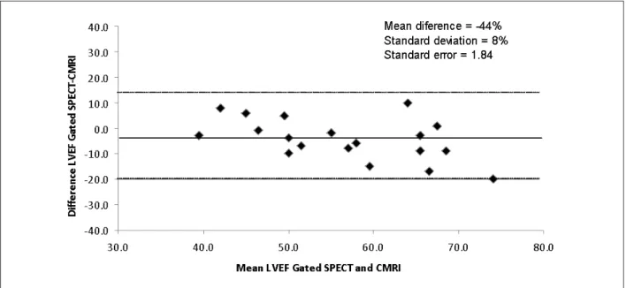
Values of ejection fraction estimates are shown in Table 1. No significant difference was found between the groups regarding the mean left ventricular ejection fraction as determined by ECG-gated SPECT (54 % ± 8%) in comparison to the mean value estimated by CMRI (58 % ± 12%, p = NS). A good correlation was observed between LVEF measurements as obtained with ECG-gated SPECT and those obtained with CMRI (ρ = 0.78, p = 0.0001). Results of linear regression analysis in the patient sample are shown in Figure 1. Results of the Bland-Altman analysis are shown in Figure 2, where it can be observed that the left ventricular ejection fraction tends to be slightly underestimated by ECG-gated SPECT in comparison to CMRI.
End-diastolic volume
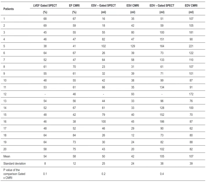
Table 1 - Values of ejection fraction (LVEF), end-diastolic volumes (EDV), end-systolic volumes (ESV), with means and standard deviation of the sample. EF-CMRI = ejection fraction on cardiac magnetic resonance imaging
Patients LVEF Gated SPECT EF CMRI
ESV – Gated SPECT ESV CMRI EDV – Gated SPECT EDV CMRI
(%) (%) (ml) (ml) (ml) (ml)
1 68 67 16 35 51 107
2 69 59 18 42 59 105
3 45 55 55 80 100 181
4 46 47 82 47 151 90
5 38 41 102 129 164 221
6 64 67 26 39 73 122
7 52 47 64 58 133 110
8 61 70 23 31 61 107
9 55 61 32 39 71 101
10 48 55 42 38 99 87
11 53 61 66 35 134 91
12 - 46 - 93 - 172
13 54 56 44 33 96 76
14 52 67 61 33 128 100
15 48 42 79 40 152 70
16 46 38 100 45 186 87
17 48 52 46 29 90 62
18 64 84 26 12 73 80
19 64 73 30 24 82 88
20 58 75 43 20 102 82
Mean 54 58 50 42 105 107
Standard deviation 8 12 25 24 38 39
P value of the comparison Gated x CMRI
0.1 0.2 0.4
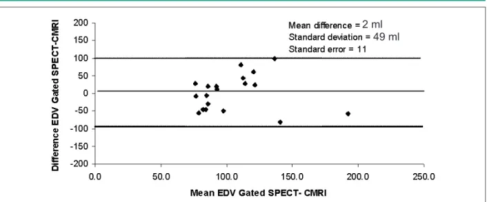
to CMRI; however, the 49-ml standard deviation indicates a greater variability of this measurement.
End-systolic volume
End-systolic volume values are shown in Table 1. No significant difference was found between the groups regarding mean ESV as determined by ECG-gated SPECT (50 ± 25 ml) in comparison to the mean value estimated by CMRI (42 ± 24 ml, p = NS). A good correlation was observed between ESV measurements as obtained with ECG-gated SPECT and those obtained with CMRI (ρ = 0.51, p = 0.02). Results of the linear regression analysis in the patient sample are shown in Figure 5. Results of the Bland-Altman analysis are shown in Figure 6, where it can be observed that ECG-gated SPECT has a better agreement with CMRI for ESV than for EDV (Figure 4).
Discussion
The present study evaluated the correlation and agreement between the measurements of left ventricular systolic function (ventricular volumes and ejection fraction) as obtained using the gated-SPECT technique in comparison to magnetic resonance imaging in patients undergoing coronary artery bypass grafting. Overall, a satisfactory correlation was found between the two techniques. However, the assessment of left ventricular end-diastolic volume showed a correlation poorer than clinically desirable. Likewise, the Bland-Altman agreement plots demonstrated broad agreement ranges between the methods, which may have practical clinical implications.
Several studies10-12 demonstrated that left ventricular systolic
long term. After myocardial infarction, increased ventricular volumes are associated with higher mortality, higher rate of adverse cardiovascular events and worse prognosis10. The
accurate analysis of ventricular volumes andejection fraction adds incremental value in the risk stratification of patients and helps guide therapy. Gated SPECT is a powerful clinical tool that permits the assessment of ventricular function and perfusion in a single acquisition, without any additional radiation exposure other than that needed for the performance of perfusion scintigraphy, with minimal costs and without patient discomfort3. Data from several studies showed that
ventricular function parameters obtained by gated SPECT are independent prognostic variables10-12. When the patient needs
scintigraphic assessment of myocardial perfusion, all additional information pertaining to the ventricular function is obtained without an increased radiation dose, which is considered an
Figure 1 -Scatter plot and Spearman’s correlation coeficient showing the correlation between LVEF as obtained by gated SPECT and by CMRI.
Figure 3 - Scatter plot and Spearman’s correlation coefficient showing the
correlation between EDV as obtained by gated SPECT and by CMRI.
increment in the benefit ratio of the test.
Gated SPECT was validated in experiments with phantoms and animals testing, as well as in patient series13. Cardiac
magnetic resonance imaging has proved to be a very accurate technique in the determination of ventricular volumes and ejection fraction, and is considered the gold-standard for the assessment of ventricular function4,6. Values obtained by gated
SPECT have satisfactorily correlated with those obtained by magnetic resonance imaging; however, there are limitations that should be known13. Quantitative gated SPECT (QGS) is
the most widely used program in the clinical practice and uses a border detection scheme to evaluate ventricular volumes and LVEF. These parameters have been demonstrated to be influenced by gender, with lower ventricular volumes and higher LVEF in women14. Quantitative programs using
information on the counting density for the determination of
Figure 5 -Scatter plot and Spearman’s correlation coeficient showing the
correlation between ESV as obtained by gated SPECT and by CMRI.
Figure 6 -Agreement plot between ESV as obtained by gated SPECT and by magnetic resonance imaging; CMRI - cardiac magnetic resonance imaging.
Figure 4 -Agreement plot between EDV as obtained by gated SPECT and by magnetic resonance imaging; CMRI - cardiac magnetic resonance imaging.
2 ml
49 ml
systolic function are less influenced by the magnitude of the left volume and by the image resolution than do algorithms based on border detection, and they generate ventricular volumes systematically greater than those calculated by the QGS program15.
In a meta-analysis on studies comparing gated SPECT with other methods for the assessment of ventricular function, excellent agreement and correlation were demonstrated between magnetic resonance imaging and gated SPECT regarding the evaluation of systolic function indexes5.
However, in the same meta-analysis, the authors concluded that further research was necessary in some subgroups of patients who were not adequately represented in the studies available up to the moment of the analysis5. One of the
subgroups that can be mentioned is that of patients with abnormal septal motion.
Abnormal interventricular septal motion following cardiac surgeries is related to the excessive anteromedial systolic translation of the heart inside the thorax due to the loss of ligament fixation and of pericardial integrity6. This
abnormal motion makes the assessment of the ventricular function difficult after cardiac surgery through the use of conventional imaging techniques13. There is a characteristic
contraction pattern seen in patients after cardiac surgery. In a study with gated SPECT that included specifically patients in the postoperative period, septal hypokinesia with preserved thickening associated with an apparently increased endocardial motion of the left ventricular lateral wall was observed, in addition to an anterior epicardial swing, different from the contraction pattern seen in normal individuals16.
Tadamura et al7 were the first to validate gated SPECT for
the evaluation of ventricular volumes and ejection fraction in 16 patients following coronary artery bypass grafting7. The
authors concluded that gated SPECT provides satisfactory functional information, including absolute ventricular volumes in patients followingcardiac surgery. However, the different temporal resolution between the techniques led to a slightly underestimated ejection fraction with gated SPECT7. The
authors suggest that further studies on this issue should be conducted in order to adequately evaluate whether gated SPECT may be accurately applied to this population, especially to patients with perfusion defects and aneurysms7.
The major difference between the present study and that mentioned in the previous paragraph is that 14 of our 20 patients presented with significant perfusion defects in comparison to only five in Tadamura et al’s study7. When
an algorithm requiring the detection of endocardial borders is used for the assessment of ventricular volumes and LVEF, it is expected that the presence of perfusion defects secondary to the infarcted areas will reduce the accuracy of the technique. This difference in the study populations may explain the disparities found between the present study and the previousone. Several studies have found similar disparities between the data obtained with gated SPECT in segments with severe reduction of radiotracer concentration and those obtained with magnetic resonance imaging17-19. The presence
of previous infarction leads to a reduction in photon counting, particularly in the end-diastole, which underestimates motion and the regional and global functions12.
Conclusion
Our data are still restricted to a limited group of patients, and we believe they should be validated in other series with other algorithms of ventricular function analysis, as well as with other radiotracers20-22 for a better understanding of the
limitations of gated SPECT in patients with abnormal septal motion. A more efficient and appropriate use of the imaging methods in cardiology is desired by all professionals who use these tests, whether they are specialists or not23; thus, we
believe our findings may be useful to them.
In conclusion, left ventricular ejection fraction obtained by gated SPECT showed a reliable correlation with that obtained by magnetic resonance imaging in patients undergoing CABG. Ventricular volumes, however, did not show an adequate correlation.
Potential Conflict of Interest
No potential conflict of interest relevant to this article was reported.
Sources of Funding
There were no external funding sources for this study.
Study Association
This article is part of the thesis of doctoral submitted by Cláudio Tinoco Mesquita, from Universidade Federal do Rio de Janeiro.
References
1. Cwajg E, Cwajg J, He ZX, Hwang WS, Keng F, Nagueh SF, et al. Gated myocardial perfusion tomography for the assessment of left ventricular function and volumes: comparison with echocardiography. J Nucl Med. 1999; 40: 1857-65.
2. DePuey E, Nichols K, Dobrinsky C. Left ventricular ejection fraction assessed from gated technetium-99m-sestamibi SPECT. J Nucl Med. 1993; 34: 1871-6.
3. Germano G, Kiat H, Kavanagh PB, Moriel M, Mazzanti M, Su HT, et al. Automatic quantification of ejection fraction from gated myocardial perfusion SPECT. J Nucl Med. 1995; 36: 2138-47.
Circulation. 1990; 82: 154-63.
5. Ioannidis J, Trikalinos T, Danias P. Electrocardiogram-gated single photon emission computed tomography versus cardiac magnetic resonance imaging for the assessment of left ventricular volumes and ejection fraction: a meta-analysis. J Am Coll Cardiol. 2002; 39: 2059-68.
6. Vignola P, Boucher C, Curfman G, Walker H, Shea W, Dinsmore R, et al. Abnormal interventricular septal motion following cardiac surgery: clinical, surgical, echocardiographic and radionuclide correlates. Am Heart J. 1979; 97: 27-34.
7. Tadamura E, Kudoh T, Motooka M, Inubushi M, Okada T, Kubo S, et al. Use of technetium-99m sestamibi ECG-gated single-photon emission tomography for the evaluation of left ventricular function following coronary artery bypass graft: comparison with three-dimensional magnetic resonance imaging. Eur J Nucl Med. 1999; 26: 705-12.
8. Taki J, Higuchi T, Nakajima K, Matsunari I, Hwang EH, Bunko H, et al. Electrocardiographic gated 99mTc-MIBI SPECT for functional assessment of patients after coronary artery bypass surgery: comparison of wall thickening and wall motion analysis. J Nucl Med. 2002; 43: 589-95.
9. Foo TK, Bernstein MA, Aisen AM, Hernandez RJ, Collick BD, Bernstein T. Improved ejection fraction and flow velocity estimates with use of view sharing and uniform repetition time excitation with fast cardiac techniques. Radiology. 1995; 195: 471-8.
10. White HD, Norris RM, Brown MA, Brandt PW, Whitlock RM, Wild CJ. Left ventricular end-systolic volume as the major determinant of survival after recovery from myocardial infarction. Circulation. 1987; 76: 44-51. 11. Matsuo S, Matsumoto T, Nakae I, Koh T, Masuda D, Takada M, et al. Prognostic
value of ECG-gated thallium-201 single-photon emission tomography in patients with coronary artery disease. Ann Nucl Med. 2004; 18: 617-22. 12. Kroll D, Farah W, McKendall G, Reinert S, Johnson L. Prognostic value of
stress-gated Tc-99m sestamibi SPECT after acute myocardial infarction. Am J Cardiol. 2001; 87: 381-6.
13. Faber TL, Vansant JP, Pettigrew R, Galt JR, Blais M, Chatzimavroudis G, et al. Evaluation of left endocardial volumes and ejection fractions computed from gated perfusion SPECT with magnetic resonance imaging: comparison of two methods. J Nucl Cardiol. 2001; 8: 645-51.
14. De Bondt P, Van de Wiele C, De Sutter J, De Winter F, De Backer G, Dierckx
RA. Age- and gender specific-differences in left ventricular cardiac function and volumes determined by gated SPECT. Eur J Nucl Med. 2001; 28: 620-4.
15. Yamada AT, Campos Neto GC, Soares J Jr, Giorgi MC, Araujo F, Meneghetti JC, et al. Diferenças relacionadas ao sexo nos volumes ventriculares e na fração de ejeção do ventrículo esquerdo estimados por cintilografia de perfusão miocárdica: comparação entre os programas Quantitative Gated SPECT (QGS) e Segami. Arq Bras Cardiol. 2007; 88: 285-90.
16. Yun J, Block M, Botvinik E. Unique contraction pattern in patients after coronary bypass graft surgery by gated SPECT myocardial perfusion imaging. Clin Nucl Med. 2003; 28: 18-24.
17. Manrique A, Faraggi M, Véra P, Vilain D, Lebtahi R, Cribier A, et al. 201Tl and 99mTc-MIBI gated SPECT in patients with large perfusion defects and left ventricular dysfunction: comparison with equilibrium radionuclide angiography. J Nucl Med. 1999; 40: 805-9.
18. Righetti A, Crawford M, O’Rourke R, Schelberg H, Daily P, Ross J Jr. Interventricular septal motion and left ventricular function after coronary bypass surgery: evaluation with echocardiography and radionuclide angiography. Am J Cardiol. 1977; 39: 372-7.
19. Stollfuss JC, Haas F, Matsunari I, Neverve J, Nekolla S, Eike JS, et al. Regional myocardial wall thickening and global ejection fraction in patients with low angiographic left ventricular ejection fraction assessed by visual and quantitative resting ECG-gated 99mTc-tetrofosmin single-photon emission tomography and magnetic resonance imaging. J Nucl Med. 1998; 25: 522-30.
20. Anagnostopoulos C, Gunning MG, Pennell DJ, Laney R, Proukakis H, Underwood SR. Regional myocardial motion and thickening assessed at rest by ECG-gated 99mTc-MIBI emission tomography and by magnetic resonance imaging. Eur J Nucl Med. 1996; 23: 909-16.
21. Nakajima K, Higuchi T, Taki J, Kawano M, Tonami N. Accuracy of ventricular volume and ejection fraction measured by gated myocardial SPECT: comparison of 4 software programs. J Nucl Med. 2001; 42: 1571-8. 22. Hyun I, Kwan J, Park KS, Lee WH. Reproducibility of Tl-201 and Tc-99m
sestamibi gated myocardial perfusion SPECT measurement of myocardial function. J Nucl Cardiol. 2001; 8: 182-7.