Instituto do Coração, University of São Paulo Medical School
Mailing address: Protásio Lemos da Luz – Incor - Divisão de Clínica - Av. Dr. Enéas C. Aguiar, 44 - 05403-000 – São Paulo, SP. Fax: 55-11-3069-5447. e-mail: [email protected]
Protásio Lemos da Luz, Desidério Favarato
São Paulo, SP - Brazil
Chronic Coronary Artery Disease
Coronary insufficiency (CI) is the state in which an imbalance occurs between the supply and demand of oxy-gen, which prevents adequate maintenance of the metabolic needs of the myocardium, resulting in ischemia of several degrees of intensity.
CI has numerous causes among which are cardiac valvular diseases (aortic stenosis), hypertrophic cardio-myopathy, microvasculature diseases (diabetes mellitus, syndrome X), anomalous origin of coronary arteries, and coronary fistulas. However, the most important cause, be-cause of its frequency and the resulting morbidity and mor-tality, is atherosclerotic obstructive CI. Therefore, this dis-cussion will be specifically about coronary atherosclerosis. The main objective is not to suggest practical applications for specific situations, but to outline concepts, based on critical analysis of data in the literature, which may provide a basis for conducting further investigation and for provi-ding treatment of coronary artery disease (CAD).
Pathophysiology
Human atherosclerosis is a chronic, progressive and systemic process characterized by an inflammatory and fibroproliferative response of the arterial wall caused by aggression against the arterial surface. As a systemic pro-cess, it frequently attacks all arterial beds, including the aorta and its main branches: carotid, renal, iliac and femoral arteries. Among the risk factors (RF) for arterial lesions are hypercholesterolemia, arterial hypertension (AH), diabetes, smoking, immunological and inflammatory reactions, and individual genetic susceptibility 1.
The coronary vascular alterations present in atheros-clerosis derive from three fundamental components: 1) en-dothelial dysfunction, which starts early, altering the reactivity of the vessel and causing paradoxical vasocons-triction following stimuli with acetylcholine, and exacerba-tion of reactivity to epinephrine and angiotensin; endothe-lial dysfunction also leads to loss of the natural antithrom-botic properties and of the selective permeability of the endothelium; 2) obstruction of the lumen of the vessel by
the atherosclerotic plaque and 3) thrombosis at the location of the lesion. Any of these could individually trigger CI, but they often occur at the same time.
Response of the arterial wall to aggressive
agents
Endothelial cells play several physiological roles in maintaining the integrity of the arterial wall and constitute a permeable barrier through which diffusion and exchanges or active transportation of several substances occur. They provide a non-thrombogenic and non-adhesive surface for platelets and leukocytes; they operate to maintain vascular tonus by releasing nitric oxide (NO), prostacyclin (PGI2) and endothelin-1; they produce and discharge growth factors and cytokines and maintain the integrity of the basal membrane rich in collagen and proteoglycans on which they are supported2. Alterations in one or more of these
functions represent initial manifestations of endothelial dysfunctions and may trigger cellular interactions with monocytes, platelets, smooth muscle cells and lympho-cytes, causing an atheromatous plaque to be formed 3.4.
One of the triggering factors of endothelial dysfunc-tionis hypercholesterolemia, followed by the increase in transportation by transcytosis of LDL to the intima; this is followed by the accumulation of such lipoproteins both in their native form and in the oxidized form, with peroxidation of the phospholipids composing them, and forming parti-cles and compact mycelia or larger vesiparti-cles named extra-cellular liposomes 5. The cytotoxic effect of the lipoproteins
causes endothelial dysfunction, which translates into hyperplasia of the basal layer, which detaches itself from the endothelium, and also the proliferation and reorganization of the extracellular matrix now containing the liposomes. Another consequence is the stimulation of the endothelium to cause chemotaxis and adherence of molecules of leuko-cytes to the endothelial surface 3.6.
Several studies 7-10 have shown the presence of
adhesion molecule-1 (VCAM-1); the intercellular adhesion molecule-1 (ICAM-1); the E-selectin, also called acute pha-se adhesion molecule; and the endothelium-leukocyte ad-hesion molecule (ELAM-1), among others. The belief is that the discharge of adhesion molecules is regulated by cytoki-nes synthesized in small concentrations by the arterial en-dothelium. Important among these are interleukin-1 (IL-1), interleukin-4 (IL-4), alpha tumor necrosis factor (TNF-α) and gamma interferon (INF-γ). During endothelial dysfunc-tion, the concentration of cytokines rises, stimulating the production of adhesion molecules, thus favoring the recrui-ting and adhesion of monocytes to the endothelial surface. Luscinskas et al 9 studied the sequence of events
trig-gered by the interaction of monocytes and endothelial cells activated by IL-4. Four distinctive phases can be noticed. The first, called the rolling phase, establishes the initial
contact between monocytes and the endothelial surface in normal hemodynamic conditions, and is mediated by L-selectin; the interaction is with the glycidic part of the glycoproteins present in the leukocyte membrane. The second phase, called the stop phase, is characterized by the
activation of monocytes, caused by chemotaxis and/or accessory molecules that activate adhesion molecules. Once activated, such molecules are connected to the mono-cytes, characterizing the strong adhesion phase, also called stop phase. This stage depends on integrins, such
as alpha4B1, present in the monocytesand endothelial VCAM-1. Adhered monocytes can spread on the apical surface of the endothelium, which represents the third phase, called spreading. This process depends on beta2
integrins synthesized by monocytes and on ICAM-1 and/ or ICAM-2 present in the endothelial cells. The monocytes that have already spread migrate to intercellular junctions and gain subendothelial space by diapedesis; this final phase is known as the diapedesis phase and seems to be
dependent on the endothelium-platelet cell adhesion molecule (EPCAM-1) expressed in leukocytes and endothe-lial cells. The adhesion molecules can foster endotheendothe-lial lesions by reducing the distance between monocytes and endothelial cells, and easing the attack of the oxygen active species, such as superoxide anion, hydrogen peroxide and hydroxyl radicals originated from activated monocytes, constituting an added factor favoring atherogenesis 11.
High concentrations of plasmatic LDL ease the pene-tration of these particles into the subendothelial area. In contact with the endothelial cells, macrophages or smooth muscle cells, the LDL particles suffer progressive oxidation; ini-tially only of their lipids (minimally oxidized LDL) and later also of their protein component (totally oxidized LDL - LDLox) 12.
The particle becomes recognizable by acetylated receptors 13
and CD-36 14 , an LDLox receptor, on the surface of
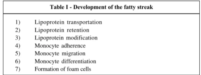
macropha-ges. Such receptors are not regulated by the intracellular cholesterol concentration; so, macrophages incorporate large particles of LDLox and become rich in lipid content. Then foam cells are formed and they are characteristic of the fatty streak, which is the earliest lesion known at the onset of atherosclerosis. Table I provides a summary of the
process of fatty streak formation. LDLox performs impor-tant actions, such as: forming foam cells, producing subs-tances that promote chemotaxis like the monocyte chemo-taxis protein - which attracts monocytes to the subendothe-lial area, reducing the recirculation of resident monocytes, which retain monocytes in the sub-endothelium; and finally, oxidized LDL is toxic to the tissues 6. This toxicity results in
a structural lesion in the endothelium.
Among the alterations caused by the presence of LDLox is also the production of interleukin-1, stimulating the migration and proliferation of smooth muscle cells of the media layer. When these migrate to the intima layer, they proliferate and start producing not only cytokines and growth factors, but also extracellular matrix, mainly compo-sed of collagen and proteoglycans, which will form part of the fibrous cap of the mature atherosclerotic plaque 3. The
endothelial erosion induced by LDLox can also cause the formation of platelet microthrombi, which will in turn produ-ce growth factors, such as the platelet-derived growth fac-tor (PDGF) 3. At this moment the endothelium may present
extensive lesions, true erosions. The interaction between platelets, endothelium, smooth muscle cells and macropha-ges will determine the degree of cellular proliferation, extracellular matrix secretion, and consequently the extension of the mature plaque. Table II presents the main phases of formation of the atherosclerotic plaque 1.
Anatomy of the atherosclerotic plaque
The evolution of the fatty streak atherosclerotic lesion into the fibrous plaque is quite slow. Pathological studies in human beings and animals have shown that the growth of the atherosclerotic plaque initially occurs towards the outer part of the vessel. Glagov et al 15 have
demonstrated remodeling of the arterial wall in atherosclero-tic plaques, with wall expansion towards the outside and lumen preservation in the beginning of the process. There-fore, the plaque can take years in this continuous remode-ling process until it causes luminal stenosis.
The mature atherosclerotic plaque shows, in addition to cells, two distinctive structural components: a slightly dense lipid nucleus and the fibrous cap, which is its fibrotic component. This fibrous component represents about 70% of the plaque’s total size 16-21, and the larger the plaque is, the
less prone it is to rupture. The fibrous cap is basically formed by smooth muscle cells, extracellular matrix and inflammatory
Table I - Development of the fatty streak
cells. The matrix consists of collagen, elastin, proteoglycans and protein microfibriles. Cytokines and growth factors regulate the synthesis of the matrix components 1.
The lipid nucleus is hypocellular and rich in
extracellu-lar lipids, mainly crystals and cholesteryl esters. Its patho-genesis is controversial, for it can derive both from lipids imprisoned in the extracellular space and from the necrosis or apoptosis of the foam cells 19. The content of this lipid
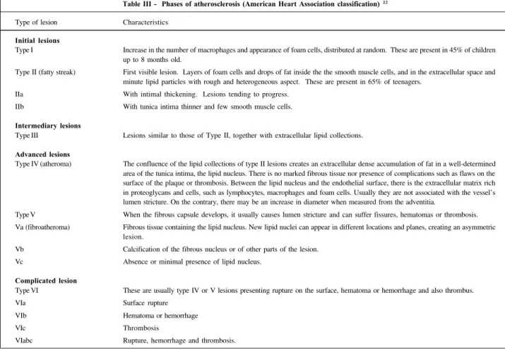
nu-cleus is highly thrombogenic. When in contact with the blood stream, due to rupture of the fibrous cap or endothe-lial erosion, the phenomena of platelet adhesion and aggregation occur, along with generation of thrombin and fibrin, with superjacent thrombus formation, representing the common starting point of acute coronary syndromes. Table III illustrates the phases of formation of atherosclero-tic plaque, according to the classification of the American Heart Association 22.
The sequence of progression of lesions, from type I to type IV is by the progressive accumulation of lipids. This evolution is slow; it starts in the first decade of life and goes until the third decade; this is the silent period of the disea-se. When it reaches phase IV, it grows by the accelerated accumulation of smooth muscle cells and collagen, going on to type V lesion; or in a more abrupt way, by instability, with the occurrence of ruptures and thrombosis or hemato-ma and then lesions type VI are formed. In the latter, the thrombi can become incorporated into the lesion, which grows and becomes again type V. This oscillation between type V and VI plaques is the main gradual occlusion mecha-nism of advanced lesions in medium caliber arteries.
Table II - Possible sequence of events in the theory of "response to lesion"
Lesion factors (lipids, hemodynamic, immunological reactions, etc)
Functional endothelial lesion
Infiltration and activation of macrophages -> foam cells
Growth factor secretion (PDGF and other peptides)
Migration and multiplication of smooth muscle cells
Plaque growth
Secondary endothelial lesion
Platelet adhesion and activation
Ulterior plaque growth
Modified from Ross R, 1986 1.
Table III - Phases of atherosclerosis (American Heart Association classification) 22
Type of lesion Characteristics
Initial lesions
Type I Increase in the number of macrophages and appearance of foam cells, distributed at random. These are present in 45% of children up to 8 months old.
Type II (fatty streak) First visible lesion. Layers of foam cells and drops of fat inside the the smooth muscle cells, and in the extracellular space and minute lipid particles with rough and heterogeneous aspect. These are present in 65% of teenagers.
IIa With intimal thickening. Lesions tending to progress. IIb With tunica intima thinner and few smooth muscle cells.
Intermediary lesions
Type III Lesions similar to those of Type II, together with extracellular lipid collections.
Advanced lesions
Type IV (atheroma) The confluence of the lipid collections of type II lesions creates an extracellular dense accumulation of fat in a well-determined area of the tunica intima, the lipid nucleus. There is no marked fibrous tissue nor presence of complications such as flaws on the surface of the plaque or thrombosis. Between the lipid nucleus and the endothelial surface, there is the extracellular matrix rich in proteoglycans and cells, such as lymphocytes, macrophages and foam cells. Usually they are not associated with the vessel’s lumen stricture. On the contrary, there may be an increase in diameter when measured from the adventitia.
Type V When the fibrous capsule develops, it usually causes lumen stricture and can suffer fissures, hematomas or thrombosis. Va (fibroatheroma) Fibrous tissue containing the lipid nucleus. New lipid nuclei can appear in different locations and planes, creating an asymmetric
lesion.
Vb Calcification of the fibrous nucleus or of other parts of the lesion. Vc Absence or minimal presence of lipid nucleus.
Complicated lesion
Type VI These are usually type IV or V lesions presenting rupture on the surface, hematoma or hemorrhage and also thrombus.
VIa Surface rupture
VIb Hematoma or hemorrhage
VIc Thrombosis
Atherosclerotic plaque instability factors: the
vulnerable plaque
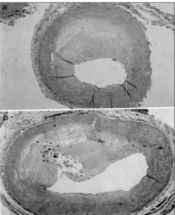
The instability of the atheromatous plaque, although unpredictable in regard to time, has reasonably defined mar-kers: the composition and relationship of the lipid nucleic mass with the total lesion mass; the thickness of the fibrous cap and the presence of inflammatory activity. Other factors are the degree of stenosis and the hemodynamic disorder related to it. Instability happens more often with plaques whose nuclei are rich in more fluid lipids 23 and with a larger mass in
relation to the lesion 19. As for the thickness of the fibrous
cap, instability occurs in thinner plaques and in the junc-tion with the normal wall of the vessel, the “shoulder” 24; in
addition, unstable plaques present smaller amounts of collagen and glycosaminoglycans compared with more stable plaques. These characteristics are more frequently associated with ruptures, fissures and thrombosis and occur more often in acute syndromes. Patients with stable angina have the accumulation of extracellular matrix as the main mechanism for arterial obstruction.
The fibrous cap of ruptured plaques shows activated macrophage infiltration, indicating inflammation where the rupture is located 21. T-lymphocytes are also present in the
fibrous cap, attracted by adhesion molecules expressed in the endothelium 25. In eccentric plaques, the inflammatory
cells are mainly located in the shoulder area, which is the place most likely to rupture. A study made with samples gathered by atherectomy, in patients with stable and unstable angina and non-Q AMI demonstrated that arteries responsible for acute syndromes contained more macro-phages than samples responsible for stable angina 26.
According to Libby 25, the IF-gamma produced by
lympho-cytes blocks collagen production by the smooth muscle cells. On the other hand, activated macrophages cause extracellular matrix degradation; both factors contribute to the weakening of the plaque’s fibrous cap.
Macrophages may degrade the extracellular matrix through two distinct mechanisms: fagocytosis or metallo-proteinases secretion; these are enzymes able to digest almost all substances present in the extracellular matrix 27,28.
The metalloproteinases operate in a neutral pH in the extra-cellular space, where they are released as proenzymes 28. So
far, twelve different enzymes from the metalloproteinase family have been identified, each with a preferential subs-trate, but not a single one. Collagen type I, a prominent com-ponent of the fibrous cap, can be degraded by metallopro-teinases 1 and 8 29. Galis et al 30 have demonstrated that it is
on the plaque’s shoulder area that there is an excess of acti-vity. Increased activity of metalloproteinases 2 to 9 has also been documented in human plasma, in unstable angina 31.
The macrophages are not the only cells able to produce and secret these enzymes. Smooth muscle cells also discharge metalloproteinases, thus also contributing to the homeos-tasis of the fibrous cap.
Recent studies have shown that patients with unstable angina and AMI have high levels of C-reactive protein and amyloid-A protein, reflecting a systemic inflammatory
process 32,33. This rise, even in normal individuals, was
related to a higher incidence of myocardial infarction and strokes 34. The same authors have shown that the reduction
of the risk of the first AMI, through the use of aspirin, seems to be related to a lower level of C-reactive protein, sugges-ting the possibility that anti-inflammatory drugs may contribute to the prevention of cardiovascular diseases.
An intriguing finding concerns the level of stenosis in unstable plaques; it was noticed that patients with acute coronary syndromes show a predominance of slight (<50%) or moderate (<70%) lesions 35-40. Table IV below
illustrates this fact.
Several explanations have been suggested for acute syndromes involving slight to moderate lesions 35. Among
these are the higher ratio of these lesions in relation to the total number of lesions and the absence of collateral circula-tion. Another theory is that the tension on the fibrous cap
Table IV - Relationship between previous stenosis and acute coronary syndrome
Author N o <50% 50%-70% >70%
Unstable angina
Ambrose et al 37 25 72 16 12
Acute infarction
Ambrose et al 37 23 48 30 22
Little et al 38 41 66 31 3
Giroud et al 39 92 78 9 13
Nobuyoshi et al 40 39 59 15 26
Average 65 20 15
is directly proportional to the diameter of the artery and to the intracoronary pressure, according to Laplace’s law 24;
therefore, the tension is higher in smaller lesions than in larger ones, due to the luminal ray. Figure 1 exemplifies stable and unstable lesions.
Coronary reserve, obstructive lesion and
ischemia
As an atherosclerotic lesion develops in an epicardial artery, luminal tightening occurs. In significant obstructions the vessel begins to greatly resist coronary flow. Within physiological limits of perfusion pressure, the coronary auto-regulation allows arteriolar resistance adjustments to maintain the proper balance between flow and demand for myocardial oxygen 41. As the resistance of the epicardial
ar-tery increases due to a higher degree of stenosis, the distal arteriolar resistance declines to keep the flow at normal levels. When the arteriolar vasodilation reserve is exhaus-ted, the myocardial perfusion is linearly dependent on the perfusion pressure. In such conditions, drops in arterial pressure (AP) can induce ischemia 42.
The decline of coronary reserve starts when lesions occupy at least 70% of the vessel diameter. In concentric lesions, the coronary flow depends on the degree of the lesion and perfusion pressure. In eccentric lesions, the degree of obstruction can be dynamic, because the part of the plaque least affected by the atherosclerotic process may suffer dilation or constriction, and thus vary the effective vessel lumen.
The reduction of the coronary reserve is progressive; initially ischemia occurs in intense oxygen demand situa-tions (tachycardia, exercises), and then in situasitua-tions of lower demand until the coronary reserve is totally impaired when the lesion occupies 90% of the vessel diameter.
Collateral circulation
Collateral circulation is blood circulation between two vessels without an intermediary capillary network. The mechanisms responsible for the development of collateral circulation are not well known. Ischemia and the pressure gradient between normal and obstructed vessels seem to be important, because the irrigation of the area supplied by collateral vessels is more deficient in endocardial layers. The functional significance of collateral circulation has been assessed in various manners. The backward flow following acute coronary occlusion in dogs varies from 8 to 36% of the normal flow. This flow may increase up to 40% with the use of drugs, such as verapamil, during acute ischemia 43. Collateral vessels that have developed slowly
also respond to vasodilating drugs and to tachycardia 44.
With collateral coronary circulation, between 28 and 50% of silent coronary occlusions in humans are not accompanied by a ventricular mechanical deficit, suggesting that the presence of pre-existing collateral circulation may protect the myocardium 45.
On the other hand, myocardial scintigraphy studies with thallium-201, during exercise, have shown that when there is an occlusion of the anterior descending artery, the anterior myocardial area shows signs of ischemia in the occlusion, even in the presence of collateral circulation. The other areas are totally protected by collateral circulation, as demonstrated by Baroldi 46, who noticed that in 44% of the
patients with up to two occluded vessels there was no histological evidence of a myocardial infarction. This suggests that collateral circulation may be adequate at rest but insufficient during intense exercise, mainly when the area irrigated by the occluded artery is large. Right after an infarction, collateral circulation may appear; however, only later does this collateral circulation influence ventricular mechanical function. For protection from the beginning, it should be present before the occlusion.
Risk factors
Advanced age, family history of CAD and male sex are commonly recognized RF47,48. As for AH, MacMahon et al 49
demonstrated in a metanalysis of more than 400,000 persons that a close correlation exists between hypertension, systo-lic or diastosysto-lic, and CAD. It is a very prevalent factor among patients with CAD. Although its control is followed by decreased mortality due to stroke, lower incidence of kid-ney insufficiency and heart failure, a great reduction in acute coronary events has not been proved. On the other hand, the metabolic alterations present in diabetes mellitus 50, the
generalized glycolysationof proteins, insulin resistance and the alteration of the metabolism of the remaining chylo-microns, lead to accentuated and diffuse atherosclerosis of all vessels. Atherosclerosis is even greater, however, in average and small caliber arteries. Thus, atherosclerosis is the cause of 80% of deaths 50,51 in persons with diabetes.
Persons with non-insulin dependent diabetes mellitus (NIDDM) (type II diabetes), although they have lower levels of glycemia, have greater cardiovascular risk than those with insulin-dependent diabetes mellitus (IDDM) (type I diabetes), because atherosclerotic alterations in their ar-teries are greater 52. These same disturbances seem to be
res-ponsible for endothelial dysfunction and microangiopathy. Control of post-prandial glycemia and the resulting metabolic disturbances that follow it has proven to be the most effective way of reducing complications in persons with diabetes 52,53.
Cigarette smoking, another risk factor, alters LDL 54,
reducing the endothelium-dependent relaxation induced by acetylcholine, in a similar way to oxidized LDL, without altering the non-endothelium dependent relaxation. Both active and passive smoking 55 are associated with the
development of several clinical forms of CAD and diffuse atherosclerosis in men and women.
individuals exposed to passive cigarette smoke. The impact of cigarette smoking was greater in persons with diabetes and hypertension. The duration and degree of exposure, but not the exposure continuation or interruption, correlated with the progression of atherosclerosis. The benefits of smoking cessation and the decrease in coronary events are well established. Hasdai et al 57 showed a higher incidence of
non-Q wave infarction and of death in patients who continued smoking compared with ex-smokers and non-smokers. In women the increase in cigarette smoking coincides with an increase in the incidence of atherosclerosis.
Dyslipidemia is perhaps the most frequent RF 58.
Coun-tless experimental and epidemiological studies 59,60 have
esta-blished the influence of hyperlipemia on the development of atherosclerosis and its cardiovascular complications. In some cases it is linked to genetic alterations (see below); but in most cases it is due to an increased intake of saturated fat. The relationship between triglycerides and coronary disease is somewhat controversial 61. In a univariate
ana-lysis, triglycerides usually predict the disease, but when other RF, mainly HDL levels, are factored into multivariate analysis, triglyceride loses its predictive power. However, a recent study 62 indicates that even within levels considered
normal, individuals with triglyceride levels >100mg/dl have more coronary events in the long-range than those with levels <100mg/dl. This is very suggestive of the long-range predictive value of hypertriglyceridemia.
A sedentary way of life and obesity go hand in hand 63,64. Regular exercise reduces considerably the incidence
of coronary events; however, a cumulative effect has not been noticed, except in preventing obesity from the fifth decade in life on. Of practical importance is the demonstration that even light intensity exercise as often as twice a week, al-though it does not lead to physical conditioning, does po-sitively influence lipid profiles 65. On the other hand, body
mass indexes >27 are associated with an increase in total and cardiovascular mortality, mainly in young people 66.
Emotional stress related to the work environment, personal and family problems 67,68 is also a RF.
It is important to note that in about 35% of the docu-mented cases of CAD, traditional RF are not found 69.
Cer-tain “genetic alterations” have already been identified 70-73
that are responsible for dyslipoproteinemia causing athe-rosclerosis; among these are familial dysbetalipoproteinemia, combined familial hyperlipemia, apolipoprotein A-I, A-I and apoC-III deficiency. One special condition is the occurrence of low HDL (<35mg/dl), especially when followed by in-creased triglycerides; it is found in approximately 4%-5% of the population. In the Prospective Cardiovascular Munster Study (PROCAM) 74 it was associated with a 6-fold increase
in the incidence of coronary events. A variation of apoA-I (delta K107) was associated with this syndrome. Increases of lipoprotein(a) (Lp(a)) 75, of fibrinogen 76 and plasma
ho-mocysteine 77-81 may contribute to some cases of CAD. The
relationship of homocysteine and atherosclerosis was first reported in 1969, when McCully 82 demonstrated precocious
atherosclerosis in children with homocystinuria. Later on,
the infusion of homocysteine in laboratory animals de-monstrated the rapid formation of typical vascular lesions. The confirmation of hyperhomocysteinemia as a RF for ar-terial disease came from clinical studies, showing the association of elevated homocysteine levels with periphe-ral vascular disease, stroke and coronary disease 80-81.
Some infectious agents, such as the bacteria Clamy-dia pneumoniae, and certain viruses have been suggested
as possible causal agents 82,83. However, it is not clear yet if
they are causal agents or simply coincide with or heighten the already on-going process. Clearly, the issue of RF is still open for discussion. In 1981, Hopkins and Williams 85
identified 246 possible coronary RF. It is possible that even more may be discovered.
Certain groups of RF are frequently found in one indi-vidual. Stress combined with smoking, hypertension or both are often found in the same individual. A sedentary life, obesity and hypercholesterolemia are also another commonly found group of RF. This grouping of RF is im-portant, because the risk of coronary events increases, not in a simple additive way, but in a geometrical progression.
Clinical syndromes in coronary artery disease
Necropsy studies in children and adolescents 86, in
soldiers who participated in the Korean 87,88 and Vietnam 89
wars, and adults approximately 25 years old, victims of sud-den death (SD) 90, clearly indicate that coronary
atheros-clerosis begins in adolescence and its initial alterations date back to childhood. However, its clinical manifestations only show up during adulthood, more frequently from the fourth decade of life on. It may then be concluded that the disease presents a long period of silent evolution, before the mani-festation of clinical CI, which can take various forms, inclu-ding the following:
a)Stable angina - Repeated anginal attacks, which
can develop over months or years are referred to as stable angina. An increase in myocardial oxygen consumption (MVO2), usually due to physical exertion or emotions, trig-gers ischemia. It is relieved by rest or the use of vasodilators. The threshold for angina may vary. When the ventricular function is normal, as it frequently is, stable angina is compatible with an acceptable quality of life.
b)Heart failure - Heart failureassociated with CAD is
mainly due to two conditions: ischemic cardiomyopathy and LV akinesia/dyskinesia (aneurysm). The ischemic cardiomyopathy is the ventricular dysfunction with diffuse hypocontractility and dilation of ischemic cause. It can be a consequence of the replacement of fibrosis in areas where multiple, small infarctions have occurred, or of global myocardial dysfunction due to chronic ischemia and hibernation. The state of myocardial hibernation 91-93 is of
The other segment of patients with heart failure se-condary to coronary disease are those with akinesia or ven-tricular aneurysm. This type of heart failure is a segmentary mechanical ventricular dysfunction following an infarction. When it is extensive, the aneurysm ends up causing dys-function of normal, not infarcted segments, leading to chro-nic CI. Aneurysms cause serious alterations in the ventricu-lar geometry, being also a cause of ventricuventricu-lar arrhythmias.
c)Silent ischemia (SI) - About 3% of the normal
popu-lation and 30% of asymptomatic patients experience silent ischemia (SI) following an infarction of the myocardium 94,95.
Most angina patients experience multiple ischemic episo-des of painless ischemia, which amounts to 75% to 92% of the total ischemic load. SI can be diagnosed by an ergometric test (ET) and Holter monitor. The prognosis depends mainly on the size of the risk area. SI is particularly troublesome because patients do not experience the classic warning sign: pain. Therefore, they may be at serious risk without knowing it. That is why it is the cause for SDin many cases.
d)Acute coronary ischemia - Unstable angina,
non-Q wave infarction, and infarction with non-Q-wave are characte-ristics of acute coronary ischemia. They are related to atherosclerotic plaque rupture and thrombosis superimpo-sition in the areas of ischemia. Thrombosis of short dura-tion or in combinadura-tion with partial arterial occlusion is asso-ciated with instances of unstable angina; more persistent thrombosis and total occlusion are associated with myocar-dial infarction. These thromboses can occur both in pa-tients with a history of prior angina or can be the first manifestation of CAD.
e)Sudden death (SD) – As indicated by the name, SD
is an event that naturally evolves to death, which is not always observed in other forms of acute ischemia. Approxi-mately 1/3 of CAD patients die of SD, which can be due to primary malignant arrhythmias, such as ventricular tachy-cardia degenerating into ventricular fibrillation or ventri-cular fibrillation associated with acute ischemia 96.
It must be stressed that in CAD there is no precise rela-tionship between anginal symptoms and the extension of the coronary or ventricular function impairment. It is quite common to find patients with recent angina who maintain practically normal physical activities until the onset of the symptoms. Coronary angiography reveals important bi- or tri-arterial lesions in such patients. It is obvious that these lesions developed long before the symptoms. On the other hand, the ventricular mechanical function may be little alte-red or even entirely normal. This can be explained because ischemia is segmentary or because collateral circulation allows for natural compensation of the coronary obstruc-tion. Hence, certain patients with serious coronary lesions can maintain practically normal physical activities for a long period of time. However, when symptoms or signs of heart failure occur, significant ventricular dysfunction is present.
Coronary disease in women - CAD in women occurs
about 10 years later than it does in men 97. This is attributed
to the protective effect of female hormones during the women’s reproductive years, because no difference exists in the prevalence of RF and type of lesion between men and women 98. However, the seriousness of the clinical
presen-tation does not differ between the sexes. Actually, it is suggested that the post-AMI evolution is worse in women than in men 99. However, more careful analyses show that this
worse evolution is mainly due to other factors, such as more advanced age and diabetes, and not the female sex 100 per se.
But cardiovascular disease is the greatest cause of death in women, even higher than total mortality due to uterine, breast, and ovarian cancers 101,102. Therefore, CAD
treatment in women must not differ from that in men, because the disease has similar evolution or prognosis in both sexes.
Prognosis
Ejection fraction of the left ventricle - Random
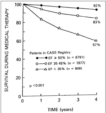
compa-rative studies of clinical and surgical treatments have pro-ved the undeniable value of measuring ejection fraction (EF) for the prognosis of CAD 103,104. In patients clinically treated
in the Coronary Artery Surgery Study (CASS) 105, the
survival rate over 4 years was 92% for EF >50%, 83% for EF 35-49%, but only 57% if EF was <35%. Hueb et al 106
repor-ted that patients with few symptoms and preserved ventri-cular function who had refused surgery showed similar re-sults as those in CASS. Therefore, EF must always be quan-tified when establishing a CAD prognosis. Figure 2 illus-trates the relationship of EF versus survival rate.
Coronary artery anatomy - Usually, the higher the
num-ber of arteries with critical lesions, the worse the long-term prognosis 103,104. The European Coronary Study Group 107,
however, has shown that critical lesions (>50%) in the left coronary trunk or the presence of proximal lesions in the two great branches of the left coronary artery, the anterior inter-ventricular branch and the circumflex branch together with lesions in the right coronary artery show a higher mortality rate than other subgroups. This mortality is reduced with myocardial revascularization. Later, patients in CASS with few symptoms and presenting uni-, bi- or tri-arterial lesions had a low mortality rate if they also had a good ventricular function, an EF over 45% visualized by contrast ventriculo-graphy or by radioisotopes.
In addition to the prognosis, knowing the coronary anatomy is fundamental in deciding about the best treat-ment, whether clinical or revascularization by angioplasty or surgery. Not only it is important to know where the lesions are located, but also the characterization of the distal beds and the presence of collateral circulation are valuable elements in determining treatment.
Risk area – The myocardial area that can be affected by
ischemia when coronary occlusion occurs is called the risk area. The knowledge of coronary artery anatomy or the ex-tension and intensity of ischemia in myocardial scintigraphy using Thallium-201 or SESTAMIBI-Tc99m allows us to quan-tify the risk area 93. However, it is also important to analyze
non-ischemic areas, because they are responsible for ven-tricular adaptation when coronary occlusion occurs. It is well documented that larger risk areas have a worse prognosis 108.
Electrophysiological status - The frequent occurrence
of ventricular arrhythmia, i.e., over 30 ventricular extrasysto-les per hour, recurrent ventricular tachycardia, the presence of low voltage potentials in the final part of the QRS complex in the high resolution electrocardiogram, in patients with left ventricular aneurysm or left ventricular dysfunction, are associated with a higher incidence of cardiovascular death and SD 109,110.
Age - In chronic CAD the influence of age is not
sim-ple. On the one hand results from CASS showed a higher survival rate in 7 years among young people (84% versus 75% for males and 90% versus 77% for females) 111.
Howe-ver, the disease is, in relative terms, more serious and has a worse prognosis in young people; perhaps because in this age group two of the most important RF are prevalent: smoking and family history of CAD. The Brazilian Medici-ne, Angioplasty, or Surgery Study (MASS) 112, which
ana-lyzed the influence of medical treatment, angioplasty and surgery in patients with critical lesions in the anterior interventricular branch of the left coronary artery, revealed a high rate of progression in non-critical lesions in younger patients, most of whom developed tri-arterial lesions at the end of the 3 years of the study. Whereas the Groupo Italiano Per lo Studio Della Streptokinase Nell’Infarto Miocardio-2 (GISSI-2) 110 reported that with acute myocardial infarction
advanced age is a bad prognostic factor.
Diagnosis
A clinical history may provide important information for the diagnosis of CAD. Anginal pain, fatigue, dyspnea, and palpitations are the most common complaints. Typical angina is the most easily diagnosed. Identification of trig-gering and relief factors, its duration and characteristic loca-tion and irradialoca-tion are indispensable for the diagnosis of typical angina. It is associated with coronary disease in 90% of men over 55 years old and 80% of women in this same age group 112a. When the pain has some, but not all of the classic
characteristics, it is called atypical angina and is associated with coronary disease in 50% of the patients over 50 years old. However, many symptoms mirror angina. SI, where the patient can be totally asymptomatic must not be overloo-ked. Family history should include history of infarction and SD, in men who are close relatives below 55 years of age, and in women also who are close relatives below 65 years of age. Life style habits can indicate significant RF. But physi-cal examination usually adds little to the diagnosis. Except in instances of heart failure, the physical examination is noticed only for being normal, which ironically contrasts with the potentially lethal situation.
Non-invasive examinations - Technical aspects will
not be discussed here, but only several peculiarities of ma-jor interest to the cardiologist in general.
Electrocardiogram at rest - Most patients with heart
failure are within normal standards. However, the presence of inactive areas suggesting a previous infarction, inverted T waves, downslope of the ST segment and left ventricular hypertrophy are associated with a higher cardiovascular mortality rate in the long- range 113. The presence of
extrasys-toles, mainly those of ventricular origin, may suggest co-ronary disease. Conduction disorders both atrial-ventricular and fascicular may occur, but these do not contribute to the detection of coronary disease because they are unspecific.
ECG Stress test (ST) – Considering the low cost,
sim-plicity, sensitivity and specificity, and prognostic value of this test, and also considering the prevalence of CAD in the population group to be studied, the ST is very useful in tra-cking and stratifying coronary atherosclerotic disease and has an excellent level of reliability 114. The interpretation of
the ST must include not only the analysis of the electrocar-diogram, but also the hemodynamic and clinical variables. It is used to track CAD in suspect cases and, as a screening tool in individuals who are beginning physical exercise programs or patients with RF. Therefore, it predicts a poor prognosis when positive in low loads, when the arterial pressure does not rise adequately and the work tolerance is less than six minutes.
Radioisotopes - The versatility of radioisotopes allows
positive and negative predictive value is considered a fun-damental exam in identifying the presence of coronary disea-se 93. Fixed uptake defects suggest areas of predominant
fi-brosis, while reversible defects suggest an ischemic process and viability of the myocardial cells. Alterations in the balan-ce between supply and demand of O2 that allow the induction of ischemia and evaluation of the viability may be triggered both by exercise and drugs such as dipyridamole. The contrast reinjection techniques, late reading and tomogra-phic cuts contribute to increase this method’s analytical capacity 115. It also allows an estimate of the extension of the
risk area. As it is well known, this has great prognostic value, for the larger the area, the worse the prognosis.
The use of deoxyfluoroglucose, with radioactive fluo-ride, of long-chain fatty acids, with C14, or of labeled ammo-nia permit the evaluation of the integrity of cellular metabo-lism, the presence of ischemia, and the quantification of the residual coronary flow in hypocontractile or akinetic areas fed by coronaries with critical lesions 93. Currently available
computer software allows simultaneous appraisal of the pre-sence of ischemia, ventricular volumes and ventricular seg-mental contraction. It is undoubtedly a method of immense value. The only restrictions are its cost and availability.
Resting and stress echocardiogram - The resting
echocardiogram has considerable application in the diag-nostic and non-invasive progdiag-nostic determination of the coronary atherosclerotic disease 116. It allows the detection
of segmental thickness and motility alterations (hypokinetic, akinetic or aneurysmal areas), which are characteristic of CAD, in addition to allowing for the evaluation of volumes and ventricular mass, and also the EF, which is of acknowled-ged prognostic value.
However, it was the use of physical stress, or rather, pharmacological stress 116a that opened great perspectives
for echocardiography. When administering dobutamine in increasing doses (up to 40µg/kg/min), three responses may be triggered: progressive improvement of the contraction, initial improvement followed by a worsening, and finally, progressive worsening 116b. The response considered
typi-cal of the presence of ischemia with viability in hypokinetic areas is the initial improvement followed by a worsening of the contraction. However, the lack of an adequate window, more common in the age bracket of coronary disease patien-ts, can prevent resting echocardiograms from being made or impair their technical quality. Physical exercise can also be used to provoke ischemia, but the increase of movements in the thorax, both due to hyperpnea and the exercise, makes this exam more difficult to conduct.
Echocardiogram with the use of micro-bubbles - The
development of stable micro-bubbles, not related to the occurrence of systemic emboli and that reflect the ultra-sound rays, causing a contrast in relation to the blood and neighboring tissues, hail a new non-invasive era in the diagnosis and determination of ischemia and ventricular function 117 . Intracavitary bubbles allow ventriculography
with contrast and, bubbles in the intratissular vessels, allow visualization of myocardial perfusion. The technical hin-drance to the use of micro-bubbles is their low commercial availability, a higher number of patients with inadequate thorax for the exam in the age bracket to be investigated, and the low specificity for perfusion defects on lateral wall. Rapid technical development in this area is expected, which will widen the use of this method.
Coronary angiography - The definitive examfor the
diagnosis of coronary obstructive atherosclerosis is coro-nary angiography. It also permits the evaluation of ventricu-lar function. It should be performed when there is reasona-ble suspicion of coronary disease, to explain precordial pain of possible ischemic origin, to establish the prognosis, and whenever any intervention is considered, whether by catheter or surgery. It should be stressed that it is an invasi-ve exam, and despite its low risk, serious complications may occur, such as systemic embolism, arterial dissections, ven-tricular fibrillation and death. Studies relating the level of stenosis with the triggering of ischemia have shown that le-sions > 70% are correlated with the decrease of coronary flow reserve. Furthermore, certain characteristics of the le-sions can be appraised. According to Ambrose et al 118, the
lesions may be concentric, eccentric type I (asymmetric ti-ghtening with smooth borders and large neck) and eccentric type II (asymmetric with thin neck or irregular borders or both). The lesions most often found in patients with unsta-ble angina are the eccentric type II. These, then, are the sions most likely to become unstable. The presence of a le-sion equal to or above 50% in the trunk or the presence of proximal lesions above 70% in the three main branches, the latter with systolic dysfunction of the left ventricle, are indica-tors of the worst prognosis with isolated clinical treatment.
Important contributions to the understanding of the physiopathology of coronary disease were the studies of endothelium-dependent coronary vasomotion, visualized during coronary angiography. Paradoxal vasoconstriction was thus documented, after the administration of acetyl-choline, in segments with stenosis and pre-stenosis 119, with
irregularities or being angiographically normal in coronary atherosclerotic patients.
Magnetic resonance imaging - Magnetic resonance
imaging (MRI) has been used in two instances to evaluate patients with coronary atherosclerotic disease: cellular metabolic study and cardiac form and function 120. The
cardiac cycle. are obtained in the T1 or anatomic relaxation phase. The result is high fidelity ventriculography. More recently, non-invasive coronary evaluation with the use of helicoidal tomography 121 is being used as a convenient way
of detecting coronary atherosclerosis, but the method is not a routine clinical application.
Treatment
The great options for CAD treatment include clinical/ medicinal treatment, angioplasty and surgical revascula-rization. The clinical treatment is obviously always used, as neither PTCA nor surgical revascularization interferes in the metabolic process of the basic disease.
Clinical treatment
Nitrates - Nitrates are prodrugs that need to be
conver-ted into nitric oxide (NO) in the endothelial and smooth muscle cells 122, in order to have their vasodilating effects.
Their basic action is the relaxation of vessel smooth muscle. Their availability in the venous, oral, sublingual and transdermic forms makes them especially versatile. Their vasodilating action starts quickly, lasting less in the arteries than in the veins, where it lasts for hours. Nitrates act through various mechanisms: they redistribute the corona-ry flow preferably to the myocardial hypoperfused zones, they counteract vasospasm, reduce the venous return, the preload, the ventricle wall tension, and therefore reduce MVO2; they also promote platelet antiaggregation. They are excellent for treating stable and unstable angina, SI, ischemic cardiomyopathy and post-AMI angina. Their most undesirable side effects are headache and hypoten-sion; the latter is more common and more of a concern with older patients. Pulmonary ventilation/perfusion defects and methemoglobinemia were also reported, but their clinical importance is questionable.
Important characteristics are the development of tole-rance and recovery of its action in a short period of time 123.
To prevent medical intolerance “free intervals” of 12 hours every 24 hours should be observed . Despite its proven action in the relief and prevention of ischemic symptoms and in the increase of tolerance to physical exercise, the decrease in mortality with the use of nitrates still has not been demonstrated 124.
Beta-blockers - The stimulatory effects of the
beta-receptors by catecholamines are upset by beta-blockers. Two large receptor classes are known: beta 1 and beta 2. The beta 1 receptors, predominantly cardiac, when stimulated respond with an increase in heart rate (HR), in contractility and A-V conduction, release renin through the juxtaglome-rular kidney cells and induce lipolysis in adipocytes. The stimulation of beta 2 receptors causes bronchodilation, vasodilation and glycogenolysis.
Because they block the action of catecholamines, beta-blockers are efficient drugs for the treatment of angina,
essentially because they reduce MVO2, due to the reduc-tion of HR. This also increases the diastolic period, increa-sing the coronary perfusion period. It has likewise been demonstrated that although beta-blockers reduce total coronary flow, they improve endocardial perfusion, there-fore favoring the myocardial region most affected by ische-mia. They are also very efficient medicines for the treatment of AH and arrhythmias dependent on the sympathetic action. It has also been demonstrated that they reduce SD and re-infarctions post-AMI 125. They also reduce pressure
increase and heart rate during effort. Therefore, they are especially useful in effort angina. However, their undesirable effects can be extremely limiting. These include weakness; sleep and mood alterations; sexual dysfunction, with impotence and retrograde ejaculation; bronchospasm; peripheral circulatory insufficiency; precipitation of heart failure; induction or intensification of cardiac blockades; and intensification of hyperglycemia induced by insulin.
Cardio-selective blockers, such as atenolol and meto-prolol, act preferably on beta 1 receptors, having less effect on beta 2; therefore, they reduce MVO2 with fewer effects on the bronchia, peripheral circulation or glycogenolysis. On the other hand, the non-selective, such as propranolol and nadolol, block the beta 1 and beta 2 receptors. Cardiosele-ctivity, however, from the clinical point of view is relative.
Some beta-blockers, such as labetalol, carvedilol and bucindolol, also cause vasodilation 126,127. Carvedilol
speci-fically has shown beneficial effects in treating chronic heart failure, partially because of its vasodilating effects. On the other hand, sotalol has a significant anti-arrhythmic effect (classes II and III types), and therefore is useful for the treat-ment of angina associated with ventricular arrhythmias.
Individual tolerance to beta-blockers can vary consi-derably. Metoprolol and propranolol can cause very intense effects (intense bradycardia) in certain people, known as “poor metabolizers”; this is due to a particular genetic polymorphism that affects about 10% of the white popula-tion. Therefore, it is always advisable to start therapeutics with low dosages of these substances. As far as metabolism is concerned, beta-blockers, especially propranolol, can increase triglycerides and reduce HDL, without altering LDL. This should be considered when patients with CAD and dyslipidemia are prescribed this medication 128.
Calcium channel antagonists - Ca++ ions are
and hence these have little effect on the conduction sys-tem. Producing coronary and systemic vasodilation, dihy-dropyridines increase the supply of oxygen to the myocar-dium and decrease MVO2 by reducing peripheral resistance and systolic stress 129. The coronary dilating action is
espe-cially important in spastic angina or with variable threshold angina. The blockade of Ca++ entrance has a negative ino-tropic effect; however, this is counterbalanced by the sympathetic stimulation induced by hypotension. Nifedipi-ne has a higher systemic vasodilating effect than verapamil and diltiazem. Nifedipine mainly reduces the systolic pressure, so its beneficial effect is primarily due to afterload reduction and also to the increase of the coronary flow. Fast action formulations have been held responsible for the increase in cardiovascular events, but this does not apply to drug slow-release formulations.
On the other hand, verapamil reduces HRand myocar-dial contractility. It can therefore cause a decrease in the cardiac output and heart failure, in patients with ventricular dysfunction, while those with normal function tolerate it well. It is contraindicated in patients with sinus node disease and in atrioventricular node disease.
Diltiazem has intermediary actions between nifedipine and verapamil. It is a systemic vasodilator, but it can also in-crease coronary flow. Its depressing action over cardiac function is less than verapamil’s. Besides being very effici-ent as an anti-anginal drug, it produces few undesirable si-de effects. When administered in the acute phase of a myo-cardial infarction it reduced mortality, except in the presence of ventricular dysfunction, when mortality increased 130.
“Second generation” Ca++ 129 antagonists
(nicardipi-ne, isradipi(nicardipi-ne, amlodipi(nicardipi-ne, and felodipine) are derived from dihydropyridine. Usually they have a higher peripheral va-sodilating action than those of the “first generation”. Amlo-dipine starts acting slowly and has a long half-life (36 hours); it causes important peripheral and coronary vasodi-lation, being especially useful in cases of hypertension and angina. It has been used even in heart failure, but in this condition its role is not yet well defined.
It was demonstrated experimentally and in men that Ca++ antagonists may have an anti-atherogenic effect 131,132.
New coronary lesions were less frequent in patients taking nifedipine than in those in the control group. However, this action needs further investigation. Despite the beneficial effects common to all Ca++ antagonists, a decrease in car-diovascular mortality with their extended use has not been demonstrated.
Calcium channel antagonists have wide use in patients who undergo coronary anastomoses with arterial conduits in order to avoid spasms of the grafts and after coronary angioplasties, and in patients for whom beta-blockers have been contraindicated. The most common undesirable side effects are lower limb edema, headache, intestinal obstipa-tion, dyspepsia, and more rarely, gum hypertrophy.
Angiotensin-converting enzyme inhibitors -
Angio-tensin-converting enzyme inhibitors prevent the
conver-sion of angiotensin I to angiotensin II in circulation. Their hemodynamic actions are due to arterial vasodilation, which increase cardiac output; they also act on the lung and venous beds, reducing the pre-load. Such effects favorably influence the cardiac remodeling following an infarction 133,134. The use of this type of medication is
bene-ficial in the acute phase of extensive infarctions, because it prevents ventricular dilation and reduces mortality both by heart failure and by SD. Theoretically, blocking the renin-angiotensin system on the atherosclerotic lesion areas has a beneficial influence on the evolution of the lesions, be-cause angiotensin, besides being a potent vasoconstrictor, is also a growth factor for smooth muscle cells 135.
There-fore, it is useful in cases of angina with heart failure or AH. Its main side effects include: dry cough due to an increase in the bradykinin half-life in the bronchial tree; and hypoten-sion by vasodilation, mainly present when associated with other hypotensive drugs.
Aspirin - Platelets play an important role in triggering
acute coronary episodes and in the growth of atherosclero-tic lesions (see “Anatomy of the Atheroscleroatherosclero-tic Plaque”). Among platelet aggregation stimuli is the production of thromboxane A2 by the platelets themselves. Aspirin blocks its formation by interfering with its synthesis from arachi-donic acid and interferes little with the production of prosta-cyclin by the endothelial cells 136.
Because aspirin operates on the key mechanism of for-mation and growth of the intra-arterial thrombus, it is capa-ble of reducing the number of fatal and non-fatal events in patients with coronary disease 137 and currently is a
funda-mental part of CAD therapeutics. Other anti-platelet medica-tions are sulfinpyrazone, ticlopidine and pentoxifylline. The great problem with antiplatelet therapies, mainly aspirin, is the tendency to promote bleeding, which can cause hemor-rhages, including strokes. Therefore, drugs should be used very carefully in individuals prone to such phenomena.
The most modern antiplatelet agents are the glycopro-tein IIb/IIIa blockers, which act in the final common pathway of platelet action in thrombus formation 138-142. The studies of
these types of drugs were made in principally acute syndro-mes. However, their role as oral agents for treatment of chro-nic coronary insufficiency is yet to be established.
Finally, it should be mentioned that in many patients combination therapy, including the simultaneous use of the medications cited above, is necessary to control symptoms; their use should always be individualized.
Treatment of the underlying disease - The treatment
therefore, be developed for each individual based on that person physiological, electrical and hemodynamic parame-ters. The recommended diet aims for a lower intake of animal fats, and favors fruits, vegetables, and legumes. The in-fluence of the global control of RF was appraised in the Stanford Coronary Risk Intervention Project (SCRIP) 143, in
the U.S.A. In SCRIP, patients underwent global control of RF for four years; it was possible to control all of them, except smoking. Even so, lower rates of new coronary lesions were noticed at coronary angiography in the control group compared with the non-control group. The reduction of fat intake had the greatest effect.
The European Action on Secondary Prevention Through Intervention (EUROASPIRE) 144 study
demons-trated that, despite the great prevalence of modifiable RF and those of genetic influence, in only 21% of the cases were the patients relatives tracked for coronary disease RF. This suggests that preventive measures are still imperfect and need to be improved.
However, the greatest contribution to the control of atherosclerosis and its manifestations has been interven-tions that reduce total serum cholesterol. Among these, statins have been the first medicines to greatly reduce hypercholesterolemia with long-term tolerance by most patients. Several clinical studies have shown that the treatment of intense or moderate hypercholesterolemia reduces coronary events, coronary mortality, global mortality, hospitalizations and the need for angioplasty or revascularization surgeries. These findings apply both to men and women, diabetics and non-diabetics, smokers and non-smokers, older or younger patients 145,146. Furthermore,
angiographic studies have documented the decrease in the progression of lesions, lower occurrence of new lesions, and even the regression of lesions in some patients 147.
Significant alterations in arterial vasomotion have also occurred with statin therapy; furthermore, this improve-ment can be reached in only one month of statin use 148. This
suggests that the lipid loweringtreatment is perhaps indicated in acute syndromes; this hypothesis, however, must be tested. A 29% reduction in strokes was also documented 149 in patients treated with statins and followed
for more than three years. More recently, such results have also been noticed in patients with only hypercholesterole-mia, but without documented CAD. The extended use of cholesterol lowering drugs has not coincided with an in-crease in incidences of cancer or violent deaths 149.
Patients with hypertriglyceridemia as the primary risk factor, followed by hypercholesterolemia or low levels of HDL-cholesterol are best treated with fibrates (benzafibrate, gemfibrozil, phenofibrate and etofibrate), which reduce triglycerides, increase HDL-cholesterol and reduce choles-terolemia. Its use in coronary disease patients was followed by a reduction in cardiovascular mortality 150,151. Therefore,
the treatment of dyslipidemia plays an essential role in the control of CAD.
Antioxidants and vitamins
Oxidative stress plays an important role in the patho-physiology of atherosclerosis 152. Antioxidants have been
experimentally shown to protect against the alterations brought on by oxidative stress. Diets rich in antioxidant substances, such as beta-carotene, alpha tocopherol (vitamin E) and ascorbic acid (vitamin C), are consumed by populations with a low incidence of coronary atheroscle-rosis, which supports the oxidation theory 147. However, only
a randomized study, the Cambridge Heart Antioxidant Study (CHAOS) 153, has demonstrated that vitamin E is capable of
reducing the incidence of new coronary events, specifically non-fatal AMI. Therefore, the diet containing these substan-ces is considered more important than supplements.
Flavonoids, substances present in grape juice, red wine and other red fruits, have an anti-inflammatory and antioxi-dant effect. In various studies they have been shown to redu-ce the incidenredu-ce of atherosclerotic lesions 154. Probucol, a
li-pid-lowering agent with a powerful antioxidant effect has been proven to reduce atherosclerosis in experimental ani-mals 155 and the coronary restenosis following angioplasty 156.
The unquestionable clinical demonstration of the beneficial effect of antioxidants in CAD treatment, however, still requires new data. Specific studies are currently underway.
Estrogens
The epidemiological evidence of the lower prevalence and incidence of coronary atherosclerosis in women in their reproductive years and of its later increase, the beneficial effects of the chronic use of estrogens in the lipid profile and of the recovery of endothelium dependent vasodilation in acute estrogen administration led to the development of studies to prove the usefulness and safety of hormone replacement therapy for women in menopause. The most recent carefully controlled study, the Heart and Estrogen/ progestin Replacement Study (HERS) 157, however, has not
proved that the use of 0.625mg of estradiol and 2.5mg of progesterone reduces the incidence of coronary events. On the contrary, the use was followed by an increase in coronary events in the first year, only decreasing in subse-quent years; furthermore, hormone therapy was followed by a threefold increase in the risk of thromboembolic phenomena and biliary lithiasis. Therefore, the use of hormone replacement therapy for the specific purpose of treating CAD does not seem justified.
Genetic predisposition and CAD treatment
Undoubtedly a family history of CAD, showing genetic predisposition to the disease, is a weighty risk factor 59,60. Genetic predisposition is present in
The fundamental concept concerning treatment is that genetic predisposition is not an isolated determinant of the occurrence or evolution of the disease. That is to say, envi-ronmental factors interact with genetic factors and they join-tly influence the course of CAD. As a consequence, even individuals with genetic predisposition for CAD should treat other RF, such as hypertension, smoking, sedentariness, etc. On the other hand, the absence of genetic factors does not grant immunity against CAD; in such cases, strictly environ-mental factors can cause atherosclerosis.
Coronary angioplasty
In several randomized studies 158,159, coronary
angio-plasty has been shown to have a similar effect as surgery and clinical treatment on the incidence of death or infarc-tion. Compared with surgery, it is associated with a higher need of new procedures, especially in the first year post-intervention. However, after this initial phase the evolution is very good. An important advance has been the use of stents, intracoronary expansible prostheses, which have in-creased the frequency of the primary success and reduced the incidence of restenosis following hospital discharge. They are presently used routinely with PTCA, being used in at least 50-70% of the cases at our institution. PTCA indica-tions have been expanded considerably; currently interven-tions in multiple vessels, in patients with ventricular dys-function and the elderly are successfully made in centers with good experience. Although initial indications were pre-ferably for proximal lesions, =3mm diameter, with extension below 2cm and in segments free of branches or curves, these indications have been periodically revised in view of the experience acquired and of new technical develop-ments. Another point to be carefully noticed is the experien-ce and competenexperien-ce of the operator and the serviexperien-ce as a who-le. The restenosis problem, about 40% in conventional PTCA(balloon) 158,159, continues without solution, despite
innumerable technical and pharmacological therapies. Two recent studies suggest that the anti-oxidant probucol 156
significantly reduces restenosis, while multivitamins do not affect it. This may seem to justify its use in PTCA. However, there is no doubt that PTCA is a remarkable advance in treating CAD. Indications for its use should always be made jointly between the clinical physician and interventionist.
Myocardial revascularization surgery
Revascularization is widely indicated, as an elective procedure, for thousands of patients due to its confirmed reduction of symptoms and increase in survival rates in certain subgroups 160. With the improvement of myocardial
protection and oxygenating techniques, the incidence of death and transoperatory myocardial infarction in elective surgeries in patients with no ventricular dysfunction is around 1% and less than 4%, respectively. Following surgeries in patients with myocardial dysfunction or aneurysms followed by a marked decrease in the EF, the mortality rate rises to about 6%.
Currently, more seriously ill and older patients are being operated upon than in the past. Despite the marked de-crease in surgical complications with improved techniques, because of the advanced age and serious illness of current surgical patients, the number of such patients has increased so much that they have influenced the global mortality rates, mainly in referral hospitals. Associated procedures, cardiac or not, repeat surgeries, renal insufficiency and age >75 years increase postsurgical morbidity and mortality.
An important development in surgical technique has been the use of grafts from the internal thoracic artery, which has a higher probability of remaining pervious in most patients, 90% in five years 161. Multiple arterial grafts
are currently frequently used.
Surgical trauma has been reduced by tactics that do not use extra-corporeal circulation and, more recently, by minimally invasive surgeries. The initial results are encouraging. However, as in all new surgical or invasive techniques, there is a learning curve and the smaller exten-sion of the surgical field can be a limitation to the wider use of these new approaches.
The frequent occurrence of disabling angina in pa-tients with preserved ventricular function, but without an appropriate coronary bed for bypass surgeries, has led to the use of the laser to perform transmyocardial passages. Initially these passages were thought of as forerunners of sinusoids that would take the blood from inside the ventri-cle to the inside of the myocardium, i.e., a reptilian-like trans-formation of the mammalian heart;however, apparently its main action is to stimulate the formation of new vessels, not always from inside the heart.
Finally, the ventricular geometrical reconstruction proposed and systematized by Jatene 162 has contributed
even more to the anatomical and functional recovery of ventricles with large aneurysmal or dyskinetic areas.