Arq Bras Cardiol 2003; 80: 561-3.
Siqueira et al Dual anterior descending coronary artery
5 6 1
Hospital Universitário Evangélico de Curitiba
Mailing address: Luciane da Luz Veríssimo Siqueira – Av. Hercílio Luz, 1425/ 1203 – 88020-001 – Florianópolis, SC, Brazil – E-mail: [email protected] English version by Stela Maris C. e Gandour
Arq Bras Cardiol, volume 80 (nº 5), 561-3, 2003
Luciane da L. V. Siqueira, Ronaldo R. L. Bueno, Ênio E. Guérios, Paulo M. P. de Andrade,
Deborah C. Nercolini, Stefan W. Negrão, Álvaro L. A. Pacheco, José C. E. Tarastchuck
Curitiba, PR - Brazil
Dual Anterior Descending Coronary Artery Associated with
Coronary Artery Disease
Case Report
The patient is a male with risk factors for coronary artery disease, who was referred for cardiac catheteriza-tion after acute myocardial infarccatheteriza-tion in the inferior wall. The patient underwent transluminal coronary angioplas-ty in the right coronary artery with successful stent im-plantation.
Anomalies of the anterior descending coronary artery are angiographic findings usually associated with congeni-tal heart diseases, such as tetralogy of Fallot and transposi-tion of great vessels 1.
The origin of the anterior descending coronary artery in the right sinus of Valsalva or in the right coronary artery is a rare anomaly with an incidence ranging from 0.03% to 0.2% of the patients undergoing routine catheterization 2.
A dual anterior descending coronary artery is an ex-ceptionally rare entity, and only 6 cases have been reported in the literature 3-7.
We report the case of a dual anterior descending coro-nary artery associated with the presence of corocoro-nary artery disease.
Case Report
The patient is a 50-year-old white male referred to under-go cardiac catheterization, because of an acute myocardial infarction in the inferior wall occurring 12 days earlier. The pa-tient had been treated in a conservative way in his city of origin. The following risk factors for coronary artery disease wer found: systemic arterial hypertension, dyslipidemia, non insulin-dependent diabetes mellitus, and positive mor-bid familial history of coronary artery disease.
On hospital admission, the patient was asymptomatic. On physical examination, he was in good general condition, his blood pressure was normal, as were his cardiac and pul-monary auscultations. At the time of admission, his chest radiogram was normal, and the electrocardiogram showed an electrically inactive zone in the inferior wall.
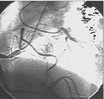
Cardiac catheterization was performed according to the Judkins technique and showed the following: a domi-nant right coronary artery with an eccentric 90% lesion and a thrombus in its initial third, and an anterior descending coronary artery partially originating from the right coronary artery and partially from the trunk of the left coronary artery (fig. 1).
The anterior descending branch originating from the left coronary artery split after a short trajectory into a septal and a diagonal branch (fig. 2).
The portion of the anterior descending branch origi-nating from the right coronary artery had an anomalous tra-jectory, passing in front of the pulmonary artery towards the anterior interventricular sulcus. From then on, the anatomic trajectory of the anterior descending branch con-tinued, which was interrupted after the first diagonal branch from the left coronary trunk (fig. 3). Neither the anterior des-cending branch originating from the left coronary trunk nor the one originating from the right coronary artery had athe-rosclerotic lesions. No lesions were observed in the left co-ronary trunk or in the circumflex branch.
The left ventricular contraction was normal.
Right coronary angioplasty was proposed to the pa-tient and was uneventfully performed, with successful pri-mary stent implantation (figs. 4 and 5).
The patient had a good in-hospital evolution and was discharged from the hospital on the second day of hospita-lization.
Discussion
5 6 2
Siqueira et al
Dual anterior descending coronary artery
Arq Bras Cardiol 2003; 80: 561-3.
sinus of Valsalva, and congenital coronary arteryatresia or stenosis 2,8,9.
In human hearts, the anterior descending branch is the vessel with the most constant origin, course, and distribu-tion 5. In the case of an anomalous origin of the anterior
des-cending branch from the right sinus of Valsalva or from the right coronary artery, its trajectory may have 2 variants: to pass between the aorta and the pulmonary artery, or to be anterior to the pulmonary trunk before reaching the anterior interventricular sulcus 2.
In the case of a dual anterior descending coronary artery, 2 branches provide the usual distribution of that co-ronary artery as follows: 1) one branch, the short anterior descending branch, ends at the beginning of the anterior
Fig. 1 – Right coronary artery with an eccentric 90% lesion in its initial third.
Fig. 2 – Left anterior descending coronary artery partially originating from the right coronary artery with an anomalous trajectory and passing in front of the pulmonary artery towards the anterior interventricular sulcus.
Fig. 3 – Anterior descending coronary artery originating from the left coronary artery and dividing after a short trajectory into a septal and a diagonal branch.
Fig. 4 – Right coronary angioplasty with stent implantation.
The most common is the origin of the circumflex artery from the right sinus of Valsalva or from the right coronary artery 4.
Traditionally, these anomalies are classified into the 6 subgroups shown in table I 2.
Classically, no clinical consequences were attributed to those findings because the blood flow in the anomalous coronary artery is usually normal.
Arq Bras Cardiol 2003; 80: 561-3.
Siqueira et al Dual anterior descending coronary artery
5 6 3
Fig. 5 – Primary success of right coronary angioplasty.
interventricular sulcus; 2) a second longer branch has a va-riable course beside the anterior interventricular sulcus and returns to this sulcus distally 4.
This anomaly was classified into 4 types by Spindola-Franco et al 10. In types I, II, and III, the anterior descending
branch originates from the trunk of the major anterior des-cending coronary artery (the anterior desdes-cending coronary artery itself). The case reported fits type IV dual anterior descending coronary artery, which is a rare anomaly. In this type, the long anterior descending branch originates in the right sinus of Valsalva or in the right coronary artery.
The clinical influence of this anomaly is still controver-sial in the literature. It was initially considered benign 2;
however, some authors associate it with stable angina, acu-te myocardial infarction, and postinfarction angina. Mello et al 6 reported 2 cases with clinical, electric, and scintigraphic
evidence of myocardial ischemia, in which the anterior des-cending branch originated from the right sinus of Valsalva.
Oliveira et al 1 reported that the right side origin of the
ante-rior descending branch is a significant anomaly that may
cause myocardial ischemia. Coyle et al 7 correlated the
origin of the anterior descending branch in the right sinus of Valsalva or in the right coronary artery with acute myo-cardial infarction and postinfarction angina.
In the literature reviewed, only 2 cases of a dual left an-terior descending coronary artery associated with obstruc-tive coronary disease were found 3, ours being the third case
reported.
Therefore, anomaly of the anterior descending corona-ry artecorona-ry may be considered as a hypothesis in the differen-tial diagnosis of angina and ischemia of the anterior wall.
References
1. Oliveira SF, Ramires JA, Meneghetti JC, et al. Anomalias congênitas de artérias coronárias: possível causa de insuficiência coronariana. Arq Bras Cardiol 1988; 50: 285-91.
2. Douglas Jr. DS, Franch RH, King III SB. Coronary artery anomalies. In: King III SB, Douglas Jr. DS. Coronary Arteriography and Angioplasty. New York: McGraw Hill Text; (Chapter 3), 1985: 33-85.
3. Tutar E, Gulec S, Pamir G, et al. A case of type IV dual left anterior descending artery anomalous origin associated with the presence of coronary atherosclero-sis. J Invas Cardiol 1999; 11: 631-4.
4. Bastos LC, Ariê S, Martins JF, et al. Dupla origem do ramo descendente anterior – das coronárias esquerda e direita – associada a origem anômala à direita do ramo circunflexo. Arq Bras Cardiol 1996; 67: 407-9.
5. Ilia R, et al. Mid left anterior descending coronary artery originating from right coronary artery. Int J Cardiol 1991; 33: 162-5.
6. Mello SC, Carvalho VB, Godoy M, et al. Origem anômala da artéria descendente anterior em artéria coronária direita: relato de dois casos. Arq Bras Cardiol 1981; 37: 467.
7. Coyle L, Thomas WJ. Anomalous left anterior descending coronary artery: malig-nant hospital course of a not so benign anomaly. Catheter Cardiovasc Interv 2000; 51: 468-70.
8. Bittel JA, Levin DC. Coronary arteriography. In: Braunwald E. Heart Disease. 5th Ed. (Chapter 8), 1980: 240-72.
9. Levin DC, et al. Hemodynamically significant primary anomalies of the coronary arteries. Angiographic aspects. Circulation 1978; 58: 25-34.
10. Spindola-Franco H, Grose R, Salomon N. Dual left anterior descending coronary artery: angiographic description of important variants and surgical implications. Am Heart J 1983; 105: 445-55.
Table I – Coronary artery anomalies
I – Anomalous origin from the aorta A - High aortic origin;
B - Origin from the sinus of Valsalva or from the contralateral coronary artery:
1. Circumflex artery originating from the right sinus of Valsalva or from the right coronary artery
2. Left anterior descending artery originating from the right sinus of Valsalva or from the right coronary artery
3. Left coronary artery originating from the right sinus of Valsalva 4. Right coronary artery originating from the left sinus of Valsalva C - Single coronary artery
II - Anomalous origin from the pulmonary artery III - Coronary arteriovenous fistula
IV - Coronary artery atresia or hypoplasia V - Coronary artery aneurysm
VI - Variations in patients with congenital heart disease: A - Tetralogy of Fallot
B - Transposition of the great arteries
C - Pulmonary atresia with intact interventricular septum D - Univentricular heart with tricuspid atresia
E - Congenitally corrected transposition of the great arteries F - Truncus arteriosus
G - Right ventricular double outflow tract H - Bicuspid aortic valve
I - Supravalvular aortic stenosis and coarctation of the aorta J - Supravalvular mitral stenosis
K - Situs inversus