UNIVERSIDADE FEDERAL DO CEARÁ
FACULDADE DE FARMÁCIA, ODONTOLOGIA E ENFERMAGEM PROGRAMA DE PÓS-GRADUAÇÃO EM ODONTOLOGIA
EFEITO DO PLASMA DE BAIXA TEMPERATURA E PRESSÃO NO BIOFILME ORAL FORMADO SOBRE ESMALTE EM UM MODELO IN SITU
FORTALEZA 2016
KARLA SHANGELA DA SILVA ALVES
EFEITO DO PLASMA DE BAIXA TEMPERATURA E PRESSÃO NO BIOFILME ORAL FORMADO SOBRE ESMALTE EM UM MODELO IN SITU
Tese apresentada ao Programa de Pós -Graduação em Odontologia da Faculdade de Farmácia, Odontologia e Enfermagem da Universidade Federal do Ceará, como requisito parcial para obtenção do título de Doutor em Odontologia. Área de concentração: Clínica Odontológica.
Orientadora: Profa. Dra. Iriana Carla Junqueira Zanin dos Santos.
KARLA SHANGELA DA SILVA ALVES
EFEITO DO PLASMA DE BAIXA TEMPERATURA E PRESSÃO NO BIOFILME ORAL FORMADO SOBRE ESMALTE EM UM MODELO IN SITU
Tese apresentada ao Programa de Pós-Graduação em Odontologia da Faculdade de Farmácia, Odontologia e Enfermagem da Universidade Federal do Ceará, como requisito parcial para obtenção do título de Doutor em Odontologia.
Aprovada em ___/___/___
BANCA EXAMINADORA
_______________________________________________________________________ Profa. Dra. Iriana Carla Junqueira Zanin dos Santos. (Orientadora)
Universidade Federal do Ceará (UFC)
___________________________________________________________________ Profa. Dra. Lidiany Karla Azevedo Rodrigues
Universidade Federal do Ceará (UFC)
___________________________________________________________________ Prof. Dr. Francisco Cesar Barroso Barbosa
Universidade Federal do Ceará (UFC)
___________________________________________________________________ Profa. Dra. Thereza Cristina Farias Botelho Dantas
Faculdade São Leopoldo Mandic
__________________________________________________________________ Profa. Dra. Juliana Paiva Marques Lima Rolim
AGRADECIMENTOS
À Universidade Federal do Ceará, na pessoa do seu Magnífico Reitor Prof. Dr. Herry de Holnada Campos; à Faculdade de Farmácia, Odontologia e Enfermagem, na pessoa da sua diretora Profa. Dra. Lidiany Karla Azevedo; ao Curso de Odontologia, na pessoa do seu coordenador Prof. Dr. Juliano Santori Mendonça; ao Programa de Pós -Graduação em Odontologia na pessoa do seu coordenador Prof. Dr. Vicente de Paulo Aragão Sabóia, pela participação desta conceituada instituição na minha formação científica, profissional e pessoal.
À CAPES, pelo apoio financeiro.
À minha querida Orientadora Profa. Dra. Iriana Carla Junqueira Zanin dos Santos, pelo incentivo, sabedoria, confiança e exemplo de competência. Obrigada por acreditar em mim no momento que mais precisei, levarei seu exemplo de orientadora, de pessoa e de profissional para o resto da minha vida.
À Profa. Dra. Lidiany Karla Azevedo Rodrigues, a esta devo a confiança em minha capacidade como pesquisadora, além da paciência e da tranquilidade para transmitir os seus ricos ensinamentos, contribuindo para minha formação profissional.
À Profa. Dra. Simone Duarte, o meu reconhecido agradecimento pelos conhecimentos transmitidos e apoio na execução experimental, sem os quais não seria possível a realização deste trabalho. Agradeço, sobretudo, o privilégio de haver trabalhado em um tema para o qual tanto vem contribuindo.
Ao professor Alejandro Pedro Ayala e à Central Analítica da UFC, pelo apoio experimental.
Aos professores do curso de Pós-Graduação em Odontologia pelos esforços para o crescimento e a melhoria do doutorado.
Aos funcionários da pós-graduação, Lucia Ribeiro Maques Lustosa, Janaíne Marques Leal, pela constante disponibilidade.
À Ramille de Araújo Lima, pelo trabalho dedicado, sempre à disposição, não poupando esforços para realização desse trabalho.
Ao Victor Pinheiro Feitosa, pela contribuição experimental e sempre à disposição auxiliando com minhas dúvidas.
Aos amigos do grupo de pesquisa: Talyta Soares, Tayara Marques, Paula Ventura e Daniela Nunes, pelas ricas trocas de experiências que tornamos possíveis durante esse percurso.
Aos amigos do Laboratório, em especial Weslanny Moraes, Marcerlo Sidou e David Queiroz, sempre companheiros, tiveram a paciência de me ensinar.
À Maria Silmara Alves de Santana, pelo auxilio experimental na espectroscopia Raman.
Aos Voluntários que participaram desta pesquisa, pois sem eles nenhuma destas páginas estaria completa.
Aos amigos Allysson Bonetti, Priscilla Rolim, Dávila Gadelha e Ranon Guilherme, por sempre estarem por perto me incentivando, durante esses últimos anos, e dando-me muitas provas de amor e de amizade.
Há muito mais a quem agradecer. A todos aqueles que, embora não nomeados, me brindaram com seus inestimáveis apoios, quero agradecer a todas as pessoas que se fizeram presentes e que se preocuparam que foram solidárias e que torceram por mim. De qualquer forma, todos os que realizam um trabalho de pesquisa sabem que não o fazem sozinhos.
O meu reconhecido e carinhoso muito obrigada!
O difícil não é iniciar uma caminhada, mas sim continuar. Ir em frente requer perseverança, persistência e humildade. Mas, semeando a boa semente, ainda que seja pela umidade das lágrimas ela germinará. Pode acontecer que os outros não valorizem os nossos trabalhos, as nossas lutas diárias. Mas os resultados dessas lutas nos fazem sentir que as experiências adquiridas na longa caminhada serviram para que acreditássemos em sonhos, mesmo sabendo que jamais deveríamos viver deles. Estas experiências, muitas vezes, nos fizeram chorar, mas jamais deixamos as lágrimas turvarem a nossa visão. Não importa também se, nesse esforço, tropeçamos e caímos, pois é aos que tombam na luta que se costuma chamar de heróis.
RESUMO
ABSTRACT
The Low temperature plasma is a promising technology that has being studied in dentistry for its various properties, among them, for their ability to destroy bacteria present in mature biofilm and also destroying the polymeric matrix of oral biofilms. The aim of this study was to evaluate the antimicrobial effect of argon plasma on oral biofilms formed in situ and also verify if this treatment damages the structure of the treated enamel. For that, twenty-two volunteers used palatine intraoral devices containing 6 to bovine enamel slabs that were dripped sucrose 10 times a day with 10% sucrose. The intraoral period was 7 days. At the 7th
day, enamel slabs were treated for 5 minutes with plasma, argon flow, 0.12% chlorhexidine, or 0.89% NaCl solution. Thus, biofilms samples were collected, weighed, serial diluted and plated in culture medium for the growth of total microorganisms, total streptococci, mutans streptococci and lactobacilli. In order to analyze whether the treatments damaged the enamel structure, the slabs were analyzed by scanning electron microscopy (SEM) and Raman spectroscopy. Low temperature plasma treatment showed a significant reduction in the viability of the total microorganisms (p <0.001), total streptococcus (p = 0.037) and mutans streptococci (p = 0.004). Argon flow also significantly reduced the mutans streptococci counts. The plasma treatment did not influence on the viability of the lactobacilli under the conditions tested (p = 0.497). SEM revealed smooth and homogeneous regions on the enamel surface, unobserved topographical differences between the glazes in biofilms subjected to different treatments. In Raman spectroscopy, we identified four major bands present in the inorganic phase: 324 (V1), 582 (V2), 960 (V3) e 1045 (V4) cm-1 and other 4 identified in the organic layer 1448 (V5), 1465 (V6), 1653 (V7) 2943(V8) cm-1. No statistical differences in
the Raman spectra were observed for enamel of different treatments. In conclusion, the plasma was effective in reducing viable bacteria present in mature oral biofilms produced in situ and not alter in the enamel surface treated biofilms.
LISTA DE FIGURAS
Figure 1 – Description of the treatments in which the enamel slabs were allocated. ... 26
Figure 2 – Experimental Design... 44
Figure 3 – Effects of the treatments PLA, AIR, SAL and CHX on viability ... 45
Figure 4 – SEM analysis respectively to PLA, AIR, CHX, SAL treatments at x5000 (1A, 2A, 3A and 4A) and x10000 (1B, 2B, 3B, and 4B) region desmineralised the enamel.. ... 46
Figure 5 – Raman band at v1=324 cm-1
characteristicof carbonate vibration, v2=582 cm-1
,v3=960 cm-1, v4= 1045 cm-1 characteristic of phosphate vibration, v5= 1448 cm -1
, v6= 1465 cm-1, v7= 1653 cm-1 and v8 = 2943 cm-1 phase organic. In (A) PLA; (B) AIR; (C) SAL; (D) CHX treatments. ... 47 Figure 6 – Raman spectra of treatments plotted together ... 48
LISTA DE TABELAS
LISTA DE ABREVIATURAS E SIGLAS
AIR Argon flow
Caae Certificado de apresentação para apreciação ética
CAPES Coordenação de Aperfeiçoamento de Pessoal de Nível Superior CFU Colony-Forming Units
CHX Chlorhexidine cm Centímetros
DNA Ácido desoxirribonucleico F Flúor
FM Future-Tech KV Quilovolt
MEV Microscópio Eletrônico de Varredura min Minuto
mm Milímetro
MSA Mitis Salivarius Agar MSB Mitis Salivarius Bacitracina NaCl Cloreto de sódio
nm Namômentro PLA Plasma
SAL Salina solution SD Standard Deviation
SEM Scanning Electron Microscopy SMH Surface Microhardness
SMHC Surface Microhardness Change TTP Tissue-Tolerable Plasma
SUMÁRIO
1 INTRODUÇÃO ... 15
2 PROPOSIÇÃO ... 19
3 CAPÍTULO... 20
3.1 Capítulo 1 ... 20
Effect of tissue-tolerable plasma on oral biofilms formed in situ ... 20
4 CONCLUSÃO ... 49
REFERÊNCIAS ... 50
ANEXO A – APROVAÇÃO DO COMITÊ DE ÉTICA ... 56
1 INTRODUÇÃO
A grande maioria dos microrganismos na natureza encontra-se ligada às superfícies, onde crescem na forma de biofilmes. Os biofilmes bacterianos são formados quando microrganismos unicelulares se tornam irreversivelmente aderidos a uma superfície sólida e envolvida por uma matriz de polissacarídeos extracelulares, podendo haver a formação de biofilmes a partir de uma ou de múltiplas espécies bacterianas (MAH & O`TOOLE, 2001, p. 34). Tem sido observado que bactérias inseridas nos biofilmes passam a exibir características fenotípicas distintas, as quais resultam no aumento de sua resistência aos agentes antimicrobianos de modo que essas comunidades microbianas têm grande importância em ambientes clínicos, ambientais e industriais (O´TOOLE, 2016, p. 5).
A placa dentária é um biofilme dinâmico composto por um ecossistema microbiano formado por centenas de espécies que se acumulam sobre a superfície dos dentes (FILOCHE, WONG, SISSONS, 2010, p. 10). O biofilme oral é único entre os vários tipos de biofilmes (HUANG, LI, GREGORY, 2011, p. 436) por requerer glicoproteínas salivares para anexar ao substrato. É composto por estruturas tridimensionais complexas, formado por comunidades de multi-espécies microbianas sobre o tecido oral (HE et al., 2015, p. 70), incorporados em uma matriz de polissacarídeos extracelulares (EPS) (HOJO et al., 2009, p. 983; YANG et al., 2011, p. 74).
O primeiro passo na formação do biofilme oral é a ligação da película adquirida, uma fina camada de glicoproteínas de origem salivar ou bacteriana que se adere à superfície dentária. Logo em seguida, inicia-se a adesão bacteriana com as bactérias pioneiras. Actinomyces spp, Streptococcus spp, Haemophilus spp, Capnocytophaga spp, Veillonella spp, Neisseria são os principais gêneros de bactérias pioneiras a aderir à superfície do dente (DIGE et.al., 2009, p. 70; HUANG, LI, GREGORY, 2011, p. 437). Ao longo do tempo, a diversidade bacteriana do biofilme cresce (RICKARD et al., 2003, p. 96), as bactérias pioneiras fornecem locais de ligações específicos às bactérias subsequentes e promovem o desenvolvimento do biofilme que aumenta gradualmente em espessura e em complexidade com a maturação da placa (HUANG, LI, GREGORY, 2011, p. 437).
os antibióticos (WELIN-NEILANDS; SVENSÄTER, 2007, p. 5635; KARYGIANNI et al., 2014b, p. 7330). Ocorre, no entanto, que o uso frequente desses agentes no tratamento de doenças crônicas pode resultar no desenvolvimento de resistência por algumas espécies, o que se tornou um problema de saúde pública mundial (SMITH et al., 2013, p. 584).
Recentemente, grandes esforços têm sido colocados na pesquisa odontológica a fim de encontrar novos métodos para eliminar microrganismos patogênicos presentes nos biofilmes orais (KARYGIANNI et al., 2016, p. 1) que não resultem no surgimento de cepas resistentes. Neste contexto, surgem os plasmas de baixa temperatura (PBT) para o tratamento antimicrobiano, sendo uma tecnologia alternativa de grande potencial e uma ferramenta promissora em uma variedade de aplicações biomédicas, com particular importância para combater infecções (MAI-PROCHNOWA et al., 2014, p. 508) por inativar bactérias (ERMOLAEVA et al., 2011, p. 80), fungos (FRICKE et al., 2012, p. 438; KLAMPFL et al., 2012, p. 5077), esporos (VENEZIA et al., 2008, p. 43), parasitas (ERMOLAEVA et al., 2012 p. 793) e vírus (ALSHRAIEDEH et al., 2013, p. 1420).
Os plasmas, que podem ser definidos como o quarto estado da matéria, são formados a partir da ionização de gases como: argônio, hélio, ozônio ou gás oxigênio. É constituído por partículas em interação permanente que incluem fótons, elétrons, íons positivos e negativos, átomos, radicais livres e moléculas excitadas e não excitadas (MOREAU; ORANGE; FEUILLOLEY, 2008, p. 611). Em geral, existem dois tipos de plasma gasoso, o plasma térmico e o não térmico. Os plasmas térmicos são obtidos a alta pressão (≥105 Pa), precisam de uma potência maior para serem formados (até 50 MW), possuem elétrons e partículas pesadas à mesma temperatura, isto é, eles estão em equilíbrio térmico entre si. Os plasmas não térmicos ou de baixa temperatura são obtidos a baixas pressões e potências sendo caracterizados por uma temperatura muito elevada dos elétrons mais do que a do gás (temperatura macroscópica), enquanto os íons e os átomos neutros são obtidos a uma temperatura muito menor (normalmente temperatura ambiente) e consequentemente não apresentam um equilíbrio termodinâmico local (MAI-PROCHNOWA et al., 2014, p. 508; SCHOLTZ et al., 2015, p. 58). Um plasma é às vezes chamado de "quente" se ele está quase totalmente ionizado ou "frio" se apenas uma pequena fração (por exemplo, 1%) das moléculas do gás estão ionizadas (MOREAU; ORANGE; FEUILLOLEY, 2008, p. 612).
vivo (FLUHR et al., 2012, p. 130, PARTECKE et al., 2012, p. 1). Espécies reativas são geradas transitoriamente pelo PBT através de diversas vias de colisões e dissociações. Estas espécies reativas oxidantes têm fortes efeitos sobre as estruturas externas das células, seja um revestimento de esporos ou de uma membrana celular. As gorduras insaturadas na bicamada lipídica das membranas celulares são susceptíveis ao ataque por radicais de hidroxila, comprometendo a função da membrana e as proteínas em membranas. Os revestimentos dos esporos são susceptíveis ao dano oxidativo por ataque químico pelas espécies reativas. Uma vez que a membrana celular e os revestimentos dos esporos foram parcialmente degradados, as espécies reativas podem danificar o material genético e as moléculas dentro do microrganismo, levando a sua destruição (HOFFMANN; BERGANZA; ZHANG, 2013, p. 21; MAI-PROCHNOWA et al., 2014, p. 508; SCHOLTZ et al., 2015, p. 53).
Adicionalmente, os tratamentos com PBTs para várias aplicações biomédicas e médicas, incluindo cicatrização de feridas, não mostraram efeitos adversos e forneceram provas da segurança da tecnologia (FLUHR et al., 2012, p. 130). Pesquisas têm demonstrado que o tratamento com o plasma a baixa temperatura (PBT) pode matar bactérias orais (YAMAZAKI et al., 2011, p. 390; GORYNIA et al., 2013, p. 1; BLUMHAGEN et al., 2014, p. 89) e inibir completamente a formação de um biofilme rico em matriz (DUARTE et al., 2011, p. 1).
Vários autores têm estudado as possíveis utilidades do PBT no campo da odontologia: no clareamento dentário (LEE et al., 2010, p. 240; SUN et al., 2010, p. 1892; JIE et al., 2010, p. 3143; PARK et al., 2011, p. 170; SEOUL et al., 2013, p. 265), na esterilização de instrumental (RUPF et al., 2012, p. 126; KOBAN et al., 2011, p. 956; SU-JIN et al., 2013, p. 2; IDLIBI et al., 2013, p. 369) e na adesão das restaurações a dentina (RITTS et al., 2010, p. 510, HIRATA et al., 2016, p.215 ). Além disso, diversos autores testaram a ação antimicrobiana do plasma em biofilmes in vitro bacterianos de S. mutans (GOORE et al., 2006, p. 1317), B. cereus e G. Stearothermophilus (MORRIS et al., 2009, p. 55), L. acidophilus e S. mutans (BO et al., 2011, p. 48), P. Gingivalis (MAHASNEH et al., 2011, p. 191), S. mutans (SUN et al., 2011, p. 143702) e em canais radiculares (JIANG et al., 2009, p. 479; LU et al., 2009, p. 668; SHAUDINN et al., 2013, p. 1).
2 PROPOSIÇÃO
3 CAPÍTULO
Esta tese está baseada no Artigo 46 do Regimento Interno do Programa de Pós -Graduação em Odontologia da Universidade Federal do Ceará, que regulamenta o formato alternativo para dissertação de mestrado e tese de doutorado e permite a inserção de artigos científicos de autoria e coautoria do candidato. Por se tratar de pesquisa envolvendo seres humanos, ou parte deles, o projeto de pesquisa deste trabalho foi submetido à apreciação do Comitê de Ética em Pesquisa da Faculdade de Medicina da Universidade Federal do Ceará via Plataforma Brasil, tendo sido aprovado sob Caae - 40975514.0.0000.5054 (ANEXO A). Assim sendo, essa tese de doutorado é composta de um capitulo que contém um artigo que será submetido para publicação no periódico “Caries Research” (ANEXO B).
3.1 Capítulo 1
Title: Effect of Tissue-Tolerable Plasma on oral biofilms formed in situ.
Authors:
Alves KSS, Faculty of Pharmacy, Dentistry and Nursing, Federal University of Ceara, Fortaleza, Brazil
Alves RHP, Faculty of Pharmacy, Dentistry and Nursing, Federal University of Ceara, Fortaleza, Brazil
Lima RA, Faculty of Pharmacy, Dentistry and Nursing, Unichristus, Fortaleza, Brazil
Santana MSA, Department of Physics, Federal University of Ceara, Fortaleza, Brazil Rodrigues LKA, Faculty of Pharmacy, Dentistry and Nursing, Federal University of Ceara, Fortaleza, Brazil
Duarte S, Department of Basic Sciences, New York University College of Dentistry, New York, USA
Zanin ICJ#, Faculty of Dentistry, Federal University of Ceara, Sobral, Brazil
Short Title: Plasma treatment on in situ biofilms
Keywords: Dental plaque, Oralbiofilms, plasma gases, in situ study.
# Corresponding Authorμ
Iriana Carla Junqueira Zanin, DDs, Ms, PhD
College of Dentistry, Federal University of Ceara, Sobral, CE, Brazil. Coronel Estanislau Frota Street, s/n
Sobral – CE- Brazil Zip codeμ 62010-560
Phone/faxμ ++ 55 88 36132603 Mobileμ ++ 55 88 97159496
E-mailμ irianaz@yahoo.com.br, iriana.zanin@pq.cnpq.br
Declaration of Interests: There are no potential conflicts of interest identified in this
The Tissue-Tolerable Plasma - TTP is a promising technology that has being studied in 2
dentistry due to not only its ability to kill bacteria present in mature biofilm and but also to 3
destroy the polymeric matrix of oral biofilms. The aim of this study was to evaluate the 4
antimicrobial effect of argon TTP on oral biofilms formed in situ and also verify if this 5
treatment damages the structure of enamel under treated biofilm. For that, twenty-two 6
volunteers used palatine intraoral devices containing bovine enamel slabs that were 7
treated 10 times per day with a 10% sucrose. After a 7 day intraoral period, the biofilm 8
formed over the enamel slabs were treated for 5 minutes, as followsμ TTP, argon flow, 9
0.12% chlorhexidine, or 0.89% NaCl solution. Thus, biofilms samples were collected, 10
diluted in decimal series and plated in culture medium for the growth of total 11
microorganisms, total streptococci, mutans streptococci and lactobacilli. In order to 12
analyze whether the treatments damaged the enamel structure, the slabs were analyzed 13
by scanning electron microscopy (SEM) and Raman spectroscopy. Tissue tolerable 14
plasma treatment showed a significant reduction in the viability of the total microorganisms 15
(p <0.001), total streptococcus (p = 0.037) and mutans streptococci (p = 0.004). Argon flow 16
also significantly reduced the mutans streptococci counts. The plasma treatment did not 17
influence on the viability of the lactobacilli under the conditions tested (p = 0.497). SEM 18
revealed smooth and homogeneous regions on the surface of the treated enamel, 19
unobserved topographical differences between the enamel subjected to different 20
treatments. In Raman spectroscopy, we were identified four major bands present in the 21
inorganic phaseμ 324 (V1), 582 (V2), 960 (V3) e 1045 (V4) cm-1 and other 4 identified in 22
the organic layer1448 (V5), 1465 (V6), 1653 (V7) 2943(V8) cm-1. No statistical differences 23
in the Raman spectra were observed for enamel of different treatments. In conclusion, the 24
plasma was effective in reducing viable bacteria present in mature oral biofilms produced 25
in situ and it did not modify the morphology and composition the enamel. 26
INTRODUCTION 28
Biofilms models may help us to accurately predict, in a controlled and simplified 29
way, a clinical outcome which can lead us to preventive actions for diseases 30
[Featherstone, 1996]. One example of biofilm model is the in situ biofilm, which may be 31
formed on different substrates that are introduced inside the oral cavity into oral devices. 32
Several models, which enable the development of non-disturbed oral biofilm, are found in 33
literature. The first report was done by Ahrens et al. [1976] and they developed a model 34
based on acrylic splints on which enamel slides were positioned. Since then, this system 35
has been widely used with a variety of substrates including glass [Arweiler et al., 2004], 36
bovine enamel [Jentsch et al., 2002], bovine dentin [Zaura-Arite et al., 2001], human 37
enamel [Teixeira et al., 2012] and dentin [Lima et al., 2009] and hydroxyapatite discs 38
[Sreenivasan et al., 2009]. 39
Antimicrobial agents are used due its ability to avoid dental biofilm formation, but 40
biofilm species may exhibit several resistance mechanisms against them [Mah&O’Toole 41
2001]. In addition, the age and the structure of a biofilm may also restrict the penetration of 42
the antimicrobial agent and deeper cells in the biofilm could not be affected [Zanin et al., 43
2005]. Disruption of the oral microflora and the difficulty of maintaining therapeutic 44
concentrations of antimicrobials in the oral cavity are also problems associated with its 45
usage [Zaura-Arite et al., 2001]. Thus an antimicrobial adjuvant would be a valuable 46
alternative in treating oral infections. 47
Regarding these alternative adjuvants, Tissue-Tolerable Plasma - TTP, that 48
represents the fourth state of matter after solid, liquid, and gaseous [Morfill et al., 2009], 49
emerges as an alternative once it is a technology that inhibits biofilm formation with the 50
potential benefit of destroying oral biofilm matrix [Duarte et al., 2011]. Plasma is a partially 51
ionized gas generated by an electrical discharge, which creates a highly reactive 52
environment with ions, electrons, excited atoms and molecules, vacuum ultraviolet and 53
ultraviolet irradiation, free radicals and chemically reactive particles [Koban et al., 2010; 54
Zamperini et al., 2010]. The production of stable plasma at atmospheric pressure has 55
attracted attention for treating living cells and tissues without thermal damage [Brun et al., 56
2012]. It is also site specific, targeting only the infected area [Goree et al., 2006], and it 57
seems to preserve the material’s bulk properties [Zamperini et al., 2010]. In addition, 58
plasma is usually produced by low-toxicity gases and elaborates its activity by producing a 59
Therefore, this approach has been suggested as environmentally friendly with no harmful 61
residues [Alkawareek et al., 2012]. 62
TTP has recently been extensively studied by researchers as a possible therapy in 63
dentistry. Researchers have mostly investigated the antimicrobial effects produced by 64
plasma as a means to remove dental biofilms and eradicate oral pathogens. It has been 65
shown that antimicrobial effects the TTP are carried out by induction reactive, photons and 66
reactive particles, ions, and molecules [Hasse et al., 2015]. The efficacy of TTP against an 67
extended spectrum of fungi and bacteria, including S. mutans, has been shown in several 68
in vitro studies [Rupf et al., 2010; Matthes et al., 2013; Bender and Kramer, 2014; 69
Preissner et al., 2015], but no studies report results in multispecies biofilms. Furthermore, 70
it has been applied effectively in antimicrobial therapy in case reports and clinical studies 71
in animals and humans, especially in otorhinolaryngology and dermatology [Isbary et al., 72
2013; Klebes et al., 2015]. Strikingly, its efficacy seems to be irrespective of microbial 73
resistance patterns [Haertel et al., 2014; Daeschlein et al., 2015] and no negative side 74
effects to viable tissues have been described, despite slight warming and temporary 75
redness [Lademann et al., 2009; Fluhr et al., 2012]. In this way, TTP has been suggested 76
for different applications in dentistry [Kim et al., 2014]. 77
Therefore, plasma is emerging as a physical treatment with microbicidal potential on 78
bacteria, parasites, fungi, spores, and viruses [ Sladek, 2005; Fridman et al., 2007; Kolb et 79
al., 2008; Von Woedtke et al., 2008; Dobrynin et al, 2009; Rupf et al., 2010]. Also, TTP 80
may allow its application inside the mouth because of it is compatible with the mammalian 81
tissues, which encourages their use in vivo [Fluhr et al., 2012; Partecke et al., 2012; 82
Delben, 2016]. The first hypothesis to be tested in this study is that TTP have 83
antimicrobial effect on microorganisms present in biofilms formed in situ. Furthermore, the 84
hypothesis that plasma treatment will damage the enamel structure will be tested. 85
MATERIALS AND METHODS 87
Ethical Aspects 88
This study was approved by the Institutional Ethical Committee and all volunteers 89
gave informed consent (Sisnep Caae - 40975514.0.0000.5054). . 90
91
Experimental design 92
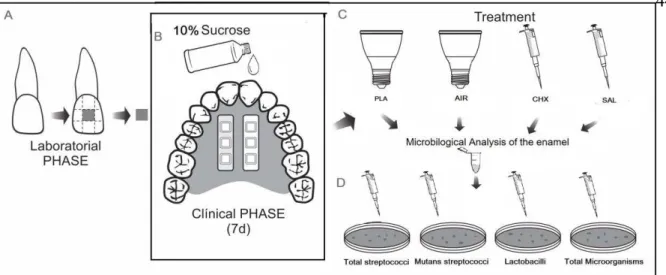
For this in situ experiment, a single-blind split mouth design was used in two intra-93
oral phases of 7 days (7d) each, in which 22 volunteers wore palatal devices containing six 94
bovine enamel slabs, positioned in pair of three. At the end of each intraoral phase, the 95
device was randomly split and each half (containing three enamel slabs) was allocated into 96
one of the following treatments: Plasma (PLA); Argon flow (AIR); Chlorhexidine 0.12% 97
(CHX) or Salina solution 0.89% (SAL) as described in Fig 1. In this way, at the end of the 98
two clinical phases, all volunteers were submitted to the four different treatments (Fig.2). 99
100
Figure 1 – Description of the treatments in which the enamel slabs were allocated. 101
Groups Treatment
PLA Plasma plume scanning enamel surface during 5 min AIR Argon gas flow scanning enamel surface during 5 min CHX A drop of 50 L on each slab during 5 min
SAL A drop of 50 L on each slab during 5 min 102
Tissue-Tolerable Plasma 103
The Tissue-Tolerable Plasma TTP source that was utilized in this study was 104
developed by the Leibniz Institute for Plasma Science and Technology (Neoplas Tools – 105
KINPen, Greifswald, Germany) and consists of a hand-held unit (length = 170 mm, 106
diameter = 20 mm, weight = 170g) connected to a high-frequency power supply (frequency 107
1.82MHz, 2–6 kV peak-to-peak, 8 W system power) for the generation of a plasma jet at 108
atmospheric pressure. The hand-held unit has a pin-type electrode (1 mm diameter) 109
surrounded by a 1.6 mm quartz capillary. An operating gas consisting of argon at a flow 110
rate of 5 standard liters per minute (slm) was used. The plasma plume emerging at the exit 111
nozzle is about 1.5 mm in diameter and extends into the surrounding air for a distance of 112
up to 10mm [Delben, 2016].The gas flow/TTP plume was targeted to all surface of enamel 113
slabs, and a surface scanning were performed manually by a single operator. 114
Specimen preparation 116
Bovine teeth were used to perform this in situ study. The teeth were stored in 0.01% 117
(v/v) thymol solution at 4°C for 30 days until be used [Cury et al., 2000]. Enamel slabs with 118
4 x 4 x 2 mm were obtained using a water-cooled diamond saw and a cutting machine 119
(IsoMet Low Speed Saw; Buehler, Lake Bluff, IL, USA). The adjustment of the enamel to 120
obtain flat plates was done with the aid of a low-speed polishing machine and 320 grit 121
paper (Carbimet Paper Discs; Bhuheler-met® II, USA), under water-cooling. Afterwards, 122
the specimens were polished using three different silicon carbide waterproof papers (320, 123
600, and 1,200-grit) as well as polishing cloths with 1µm diamond paste (Buehler®, lllinois, 124 USA). 125 126 Microhardness analysis 127
Surface Microhardness (SMH) was performed using a Knoop indenter with a 50g 128
load for 5s in a Future-Tech FM Microhardness Tester coupled to the FM-ARS software 129
(Future-Tech Corp., Tokyo, Japan). Initial SMH measurements were made in the center of 130
the enamel surface making one row of five indentations. For selection purposes, enamel 131
slabs with SMH values ranging from 272- 440 Knoop hardness number were selected in a 132
total of two-hundred and sixty-four slabs that were used in this experiment [Meredith et al., 133
1996]. After the intra-oral phase and treatments, SMH measurements were repeated 134
spaced at 100 µm on the side right of the five baseline indentations. Also, the percentage 135
of surface microhardness change (%SMC) was calculated (%SMC = SMH after treatment 136
– SMH baseline x 100/ SMH baseline). 137
138
In situ palatal devices 139
After baseline SMH, the slabs were autoclaved (121 ºC, 15 min) [Yamamoto et al., 140
2005] and stored in 100% humidity until being inserted into the palatal appliances. For 141
each subject, two acrylic palatal devices were prepared, in which two cavities (18 x 6 x 3 142
mm) were made on the left and right sides; and three slabs were attached with wax in 143
each cavity. In order to allow biofilm accumulation, and to protect it from mechanical 144
disturbance, a plastic mesh was positioned on the acrylic resin, leaving a 1 mm space 145
from the slab surface [Hara et al, 2003]. 146
147
In situ study Population 148
Twenty-two healthy volunteers (16 women and 6 men), aged from 19 to 34 years, 149
participants received oral and written instructions about the experimental design. The 151
inclusion criteria were normal salivary flow rate, normal buffering capacity of saliva and S. 152
mutans colony-forming units (CFUmg-1) in biofilms of at least 105 after 36 h of oral hygiene 153
suspension. Exclusion criteria included active caries lesions, use of antibiotics within the 154
past 3 months prior to the study, and the use of fixed or removable orthodontic devices. 155
156
In situ biofilm formation 157
During the lead-in period (7d) and throughout the clinical 2 phases (7d each), the 158
volunteers brushed their teeth with a fluoridated dentifrice [Sorriso Super Refrescante – a 159
calcium carbonate based dentifrice, 1,450 µg fluoride (F) g-1, as monofluorophosphate 160
[MFP, Colgate-Palmolive, São Paulo, SP, Brazil]. Also, the volunteers received oral and 161
written instructions to wear the appliances at all times, including at night. They were 162
allowed to remove the appliances only during meals, when consuming acid drinks, and 163
when performing oral hygiene. When removed, the devices were kept moist in plastic 164
boxes to keep the bacterial biofilm viable [Cury et al., 2000]. The cariogenic challenge was 165
provided by the volunteers who dripped a 10% sucrose solution onto all the enamel slabs, 166
10 times a day, according to a predetermined schedule (at 08:00, 09:30, 11:00, 12:30, 167
14:00, 15:30, 17:00, 18:30, 20:00, and 21: 30 h) [Duggal et al., 2001]. Before replacing the 168
palatal appliance in the mouth, a 5-min waiting time was standardized to allow diffusion of 169
the sucrose into the dental biofilm. Brushing with the dentifrice was performed three times 170
a day, after mealtimes when the volunteers habitually carried out their oral hygiene 171
procedures. The appliances were brushed extra-orally, except for the slab area, to avoid 172
disturbing the biofilm. All volunteers consumed fluoridated water (0.70 mg F 1-1), and no 173
restriction was made with regard to the volunteers’ dietary habits. 174
175
Treatment of in situ biofilms 176
Microbiological analyses of the oral biofilm were performed on the 7th day of the 177
experiment immediately after performing the treatments. The distribution of treatments on 178
the palatal device in each intra-oral phase was determined randomly by raffle. All 179
volunteers attended to laboratory in fasting and one drop of 10% sucrose was added to 180
each slabs before the device was removed from mouth. Thirty minutes after, the plastic 181
meshes of the devices were removed with a scalpel blade (#15C), the biofilm formed in 182
situ were exposed, and the treatments with PLA, AIR, CHX or SAL were performed. 183
Biofilms were then scraped carefully and weighed using microcentrifuge tubes, to which 184
sonicated at an output of 7W (Sonifier450D, São Bernado do Campo,SP, Brazil) using 3 186
pulses of 15 s with interval of 15 s of rest on ice. Afterwards, the suspension was serially 187
diluted (1:10, 1:100, 1:1.000, 1:10.000, 1:100.000, and 1:1.000.000) in 0.89% NaCl 188
solution. In order to assess microorganism viability, samples were plated in triplicate on 189
the following culture media: mitis salivarius agar (MSA), containing 15% sucrose, to 190
determine total streptococci; MSA agar plus 0.2 units of bacitracin ml-1 to determine 191
mutans streptococci [Gold et al., 1973], rogosa agar supplemented with 0.13% glacial 192
acetic acid to assess the number of lactobacilli [Rogosa et al., 1951] and blood agar to 193
determine total microorganisms. The plates were incubated for 48 h at 37°C in a partial 194
atmosphere of 5% CO2. Representative colonies of mutans streptococci, total streptococci, 195
lactobacilli, and total microorganisms were counted using a colony counter, and the results 196
were expressed as colony-forming units (CFU) mg-1 of biofilm. 197
198
Scanning Electron Microscopy (SEM) 199
For SEM, samples from bovine enamel slabs rinsed with distilled water, were 200
vacuum dried for 24h and then mounted on a SEM stub (aluminum discs), fixed with 201
double sided adhesive carbon tape, coded and mounted onto aluminium stubs with 202
Acheson silver DAG (Agar Scientific, U.K.) and then coated with a 15 nm thick layer of 203
gold, using a Polaron SEM coating unit. The specimens were then examined using an 204
SEM (Inspect™ S50, Jeol, Tokyo, Japan), operating at 20 KV and working distance 205
10mm. Images were taken at 2 levels of magnification (×5000 and ×10000) in order to 206
assess for changes in surface structure and captured using an specific software (EDS 207
Software for SEM, Oxford instruments).The images were analyzed visually. 208
209
Raman Spectroscopy 210
The Raman spectra were acquired on a Raman spectrometer (Xplora HORIBA, 211
Kyoto, Japan). For excitation of the samples it was used a HeNe laser operating at 632.8 212
nm wavelength. The enamel slab sample was placed onto a substrate, the laser beam was 213
focused on the sample surface using a microscope (OLYMPUS, Japan) with lens of 10x 214
and numerical aperture 0.75 forming a spot approximately 4 m over the surface of the 215
sample, and three points were chosen for Raman measurement with an exposure time of 216
30 s. The first point was located in the central region of the dental slab, and the others 217
were located to the right and to the left of this first point. The distance between the points 218
was 1 mm. The Raman system was calibrated with a silicon semiconductor using the 219
3600 cm−1 and allowed a characterization of both mineral content (hydroxyapatite) and 221
organic (essentially collagen) constituents. The curve was identified for each band using 222
an OriginPro 8.6 32 bit software system (Operating System: 7, Copyright 2012 by 223
OriginLab Corporation, Northampton:MA. USA). 224
225
Statistical analysis 226
The sample size was calculated by using data from an initial study, assuming on a 227
medium effect size of 0.5, significance level 5%, and statistical power 95%, it was found 228
that the study required 16 participants. Twenty- two participants were screened to ensure 229
that at least 16 subjects would successfully complete the study. Each participant wore an 230
upper removable appliance containing six enamel slabs. The study consisted of two 231
phases and each phase lasted for 7-days. Therefore, a total number of 264 enamel slabs 232
were required for the whole study. 233
The normality distribution of the data was checked using the Kolmogorov-Smirnov 234
statistical test. The mean and the standard deviation (SD) of the numbers of surviving 235
microorganisms for each treatment were calculated. Colony-forming units were 236
transformed into log 10 colony-forming units in order to reduce variance heterogeneity. To 237
determine the differences between test and control values, the unpaired one-way analysis 238
of variance followed by a Holm-Sidak test or non-parametric Kruskal-Wallis followed by a 239
Dunn's Multiple Comparison Test were used when appropriate. Paired- t test was used to 240
compare baseline and after intraoral phase SMH. Differences among the experimental 241
periods in relation to %SMC were tested by Kruskal-Wallis One Way Analysis of Variance 242
on Ranks. For Raman in order to normalize measurements and allow their comparison, 243
the band height parameter was used. The mean of the peaks height were compared using 244
test one-way analysis of variance (ANOVA). Significance level was set at 5% (p < 0.05) 245
using the software SigmaPlot 11.0 (La Jolla, CA, USA). 246 247 248 RESULTS 249 250 Microhardness analysis 251
The results for enamel surface microhardness before and after treatment and also 252
the percentage of microhardness change can be found in table 1. The comparison of 253
different treatments showed no statistical significant difference among them (p= 0.36). 254
Microbiological viability 256
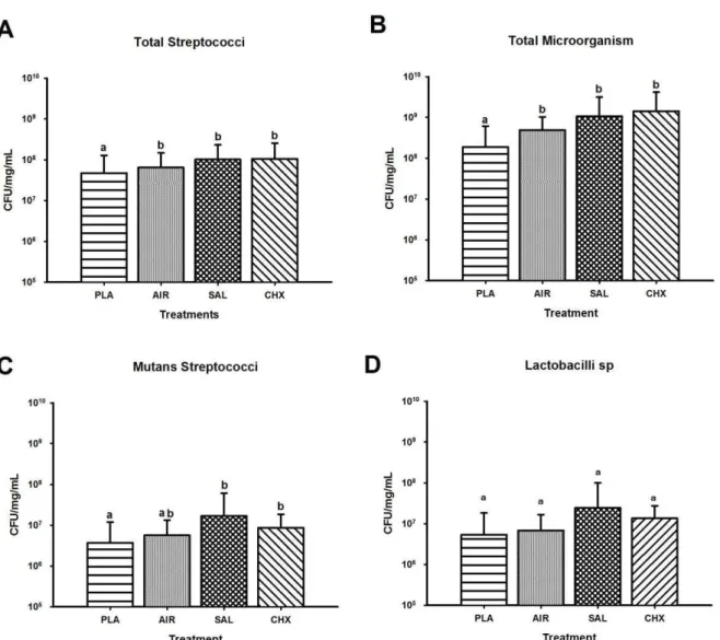
All volunteers showed normal patterns in the salivar tests with mean values of 1.56 257
ml/min salivary flow rate, normal buffering capacity (pH= 5.13) and 7.37 x 10⁶ CFU ml–1 of 258
mutans streptococci in biofilms formed after 36 h of oral hygiene suspension. The effect of 259
plasma treatment on viability of in situ biofilms can be found on Figure 3. Plasma treatment 260
showed a significant reduction for total microorganism (p<0.001), total streptococci (p= 261
0.037), and mutans streptococci (p= 0.004) counts. Mutans streptococci counts were also 262
reduced by argon gas treatment. Thus, plasma treatment did not demonstrate influence on 263
lactobacilli viability (p= 0.497). For the other tested microorganisms there were no 264
statistical difference among salina solution, argon gas and CHX treatments. The 265
percentage of viable mutans streptococci related to total streptococci was 7.86% in plasma 266
treatment, 10.23% in argon treatment, 15.34% in salina group and 8.10% in chlorhexidine 267
treatment. 268
269
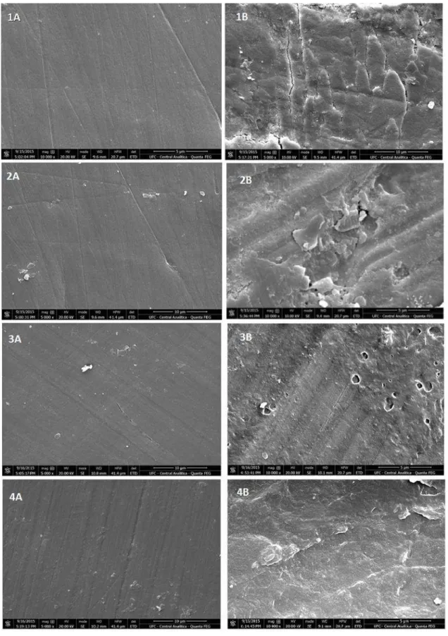
Scanning Electron Microscopy (SEM) 270
SEM analysis revealed smooth and homogenous surface topography of enamel at 271
both x5000 (Figure 4: 1A, 2A, 3A and 4A) and x10000 whereas in region enamel the 272
demineralized showed irregularities and stepped lamination appearance (Figure 4: 1B, 2B, 273
3B, and 4B) magnification. No difference was observed among the surfaces of different 274 treatment groups. 275 276 277 Raman Spectroscopy 278
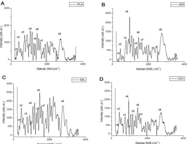
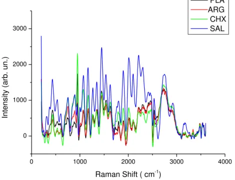
The average Raman spectra for the four treatment groups in the region of 200- 279
3600 cm-1 are shown in Fig. 5. All spectra were normalized based on the 960 cm-1 280
phosphate peak, which was the most intense peak. The region spanning from 240 to 1.045 281
cm-1 was characteristic of phosphate and carbonate groups and representative of the 282
mineral phase of enamel and the region spanning from 1448 to 3080 cm-1 was 283
characteristic of C-H bond stretching and amide bending vibrational modes from proteins 284
in the organic matrix of the enamel. 285
The band in the range of 1 (240-330 cm-1) represents the carbonate vibration, 286
bands at 2 (582 cm-1), 3 (960 cm-1), 4 (1045 cm-1), represent the phosphate vibrations 287
in hydroxyapatite, and bands v5 (1448 cm-1), v7 (1653 cm-1) amide bending vibrational 288
modes from proteins in the organic matrix of the enamel and v6 (1465cm-1), v8 (2943cm -289
In Fig. 06, Raman-spectra from enamel slabs with treatment plasma (in black), 291
argon flow (in red), 0.89% NaCl saline solution (in blue) and chlorhexidine (in green) are 292
shown with similar vibrational modes. Although in the peaks intensity of the SAL group 293
was higher than the other groups, no statistically significant differences were detected in 294
the peak intensity observed for all bands in all tested groups (p= 0.123). 295
296
DISCUSSION 297
The main finding of the present study is that Tissue-Tolerable Plasma was effective 298
against mature oral biofilm formed in situ, being more effective in reduce bacterial viability 299
than chlorhexidine, a well-known antimicrobial substance. Additionally, this is the first in 300
situ study evaluating the antimicrobial effectiveness of Tissue-Tolerable Plasma treatment 301
on oral biofilms. The oral biofilm containing pathogenic bacteria communities is one of the 302
major factor associated with dental caries [Guo et al., 2015]. Also, microorganisms 303
growing in biofilms may be up to 1000 times more resistant to antimicrobial agents than 304
their planktonic counterparts [Costerton et al., 1999; de Melo et al., 2013]. In this way, the 305
interest in new strategies to effectively inactivate pathogenic bacteria in oral biofilms has 306
emerged in the scientific community and the use of the Tissue-Tolerable Plasmas are one 307
of these new therapies because of its effectiveness against oral microorganisms [Gorynia 308
et al., 2013; Blumhagen et al., 2014; Sladek et al., 2014]. 309
This study compared the antimicrobial effect of several treatments on oral biofilms. 310
CHX solution is a golden standard to inactivate or prevent dental plaque formation 311
[Matthews, 2011] when compared to other chemical agents used in dentistry [Filoche et 312
al., 2005]. However, a 0.1% CHX solution seems to be inefficient against mature oral 313
biofilms [Vitkov et al., 2005]. The main advantage of using CHX is its wide antimicrobial 314
spectrum, acting on both Gram-positive and Gram-negative microorganisms and its 315
prolonged and continuous effect even in the presence of blood and other body fluids 316
[Rosenthal et al., 2004]. However, the prolonged use of CHX can cause mucous peeling, 317
stains on the teeth, alterations in the sense of taste, compromising of the wounds healing 318
and reduction of fibroblast adhesion to radicular surfaces [Zheng and Wang, 2011]. Thus a 319
potential antimicrobial adjuvant alternative to CHX with less side-effect would be of great 320
value in dentistry. In our study, plasma treatment showed better results than CHX in 321
by the Koban et al. [2010] in vitro using TTP treatment against oral biofilms formed on 323
titanium discs. 324
In our study, complex in situ multispecies biofilms were significantly affected by TTP 325
after 5 min treatment. Although we did not investigate the mechanism of action of TTP on 326
bacterial species, there are many possible mechanisms that may explain its bactericidal 327
activity, such as electron bombardment of cell membranes, charge accumulation, chemical 328
reactions such as oxidation, destruction of nucleic acids by ultraviolet radiation, and 329
ablation of the cell membrane [Laroussi, 2005]. In all cases, the site of action for the 330
plasma deactivating mechanisms is either at the cell membrane or the DNA. Argon plasma 331
acts via a complicated mechanism that includes a synergetic action of reactive species of 332
different kinds, including ionized argon gas molecules [Shimizu et al., 2008]. TTP reactive 333
particles produce a general mechanical effect on the surface of living organisms, which 334
has been called etching [Lerouge et al., 2000a; Moisan et al., 2001; Moreau et al., 2008]. 335
Etching is due to the reaction of highly reactive gas radicals with organic materials, 336
generating products that disrupt membrane surface. It may cause perforations in the 337
membranes of microorganisms, which, in turn, remove the obstacle to secondary reactive 338
species that might be formed in the medium. The effectiveness of etching depends on 339
plasma composition [Lerouge et al., 2000b]. Park et al. [2014], related that it is possible 340
that several components work together to produce a synergistic effect rather than a single 341
component contributing to the sterilization. TTPs may produce a large amount of reactive 342
oxygen species (ROS) when it passes through air, and in particular, high levels of OH are 343
generated when plasma reacts with water or tissue fluid. It is well known that OH 344
effectively kills bacteria. Furthermore, the half-life of plasma-generated ROS is very short, 345
and hence, its retention in the oral cavity is short and less likely to induce harmful effects 346
on tissues. 347
The TTP treatment was more effective to reduce total microorganisms than 348
chlorhexidine treatment. Ermolaeva et al. [2011] suggested that, in general, TTP argon 349
plasma is more effective against Gram-negative than Gram-positive bacteria because of 350
the differences in cell-wall structure. In our study, we did not test plasma treatment in 351
gram-negative bacteria disconnected of other microorganism, but our oral biofilms contain 352
both gram-negative and positive bacteria and the treatment was effective in reduce them. 353
So, further experiments are needed to evaluate the effectiveness of plasma treatment on 354
As observed to total microorganisms, the counts of total streptococci after TTP 356
treatment was inferior than observed to chlorhexidine. The degree of inhibition is probably 357
related to the thickness of the biofilm once thicker biofilm, such as those formed in the 358
presence of sucrose [Roberts et al. 2002], would present a greater challenge for the 359
penetration of plasma free radicals [Sladek and Stoffels, 2005; Vleugels et al. 2005]. This 360
could also inhibit the antibacterial activity of the TTP, killing cells only at the surface of 361
biofilms and inducing a sublethal response to cells attached to the substrate [Sladek and 362
Stoffels 2005]. Also, the partial antimicrobial effect achieved in our study may be explained 363
by characteristics of different bacteria present in biofilms that turn them more resistant to 364
antimicrobial treatments. In this way, Gorynia et al., [2013] tested argon plasma in S. 365
sanguinis biofilms and found that TTP was not effective on this bacteria which may be 366
attributed to its fast metabolic ratio and also to its ability of attaches to surfaces 10 to 100 367
times stronger than S. mutans, S. mitis and S. salivarius, generating more stable biofilms 368
[Rosan et al., 1982]. 369
Despite the majority of the studies test TTP treatment against planktonic cultures, 370
Goree et al. [2006] investigated the use of a plasma needle in mutans streptococci 371
colonies and observed that TTP was able to kill bacteria after 10 seconds of treatment. 372
They concluded the plasma could provide an attractive alternative treatment for dental 373
clinic. Yang et al. [2011] and Blumhagen et al. [2014] used the TTP argon plasma against 374
mutans streptococci and Lactobacillus acidophilus and observed that TTP was effective in 375
reducing the number of S. mutans after 11-15 sec and that results were dependent of the 376
bacterial supporting media. Similar results were also observed on monospecies biofilms 377
with different types of plasma source such as helium and oxigen gas [Sladek et al., 2004; 378
Yamazaki, 2011]. Also, the percentage of viable cariogenic mutans streptococci related to 379
total streptococci was almost half in TTP treatment compared to negative control. 380
In our study there was not observed statistically significant reductions on viability of 381
Lactobacilli sp among the tested treatments. Yang et al., [2011] found that longer plasma 382
treatment was required for deactivating L. acidophilus than used to kill S. mutans. The 383
sterilization mechanisms of TTP indicate that bacterial sizes and structure would affect the 384
plasma effectiveness and efficiency in bacterial deactivation. The bigger cell size of L. 385
acidophilus (~ 1 × 3 m in diameter) would receive less free radicals per unit of cell making 386
than harder to kill than the smaller S. mutans (~ 1 m in diameter). In order to achieve the 387
several minutes of exposure time were needed to kill L. acdophilus, while only tens of 389
seconds of exposure time was needed for S. mutans. 390
In the present study, enamel demineralization was observed after 7 days of plaque 391
accumulation at high frequency of exposure to sugar (Table 1). It was observed a 392
reduction in superficial enamel microhardness (SMH) due to the cariogenic challenger to 393
which enamel blocks were submitted. The SMH is considered a very sensitive method of 394
evaluating early caries lesion [Zero, 1995]. Tenuta et al. (2003) suggest that it takes 395
around 3-4 days for dental plaque to manifest its cariogenicity on dental substrate but this 396
period could be shorter for bovine enamel [Featherstone and Mellberg, 1981]., Thus, the 397
mineral loss found in our study is similar to those founded for human enamel by other 398
authors [Park et al., 2004]. Also, the treatment with argon plasma, argon flow, 0.12 % 399
chlorhexidine, and 0.89% NaCl solution had no effect on SMC%, suggesting that the 400
plasma treatment do not interfere in enamel hardness under tested conditions. These 401
results indicated that plasma treatment did not induce damage in the enamel surface 402
despite enamels slabs have suffered cariogenic challenge. Chen et al. [2013] also did not 403
observed surface modification of several dental substrates submitted to by Tissue-404
Tolerable Plasma treatment. 405
The raman spectroscopy allows a molecular analysis of mineralized dental tissues. 406
The information is provided as curves representing the intensity of the signal according to 407
the frequency and mathematical analyses may be performed in order to provide 408
comparative and quantitative analysis [Tramini et al., 2000]. To our knowledge, this is the 409
first study that has characterized and compared the molecular structure of the enamel 410
submitted to the plasma treatment. The reading of the slab in three different points allows 411
us to compare statistically all the treatments, however, no significant difference in mean 412
intensity of the peaks. 413
In conclusion, our results demonstrate that the argon TTP under tested conditions 414
was effective in reducing the viability of mutans streptococci, total streptococci and total 415
microorganism, without damage the surface of enamel. 416
417
418
This project received financial support from CAPES 88881.062159̷ 2014-01 PVE̷ CAPES. 420
We thank the volunteers for their valuable participation. The first author received a 421
scholarship during this study from CAPES. 422
423
REFERENCES 425
Ahrens G. Effect of fluoride tablets on uptake and loss of fluoride in superficial enamel in 426
vivo. Caries Res. 1976; 10:85-95 427
Alkawareek MY, Algwari QT, Gorman SP, Graham WG, O’Connell D, Gilmore BF. 428
Application of atmospheric pressure nonthermal plasma for the in vitro eradication of 429
bacterial biofilms. FEMS Immunol Med Microbiol. 2012;65(2):381–4. 430
Arweiler NB, Hellwig E, Sculean A, Hein N, Auschill TM. Individual vitality pattern of in situ 431
dental biofilms at different locations in the oral cavity. Caries Res. 2004;38:442-7. 432
Auschill TM, Arweiler NB, Netuschil L, Brecx M, Reich E, Sculean A. Spatial distribution of 433
vital and dead microorganisms in dental bio -films. Arch Oral Biol. 2001;46:471-6. 434
Bender C, Kramer A. Efficacy of Tissue Tolerable Plasma(TTP) against Ixodes ricinus. 435
GMS Hyg Infect Control 9(1), 2014. 436
Benelli EM, Serra MC, Rodrigues AL Jr, Cury JA. In situ anticariogenic potential of glass 437
ionomer cement. Caries Res 1993; 27: 280–284. 438
Bin, L.; Goree, J.; Drake, D; Stoffels, E. Killing of S. mutans bacteria using a plasma 439
needle at atmospheric pressure. IEEE Trans Plasma Sci 34, 1317–1324, 2006. 440
Blumhagen A, Singh P, Mustapha A, Chen M, Wang Y, and Yu Q, Plasma Deactivation 441
of Oral Bacteria Seeded on Hydroxyapatite Disks as Tooth Enamel Analogue. Am J Dent. 442
2014 April ; 27(2): 84–90. 443
Brun P, Brun P, Vono M, Venier P, Tarricone E, Deligianni V, Martines E, Zuin M, 444
Spagnolo S, Cavazzana R, Cardin R, Castagliuolo I, Valerio Al, Leonardi A. Disinfection of 445
ocular cells and tissues by atmospheric-pressure cold plasma. PloS One. 446
2012;7(3):e33245. 447
Ccahuana-Vásquez RA, Tabchoury CP, Tenuta LM, Del Bel Cury AA, Vale GC, Cury JA. 448
Effect of frequency of sucrose exposure on dental biofilm composition and enamel 449
demineralization in the presence of fluoride. Caries Res. 2007;41(1):9-15. 450
Costerton JW, Stewart PS, Greenberg EP. Bacterial biofilms a common cause of 451
persistent infections. Science. 1999;284:1318–22. 452
Cury JA, Rebello MA, Del Bel Cury AA. In situ relationship between sucrose exposure and 453
the composition of dental plaque. Caries Res 1997; 31: 356–360. 454
Cury, J. A., Rebelo, M. A., Del Bel Cury, A. A., Derbyshire, M. T., Tabchoury, C. 455
P.Biochemical composition and cariogenicity of dental plaque formed in the presence of 456
sucrose or glucose and fructose. Caries Res, 2000; 34, 491-7. 457
Daeschlein G, Napp M, Lutze S, Arnold A, von Podewils S, Guembel D, Junger M (2015) 458
Skin and wound decontamination of multidrug-resistant bacteria by cold atmospheric 459
plasma coagulation. J Dtsch Dermatol Ges. 2015 13(2):143–150 460
Delben, J.A., MurataM R.M, Wei, X., Castro, M.L,. Assunção,W.G., da Silva,N.R.F.A., 461
Duarte S. Low-Temperature Plasma: An Effective Approach Against Candida albicans 462
De Melo WC, Avci P, de Oliveira MN, Gupta A, Vecchio D, Sadasivam M, Chandran R, 464
Huang YY, Yin R, Perussi LR, Tegos GP, Perussi JR, Dai T, Hamblin MR. Photodynamic 465
inactivation of biofilm: taking a lightly colored approach to stubborn infection. Expert Rev 466
Anti Infect Ther. 2013 Jul;11(7):669-93. 467
Dobrynin D, Fridman G, Friedman G, Fridman A. Physical and biological mechanisms of 468
direct plasma interaction with living tissue. New J Phys. 2009;11:115020. 469
Duarte, S., Kuo, S. P., Murata, R. M., Chen, C. Y., Saxena, D., Huang, K. J. & Popovic, S. 470
Air plasma effect on dental disinfection. Physics of Plasmas, 2011. 18. 471
Duggal MS, Toumba KJ, Amaechi BT, Kowash MB, Higham SM. Enamel demineralization 472
in situ with various frequencies of carbohydrate consumption with and without fluoride 473
toothpaste. J Dent Res. 2001 Aug;80(8):1721-4. 474
Ermolaeva SA, Varfolomeev AF, Chernukha MY, Yurov DS, Vasiliev MM, Kaminskaya 475
AA, Moisenovich MM , Romanova JM, Murashev AN, SeleznevaII, Shimizu T, 476
Sysolyatina EV,. Shaginyan IA, Petrov OF, Mayevsky EI, Fortov VE. Morfill GE, 477
Naroditsky BS and Gintsburg AL. Bactericidal effects of non-thermal argon plasma in vitro, 478
in biofilms and in the animal model of infected wounds. Journal of Medical Microbiology. 479
2011; 60, 75–83. 480
Featherstone JDB, Mellberg JR. Relative rates of progress of artificial carious lesions in 481
bovine, ovine and human enamel. Caries Res 1981;15:109-14. 482
Featherstone JD. Modeling the caries-inhibitory effects ofdental materials. Dent 483
Mater 1996; 12: 194, 7. 484
Filoche SK, Soma K, Sissons CH. Antimicrobial effects of essencial oils in combination 485
with chlorexidine digluconate. Oral Microbiol Immunol. 2005;20(Suppl 4):221–225. 486
Fluhr JW, Sassning S, Lademann O, Darvin ME, Schanzer S, Kramer A, Richter H, Sterry 487
W, Lademann J. In vivo skin treatment with tissue-tolerable plasma influences skin 488
physiology and antioxidant profile in human stratum corneum. Exp Dermatol. 2012 489
Feb;21(2):130-4. 490
Fridman, G., Brooks, A. D., Balasubramanian, M., Fridman, A., Gutsol, A., Vasilets, V. N., 491
Ayan, H.,Friedman, G. Comparison of direct and indirect effects of non-thermal 492
atmospheric-pressure plasma on bacteria. Plasma Processes and Polymers. 2007; 4, 493
370–375. 494
Gilmour ASM, Edmunds DH, Newcombe RG. Prevalence and depth of artificial caries-like 495
lesions adjacent to cavities prepared in roots and restored with a glass ionomer or a 496
dentinbonded composite material. J Dent Res 1997; 16: 1854–1861. 497
Gold OG, Jordan HV, Van Houte J. A selective medium for Streptococcus mutans. Arch 498
Oral Biol 1973; 18: 1357–1364. 499
Goree J, Liu B, Drake D, Stoffels E: Killing of S-mutans bacteria using a plasma needle at 500
atmospheric pressure. IEEE Transactions on Plasma Science 2006, 34:1317–1324. 501
Gorynia S, Koban I, Matthes R, Welk A, Gorynia S, Hübne NO, Kocher T, Kramer A. In 502
vitro efficacy of cold atmospheric pressure plasma on S. sanguinis biofilms in comparison 503
Haertel B, von Woedtke T, Weltmann KD, Lindequist U.Non-thermal atmospheric-pressure 505
plasma possible application in wound healing. Biomol Ther (Seoul). 2014. 22(6):477 –490. 506
Hasse S, Duong Tran T, Hahn O, Kindler S, Metelmann HR, von Woedtke T, Masur K. 507
Induction of proliferation of basal epidermal keratinocytes by cold atmospheric-pressure 508
plasma. Clin Exp Dermatol. 2015:13(2):83-92.,. 509
Hara AT, Queiroz CS, Paes Leme AF, Serra MC, Cury JA. Caries progression and 510
inhibition in human and bovine root dentine in situ. Caries Res 2003; 37: 339–344. 511
Hirata R, Sampaio C, Machado LS, Coelho PG, Thompson VP, Durte S, Ayres APA, 512
Giannini M.Short- and Long-term Evaluation of Dentin-Resin Interfaces Formed by Etch-513
and-Rinse Adhesives on Plasma-treated Dentin.J Adhes Dent 2016; 18: 215–222 514
Isbary G, Morfill G, Schmidt HU, Georgi M, Ramrath K, Heinlin J, Karrer S, Landthaler M, 515
Shimizu T, Steffes B, Bunk W, Monetti R, Zimmermann JL, Pompl R, Stolz W. A first 516
prospective randomized controlled trial to decrease bacterial load using cold atmospheric 517
argon plasma on chronic wounds in patients. Br J Dermatol 2010 163(1):78–82. 518
Isbary G, Shimizu T, Zimmermann JL, Thomas HM, Morfill GE, Stolz W. Cold atmospheric 519
plasma for local infection control and subsequent pain reduction in a patient with chronic 520
post-operative ear infection. New Microbes New Infect. 2013 1(3):41–43. 521
Jentsch H, Hombach A, Beetke E, Jonas L. Quantitative transmission electron microscopic 522
study of dental plaque--an in vivo study with different mouthrinses. Ultrastruct Pathol. 523
2002;26:309-13. 524
Kim JH, Lee MA, Han GJ, Cho BH. Plasma in dentistry: a review of basic concepts and 525
applications in dentistry. Acta OdontolScand. 2014, 72(1):1–12 526
Kolb, J. F., Mohamed, A. A. H., Price, R. O., Swanson,R. J., Bowman, A., Chiavarini, R. L., 527
Stacey, M. , Schoenbach, K. H. Cold atmospheric pressure air plasma jet for medical 528
applications. Applied Physics Letters. 2008; 92, 241501–241503. 529
Klebes M, Ulrich C, Kluschke F, Patzelt A, Vandersee S, Richter H, Bob A, von Hutten J, 530
Krediet JT, Kramer A, Lademann J, Lange-Asschenfeld B. Combined antibacterial effects 531
of tissue- tolerable plasma and a modern conventional liquid antiseptic on chronic wound 532
treatment. J Biophotonics. 2015. 8(5):382–391. 533
Koban I, Matthes R, Hubner NO, Welk A, Meisel P, Holtfreter B, Sietmann R, Kindel E, 534
Weltmann KD, Kramer A, Kocher T. Treatment of Candida albicans biofilms with low-535
temperature plasma induced by dielectric barrier discharge and atmospheric pressure 536
plasma jet. New J Phys. 2010;12:073039. 537
Lademann J, Richter H, Alborova A, Humme D, Patzelt A, Kramer A, Weltmann KD, 538
Hartmann B, Ottomann C, Fluhr JW, Hinz P, Hubner G, Lademann O. Risk assessment of 539
the application Clin Oral Invest of a plasma jet in dermatology. J Biomed Opt. 2009. 540
14(5):054025. 541
Lademann J, Richter H, Schanzer S, Patzelt A, Thiede G, Kramer A, Weltmann KD, 542
tolerable plasma and an octenidine hydrochloride-based wound antiseptic on human skin. 544
Skin Pharmacol Physiol. 2012. 25(2):100–106 545
Laroussi M. Low temperature plasma-based deactivation: overview and state-of-the-art. 546
Plasma. Process Polym. 2005; 2:391–400. 547
Lee H, Park G, Seo Y, Im Y, Shim S, Lee H. Modeling of atmospheric plasmas for 548
biomedical applications. J Phys D: Appl Phys. 2011; 44:1–27. 549
Lerouge, S., Guignot, C., Tabrizian, M., Ferrier, D., Yagoubi, N. and Yahia, L. Plasma-550
based sterilization: effect on surface and bulk properties and hydrolytic stability of 551
reprocessed polyurethane electrophysiology catheters. J Biomed Mater Res. 2000a ; 52, 552
774–782. 553
Lerouge, S., Wertheimer, M. R., Marchand, R., Tabrizian, M. and Yahia, L. Effect of gas 554
composition on spore mortality and etching during low-pressure plasma sterilization. J 555
Biomed Mater Res. 2000b; 51, 128–135. 556
Mah Tf.; O'toole Ga. Mechanisms of biofilm resistance to antimicrobial agents. Trends 557
Microbiol.;9(1):34-9, 2001 558
Matthews D . No difference between 0.12 and 0.2% chlorhexidine mouthrinse on reduction 559
of gingivitis. Evid Based Dent. 2011. 12(1):8–9. 560
Matthes R, Bender C, Schluter R, Koban I, Bussiahn R, Reuter S, Lademann J, Weltmann 561
KD, Kramer A. Antimicrobial effi- cacy of two surface barrier discharges with air plasma 562
against in vitro biofilms. PLoS One. 2013. 8(7):e70462 563
Meredith N, Sherriff M, Setchell DJ, Swanson SA.Measurement of the microhardness and 564
Young's modulus of human enamel and dentine using an indentation technique.Arch Oral 565
Biol. 1996; 41(6):539-45. 566
Moisan, M., Barbeau, J., Moreau, S., Pelletier, J., Tabrizian, M. & Yahia, L. H. Low-567
temperature sterilization using gas plasmas: a review of the experiments and an analysis 568
of the inactivation mechanisms. Int J Pharm. 2001; 226, 1–21. 569
Morfill, G. E., Kong, M. G. & Zimmermann, J. L. Focus on plasma medicine. New Journal 570
of Physics 2009;11, 1150-1159. 571
Moreau, M., Orange, N. & Feuilloley, M. G. J. Nonthermal plasma technologies: new tools 572
for bio-decontamination. Biotechnol Adv. 2008; 26, 610–617. 573
Netuschil L, Reich E, Unteregger G, Sculean A, Brecx M. A pilot study of confocal laser 574
scanning microscopy for the assessment of undisturbed dental plaque vitality and 575
topography. Arch Oral Biol. 1998;43:277-85. 576
Park BG, Kim KS, Jung KH, Jung SB.Effect of atmospheric-pressure plasma treatment on 577
the adhesion characteristics of screen-printed Ag nanoparticles on polyimide. J Nanosci 578
Nanotechnol. 2014 Dec;14(12):9448-53. 579
Partecke LI, Evert K, Haugk J, Doering F, Normann L, Diedrich S, Weiss FU, Evert M, 580
Huebner NO, Guenther C, Heidecke CD, Kramer A, Bussiahn R, Weltmann KD, Pati O, 581
Bender C, von Bernstorff W. Tissue tolerable plasma (TTP) induces apoptosis in 582