ABSTRACT
http://dx.doi.org/10.1590/1678-775720150139
Acinar autolysis and mucous extravasation
in human sublingual glands: a microscopic
postmortem study
Luciana Reis AZEVEDO-ALANIS1, Elen de Souza TOLENTINO2, Gerson Francisco de ASSIS3, Tânia Mary CESTARI3,
Vanessa Soares LARA4, José Humberto DAMANTE4
1- Pontifícia Universidade Católica do Paraná, Escola de Saúde e Biociências, Programa de Graduação em Odontologia, Curitiba, PR, Brazil. 2- Universidade Estadual de Maringá, Departamento de Odontologia, Maringá, PR, Brazil.
3- Universidade de São Paulo, Faculdade de Odontologia de Bauru, Departamento de Ciências Biológicas, Bauru, SP, Brazil. 4- Universidade de São Paulo, Faculdade de Odontologia de Bauru, Departamento de Estomatologia, Bauru, SP, Brazil.
Corresponding address: Elen de Souza Tolentino - Av. Mandacaru, 1550 - Bloco S08 - Maringá - PR - Brazil - 87083-170 - Phone/Fax: +55 (44) 2101-9051 - e-mail: [email protected]
6XEPLWWHG$SULO0RGL¿FDWLRQ-XQH$FFHSWHG-XQH
A
lthough some morphological investigations on aged human sublingual glands (HSG) ound eventual phenomena identi ed as autolysis and mucous extravasation the exact meaning o these ndings has not been elucidated b ective: he aim o this or is to investigate whether acinar autolysis and mucous extravasation are related to the aging process in human sublingual glands. We also speculate if autolytic changes may assist forensic pathologists in determining time of death. Material and Methods: 186 cadavers’ glands were allocated to age groups: I (0–30 years); II (31–60), and III (61–90). Time and mode of death were also recorded. Acinar autolysis and mucous extravasation were classi ed as present or absent. Ultrastructural analysis was performed using transmission electron microscopy (TEM). Data were compared using Mann-Whitney U, Spearman’s correlation coef cient, rus al-Wallis, and Dunn tests (p 0.0 ). esults: There was correlation between age and acinar autolysis (r=0.38; p=0.0001). However, there was no correlation between autolysis and time of death. No differences were observed between genders. TEM showed mucous and serous cells presenting nuclear and membrane alterations and mucous cells were more susceptible to autolysis. Conclusion: Acinar autolysis occurred in all age groups and increased with age while mucous extravasation was rarely found. oth ndings are independent. Autolysis degrees in HSG could not be used to determine time of death.Ke yw or ds: Aging. Salivary glands. Histology. Autolysis. Forensic Dentistry.
I N TROD UCTI ON
Age-related microscopic changes have been reported for the major2,5,6,8,11,19 and minor15,18,21,22 salivary glands of humans2,5,8,14,15,18,19,21,22 and rats6,11. Main microscopic aspects described were: replacement of parenchyma by fat and connective tissue2,8,15,21, acinar atrophy and increase in the number of duct/duct-like structures2,6,8,15,19,21, focal and diffuse mononuclear infiltration2, oncocytosis2,8,15, and congested blood vessels2. In a previous study of age-related changes in human sublingual glands (HSG)2, in addition to the main microscopic aspects described, occasional
phenomena such as acinar autolysis and mucous extravasation were also observed.
autolysis. Postmortem autolytic changes have also been described in heart9,20, pancreas16,20, kidneys20, liver20, skeletal muscle20, temporal bones10, blood cells13, blood vessels12, sweat glands3, and heart muscle1.
Although some previous investigations on HSG found contingent phenomena identi ed as autolysis and/or mucous extravasation2,5,8,11, the exact meaning of these ndings has not been elucidated yet. Information is scarce concerning postmortem autolytic changes in the acini of SG, especially to distinguish these from pathological causes before death or others artifacts. According to some authors, postmortem autolysis depends on various factors and the most important is postmortem interval9,10,16,17. However, some of these postmortem autolytic changes are similar to alterations also described as apoptosis20 and inadequate/delayed fixation7,10,12. It explains why these occasional ndings can be misdiagnosed as xation failures. Additionally, it is speculated if these phenomena may be a result of surgical dissection trauma during glandular removal. It seems evident that these contingent microscopic alterations may theoretically represent a potential problem for pathologists and surgeons4. Therefore, the aim of the present study is to document age-related changes of HSG of cadavers with regard to acinar autolysis and mucous extravasation. Elucidation of these alterations could assist researchers and pathologists in recognizing the phenomena and discard pathological areas and other artifacts. Moreover, autolytic changes may assist forensic pathologists in determining time of death.
M ATERI AL AN D M ETH OD S
The Human Research Ethics Committee of the Bauru School of Dentistry – University of São Paulo (Process No. 059/2010) approved this study, which followed the guidelines of the Helsinki Declaration.
A total of 186 HSG were obtained bilaterally from 93 cadavers during necropsies at the São Paulo Death eri cation Service (School of Medicine, University of São Paulo) using the methods and the exclusion criteria of a previous study2. Glands were dissected intact via a wide neck ap. Following the inferior cortical of the body of the mandible, soft tissues were dissected and the lower edge of the sublingual gland was accessed. Dissection extended into the oor of the mouth, without perforating it. Geniohyoid and genioglossus muscles were released with scissors and the tongue and suprahyoid muscle were pulled to the opposite side with an Allis forceps. Then, the gland was completely visible, attached to the tongue muscles, and was dissected with blunt scissors, from its inferior edge to the
oor of the mouth, which was nally incised with a scalpel. Each piece carried a longitudinal strip of the oor of the mouth, approximately 1 cm wide. This strip worked as a reference for handling the sample. The same surgeon collected all glands and no differences or dif culties were found according to the postmortem interval. Glands with macroscopic autolysis were excluded2.
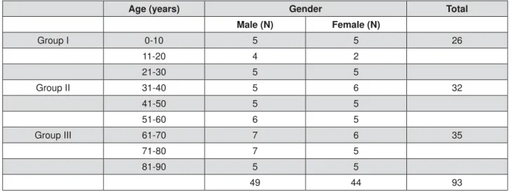
Cadavers (49 men/44 women) were divided into 9 decades of life, with age ranging from 7 months to 90 years. Considering that the present work is a continuation of a previous study2, the model of sample’s distribution is the same used before (Figure 1).
Approximately 5 cadavers of each gender per decade of life were studied. The sample was divided into 3 age groups: group I ( 30 years): 26 cadavers - 14 men (mean age: 14.8 years) and 12 women (mean age: 13.4 years); group II (31–60 years): 32 cadavers - 16 men (mean age: 47.0 years) and 16 women (mean age: 44.6 years); group III ( 61 years): 35 cadavers - 19 men (mean age: 74.5 years) and 16 women (mean age: 73.7 years). Data regarding cause of death were obtained from autopsy reports. According to internal rules of the
Age (years) Gender Total
Male (N) Female (N)
Group I 0-10 5 5 26
11-20 4 2
21-30 5 5
Group II 31-40 5 6 32
41-50 5 5
51-60 6 5
Group III 61-70 7 6 35
71-80 7 5
81-90 5 5
49 44 93
Death eri cation Service, no necropsies could be processed before 6 hours of arrival of the body. Interval between time of death and necropsy ranged from 6:05 to 92:55 h, with a mean of 16:46 h. The variable “time of death” was then considered for the purpose of elucidating the in uence of postmortem interval on morphological appearance, which could re ect the original physiological state before death.
After xation with phosphate-buffered formalin solution for 1 week at room temperature, specimens were cut into 5 mm thick slices. Regardless of the size of the gland, only 3 slices (anterior, middle, and posterior) were processed histologically. Alternate 5 μm thick sections were stained with hematoxylin and eosin (H.E.) and a single pathologist performed the microscopic examinations2.
Microscopic examination was performed in a light microscope Carl Zeiss MicroImaging GmbH (Carl Zeiss MicroImaging, Jena, Thuringia, Germany) using x5, x10, x40, and x100 sequential objectives, which resulted in original magni cations of x12.5, x25, x100, and x250, respectively. The whole microscopic slide was examined from the super cial portion of the gland section subjacent to the oral mucosa to the depth, and from anterior to posterior borders2.
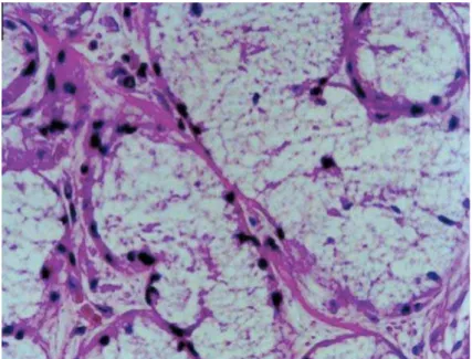
An acinus was counted as autolyzed when it presented the minimum sign of loss of cell limit, regardless of the degree11 (Figure 2A). Mucous extravasation was sparsely distributed in the glandular parenchyma (Figure 2B).
Acinar autolysis and mucous extravasation were occasional ndings classi ed as present or absent. When present, a degree of severity was attributed to them15. Microscopic ndings (sign of loss of cell limits and/or presence of mucous sparsely distributed in the glandular parenchyma) were classi ed as discrete when observed in up to 1/3 of the section, moderate when involving 1/3–2/3 of the section, and intense when more than 2/3 of the section was affected. Scores were attributed to those microscopic aspects, ranging from 0 to 3, with
0: absent, 1: discrete, 2: moderate, and 3: intense. As 3 initial scores were obtained for right gland and 3 initial scores for left gland, we had to obtain one intermediate score to represent the right gland and one intermediate score to represent the left one. Thus, the 3 initial scores of right gland were added, resulting in only one value (Intermediate score I), which varied from 0 (if all the initial scores were 0 - absent) to 9 (if the 3 initial scores were 3 - intense). This value was submitted to a conversion scale, so that, if it were 0, the intermediate score would be 0. If it were 1–3, the intermediate score would be 1; if it was 4–6, the intermediate score would be 2, and if it was 7–9, the intermediate score would be 3. The same steps were repeated for the left gland, and nally only two intermediate scores were obtained (Intermediate score II). When both of them were added, we obtained one value that varied from 0 (if both scores were absent) to 6 (if both scores were intense). Then, a second conversion scale was applied, in the same way as the latter, so that just one value ( nal score) could be obtained, representative of the degree of severity of each microscopic nding2. An example of this score’s conversion is illustrated in the scheme bellow.
Observer reproducibility was assured as follows: the slides of 24 HSG from 12 individuals were presented blindly and randomly to the same observer. After 2 months, microscopic analysis was re-initiated and the same slides were presented to the observer again. Kappa statistics measured observer reproducibility. Results obtained from each of the microscopic aspects were: acinar autolysis (K=0.72) and mucous extravasation (K=0.85).
One sublingual gland of a male cadaver (71 years) with advanced macroscopic autolysis was removed and processed for transmission electron microscopy (TEM). Guided by the light microscopy, the points of possible autolysis were captured for examination.
The correlation between acinar autolysis/
mucous extravasation and chronological age was carried out by Spearman’s correlation coef cient. The microscopic aspects that showed a medium and low correlation (r 0.60) and were signi cant were submitted to Kruskal-Wallis and Dunn tests (for comparison between age groups I, II, and III). The Mann-Whitney U test was used to analyze the relationship between microscopic aspects and gender. evel of signi cance was set at 5 for all tests and the software Pacotico v.0.5, Microsoft Visual FoxPro, was used.
RESULTS
An acinus was counted as autolyzed when it presented the minimum sign of loss of cell limit, regardless of the degree11. The loss of integrity of the external limits was observed particularly in the mucous acini (Figure 3).
Acinar autolysis was observed on nearly the
entire sample (Figure 3). The phenomenon was not found in only 10 cadavers (8 in Group I, 1 in Group II, and 1 in Group III) (Table 1). While, in Group I, 18 individuals (69.23%) presented acinar autolysis with degrees 0 or 1, in group III 26 individuals (74.29%) presented degrees 2 or 3. The correlation between age and the acinar autolysis was low, but signi cant (r=0.38; p=0.0001). There were signi cant difference between groups I and II, and between groups I and III (H=16.81; p=0.0002) (Table 1).
Figure 4 shows the acinar autolysis phenomenon distribution and the different degrees of severity in the groups. It is important to emphasize the absence of autolysis (degree 0) and the low degrees of severity in the young individuals.
TEM examination showed mucous cells presenting granulations and loss of membrane integrity, with their contents partially degraded, and absence of nucleus and organelles (Figure 5). In the serous
Figure 3-$FLQDUDXWRO\VLV/RVVRIDFLQDUFHOOOLPLWVDQGGLVSHUVHQXFOHL0DOH\HDUVROG+(2ULJLQDOPDJQL¿FDWLRQ
x400). H.E.=Hematoxylin & eosin
acini, secretion granules were partially preserved and cells still presented cytoplasmic organelles and nuclei integrity despite the loss of membrane integrity (Figure 6).
D e s p i t e t h e r e w e r e c a s e s o f m u c o u s extravasation, these phenomena were rarely found. They occurred mainly at degree 1 (Figure 2B), with the values dispersed in the different ages (Table 2). There was no signi cant correlation between age and mucous extravasation (p=0.6976).
Variables acinar autolysis and mucous
extravasation showed no signi cant differences between male and female genders (p>0.05). Considering the variable “time of death”, there was no signi cant correlation between autolysis and postmortem interval (p>0.05). Main causes of death included acute and chronic diseases, such as pulmonary edema, bronchopneumonia, acute myocardial infarction, cerebral vascular accident, ischemic heart disease, cerebral edema, and congestive heart failure.
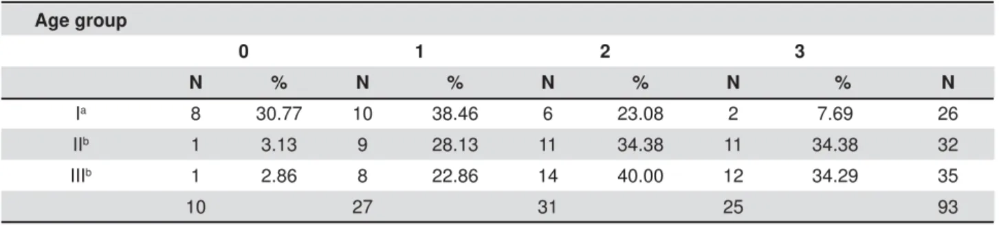
Age group
0 1 2 3
N % N % N % N % N
I 9 34.62 10 38.46 6 23.08 1 3.85 26
II 4 12.50 17 53.13 8 25.00 3 9.38 32
III 11 31.43 15 42.86 9 25.71 0 0.00 35
24 42 23 4 93
Table 2- Distribution of the different degrees of diffuse mucous extravasation according to the aging Age group
0 1 2 3
N % N % N % N % N
Ia 8 30.77 10 38.46 6 23.08 2 7.69 26
IIb 1 3.13 9 28.13 11 34.38 11 34.38 32
IIIb 1 2.86 8 22.86 14 40.00 12 34.29 35
10 27 31 25 93
H=16.81; p=0.0002 * Groups followed by the same letter did not differ from one another. Table 1- Distribution of the different degrees of acinar autolysis according to the age group
D I SCUSSI ON
When an individual dies, cells and their structures lose integrity, and endogenous enzymes begin to break down the body tissue by autolysis17. Postmortem autolytic changes have been described in many organs and tissues3,9,10,12,13,16,20. We did not nd studies related to the acinar autolysis process in HSG. It is known that many microscopic changes in SG occur with the aging process2,8,19. Autolysis and mucous extravasation are only mentioned in some postmortem2,8,11 and in vivo studies5 in human2,5,8,19 and animal11 SG as occasional ndings. However, these phenomena are not clari ed or even described in the literature.
Shimizu, et al.16 (1990) found a high correlation between autolysis and postmortem interval in the pancreas. According to the authors, in general, it is true that the longer the postmortem interval, the more extensive the autolysis16. Nevertheless, they agree that there were some cases in which the pancreas was unexpectedly well preserved irrespective of the postmortem interval (i.e., no autolysis at a postmortem interval of 40 h and, in some cases, poorly preserved at a postmortem interval of 2 h)16. In the present investigation, there was no significant correlation between autolysis and postmortem interval. It is known that autolysis depends on various factors10,20, such as body temperature and environmental temperature, as well as mode of death16. The study of Shimizu, et al.16 (1990) suggests that mode of death, temperature at time of death, as well as various causes of death and clinical conditions (i.e., alcoholism, hypertension, smoking, allergy) need to be taken into account with respect to those cases of unexpectedly severe or mild postmortem
autolysis, which can occur regardless of a short or long duration of the postmortem interval. In forensic practice, factors in uencing postmortem degeneration are extremely diverse20. Further studies would be helpful to clarify the precise effects of various conditions before death, and of environmental conditions after death, on the time course of postmortem ultrastructural changes20.
Considering mode of death, individuals that die by chronic processes (as malignant neoplasias, cardiovascular and respiratory chronic diseases, diabetes mellitus, etc.) have less enzymes or less enzymatic activity than those who die due by acute processes16. Therefore, when death is acute, autolysis is severe, and when death is chronic, autolysis is moderated16. Our sample was mixed, with deaths caused by acute and chronic diseases, which may explain the differences in the degrees of autolysis in the different individuals (Table 1).
Regarding ultrastructural cellular alterations, our ndings were similar to the changes described in SG of rats11 and in other cell types3,10,12,13,20. In the present study, TEM examination was also conducted in order to give information regarding the autolytic process in both serous and mucous acini and whether the autolytic process was different between both types of acini. SG are mixed glands, composed by both serous and mucous acini. Maybe one type of acini would be more susceptible to the autolytic process than the other. TEM examination showed mucous cells presenting partial or complete loss of limits and a disorganized cytoplasm, with their contents partially degraded. The nuclei and organelles were absent (Figure 5). In the serous acini, the secretion granules were partially preserved and the cells still presented cytoplasmic organelles. Nuclei integrity was preserved, despite the loss of
membrane integrity (Figure 6). In other words, we observed that a greater resistance to autolysis occurred in serous cells when compared to mucous cells, corroborating studies in both humans2,8,21 and rats11. Contrary to previous reports, Tandler, et al.18 (1969), who studied human labial glands that are pure mucus in nature, considered that serous acinar cells may represent immature mucous cells, while mucous cells are mature and less susceptible to morphological age-related changes.
Some of these postmortem autolytic changes are similar to alterations also described as apoptosis20 and inadequate/delayed xation7,10,12. As stated by Margarone, et al.7 (1985), acini of minor salivary glands are especially susceptible to delay in xation, therefore presenting artifacts similar to postmortem autolytic changes. Nuclear alterations similar to apoptosis20 or xation artefact1 have been described for different organs of the body, including the SG11. It is worth pointing out that nuclear changes were also observed in the mucous acinar cells of the present study.
In our study, the SG were xed in phosphate-buffered formalin solution for 1 week at room temperature respecting all technical principles of xation. We do not believe that the occasional events of autolysis were caused by xation due to the following reasons: 1 - autolysis should be greater in the center of the gland because penetration of the solution is centripetal. However, the areas of autolysis were scattered in the glandular parenchyma and did not obey any direction or arrangement; 2 - the SG of the elderly were smaller macroscopically. Thus, the solution penetrated the whole gland more easily, avoiding the autolysis process; 3- Autolysis can be prevented by xation of the tissue with formalin because the enzymes in the cells are thus inactivated16. There was signi cant correlation between autolysis and chronological age of the cadavers. In addition, 3 glands remained in saline solution for 24, 48, and 72 h before xing and processing them with the same criteria of the studied material. To our surprise, the gland preserved its histological architecture up to 48 h, allowing the histological study. At 72 h, the architecture was microscopically damaged. Thus, xation fails were not responsible for the autolysis artifacts occasionally found.
We did not nd studies that associate autolysis and/or mucous extravasation with age. In our investigation, there was signi cant correlation between acinar autolysis and the aging process (r=0.38; p=0.0001), despite some degree of autolysis being present in all ages. The high degrees of autolysis in our material were found in glands of adult and old individuals (Table 1). This is an unprecedented nding. We found no studies relating the autolysis process and the aging, even
because this phenomenon is usually investigated in postmortem studies and not in works related to senile changes. It seems evident that the process of autolysis is not exclusively a postmortem event, since Sá, et al.14 (2013) found this phenomenon in patients in vivo. However, association with the aging was not investigated by the authors14. We believe that autolysis integrates a set of changes already proven in the salivary glands of the elderly. However, the explanation for these changes has not been elucidated yet. We can infer that the reduction in the production of saliva with aging plays a role in the glandular physiology, resulting in various morphological changes. On the other hand, the opposite could also be considered. Extrapolating this data to the clinic, we understand that the junction of all age-related microscopic changes may explain the decrease in glandular function19.
There was no signi cant correlation between mucous extravasation and age (Table 2). Therefore, although mucous extravasation and acinar autolysis often appear together2, the contradictory results when both were correlated to age discards the speculation that mucous extravasation is a result of acinar autolysis. That is why we consider both as “distinct occasional ndings”.
Despite autolysis increasing with age, young glands also presented this alteration, as well as mucous extravasation. We hypothesized that this phenomenon may be a result of surgical dissection trauma during glandular removal, even with the surgical principles being respected. We highlight the possible association of acinar autolysis and mucous extravasation with surgical dissection because, in microscopic studies of HSG in vivo5, these alterations were also found. In the study of Nery, et al.11 (2010), isolated autolytic changes were found even in the 0 h group. Pallot, et al.12 (1992) stated that some cells show autolytic changes within the few minutes required to complete the organ dissection. We agree with these authors and speculate that SG manipulation during dissection could promote the occurrence of this condition. An investigation between surgical trauma and postmortem interval should be conducted to highlight the phenomenon.
In addition to the other microscopic age-related changes in HSG2,5,8, acinar autolysis and mucous extravasation had no significant differences between males and females.
diagnosis and decision.
CON CLUSI ON
Aged human sublingual glands are more susceptible to acinar autolysis. However, mucous extravasation is an age-independent and rare nding that can be a result of the surgical trauma when the glands are removed. This trauma may also contribute to autolysis, mainly when this phenomenon is sectorial. The autolysis degrees in HSG could not be used to determine time of death. Although postmortem interval is an important factor, autolysis depends on various factors.
CON FLI CT OF I N TEREST STATEM EN T
The authors declare no con icts of interest with respect to the authorship and publication of this article.
REFEREN CES
1- Armiger LC, Seelye RN, Carnell VM, Smith CU, Gavin JB, Herdson PB. Morphologic and biochemical changes in autolysing dog heart muscle. Lab Invest. 1976;34(4):357-62.
2- Azevedo LR, Damante JH, Lara VS, Lauris JR. Age-related changes in human sublingual glands: a post mortem study. Arch Oral Biol. 2005;50(6):565-74.
3- Cingolani M, Osculati A, Tombolini A, Tagliabracci A, Ghimenton C, Ferrara SD. Morphology of sweat glands in determining time of death. Int J Legal Med. 1994;107(3):132-40.
4- Hyunn JJ, Chun HJ, Keum B, Seo YS, Kim YS, Jeen YT, et al. Autolysis: a plausible nding suggestive of long ESD procedure time. Surg Laparosc Endosc Percutan Tech. 2012;22(2):e115-7. 5- Iwaki Filho L, Damante JH, Consolaro A, Bonachela WC, Damante CA. Mouth oor enlargements related to the sublingual glands in edentulous or partially edentulous patients: a microscopic study. J Appl Oral Sci. 2006;14(4):264-9.
6- Liu P, Denny PA, Denny P. The effect of ageing on parenchymal cell populations in adult female mouse submandibular gland. Arch Oral Biol. 2000;45(7):585-92.
7- Margarone JE, Natiella JR, Vaughan CD. Artifacts in oral biopsy specimens. J Oral Maxillofac Surg. 1985;43(3):163-72.
8- Moreira CR, Azevedo LR, Lauris JR, Taga R, Damante JH. Quantitative age-related differences in human sublingual gland. Arch Oral Biol. 2006;51(11):960-6.
9- Muñoz DR, Almeida M, Lopes EA, Iwamura ES. Potential de nition of the time of death from autolytic myocardial cells: a morphometric study. Forensic Sci Int. 1999;104(2-3):81-9. 10- Nadol JB Jr, Burgess B. A study of postmortem autolysis in the human organ of Corti. J Comp Neurol. 1985;237(3):333-42. 11- Nery LR, Moreira CR, Cestari TM, Taga R, Damante JH. Postmortem acinar autolysis in rat sublingual gland: a morphometric study. J Appl Oral Sci. 2010;18(5):509-14. 12- Pallot DJ, Seker M, Abramovici A. Post-mortem changes in the normal rat carotid body: possible implications for human histopathology. Virchows Arch A Pathol Anat Histopathol. 1992;420(1):31-5.
13- Penttilä A, Laiho K. Autolytic changes in blood cells of human cadavers. II. Morphological studies. Forensic Sci Int. 1981;17(2):121-32.
14- Sá JC, Tolentino ES, Azevedo-Alanis LR, Iwaki-Filho L, Lara VS, Damante JH. Morphology and morphometry of the human sublingual glands in mouth oor enlargements of edentulous patients. J Appl Oral Sci. 2013;21(6):540-6.
15- Scott J. Qualitative and quantitative observations on the histology of human labial salivary glands obtained post mortem. J Biol Buccale. 1980;8(3):187-200.
16- Shimizu M, Hayashi T, Saitoh Y, Ohta K, Itoh H. Postmortem autolysis in the pancreas: multivariate statistical study. The influence of clinicopathological conditions. Pancreas. 1990;5(1):91-4.
17- Sterzik V, Belenkaia L, Liehr AW, Bohnert M. Spectrometric evaluation of post-mortem optical skin changes. Int J Legal Med. 2014;128(2):361-7.
18- Tandler B, Denning CR, Mandel ID, Kutscher AH. Ultrastructure of human labial salivary glands. I. Acinar secretory cells. J Morphol. 1969;127(4):383-407.
19- Tolentino ES, Soares CT, Honório HM, Damante JH. Phenotype and cell proliferation activity of duct-like structures in human sublingual glands: a histological and immunohistochemical study. J Appl Oral Sci. 2015. In press
20- Tomita Y, Nihira M, Ohno Y, Sato S. Ultrastructural changes during in situ early postmortem autolysis in kidney, pancreas, liver, heart and skeletal muscle of rats. Legal Med. 2004;6(1):25-31. 21- Vered M, Buchner A, Boldon P, Dayan D. Age-related histomorphometric changes in labial salivary glands with special reference to the acinar component. Exp Gerodontol. 2000;35(8):1075-84.
22- Vered M, Buchner A, Haimovici E, Hiss Y, Dayan D. Focal lymphocytic in ltration in aging human palatal salivary glands: a comparative study with labial salivary glands. J Oral Pathol Med. 2001;30(1):7-11.