ABSTRACT
Biocompatibility and bioactivity of calcium
silicate-based endodontic sealers in human dental pulp
cells
Leticia Boldrin MESTIERI1, Ana Lívia GOMES-CORNÉLIO1, Elisandra Márcia RODRIGUES1, Loise Pedrosa SALLES2, Roberta BOSSO-MARTELO1, Juliane Maria GUERREIRO-TANOMARU1, Mário TANOMARU-FILHO1
1- Universidade Estadual Paulista, Escola de Odontologia, Departamento de Odontologia Restauradora, Araraquara, SP, Brazil. 2- Universidade de Brasília, Instituto de Biologia, Departamento de Biologia Celular, Brasília, DF, Brazil.
Corresponding address: Mário Tanomaru-Filho - Rua Humaitá, 1680 - Caixa Postal 331 - Centro - 14801-903 - Araraquara - SP - Brazil - Phone: +55-16-3301-6390 - Fax: +55-16-3301-6392 - e-mail: [email protected]
6XEPLWWHG$SULO0RGL¿FDWLRQ-XO\$FFHSWHG-XO\
M
ineral Trioxide Aggregate (MTA) is a calcium silicate-based material. New sealers havebeen developed based on calcium silicate as MTA Fillapex and MTA Plus. Objective: The aim of this study was to evaluate biocompatibility and bioactivity of these two calcium silicate-based sealers in culture of human dental pulp cells (hDPCs). Material and Methods: The cells were isolated from third molars extracted from a 16-year-old patient. Pulp tissue
was sectioned into fragments with approximately 1 mm3 and kept in supplemented medium
to obtain hDPCs adherent cultures. Cell characterization assays were performed to prove the osteogenic potential. The evaluated materials were: MTA Plus (MTAP); MTA Fillapex (MTAF) and FillCanal (FC). Biocompatibility was evaluated with MTT and Neutral Red (NR) assays, after hDPCs exposure for 24 h to different dilutions of each sealer extract (1:2, 1:3 and 1:4). Unexposed cells were the positive control (CT). Bioactivity was assessed by alkaline phosphatase (ALP) enzymatic assay in cells exposed for one and three days to sealer
extracts (1:4 dilution). All data were analyzed by ANO A and Tukey post-test (p . ).
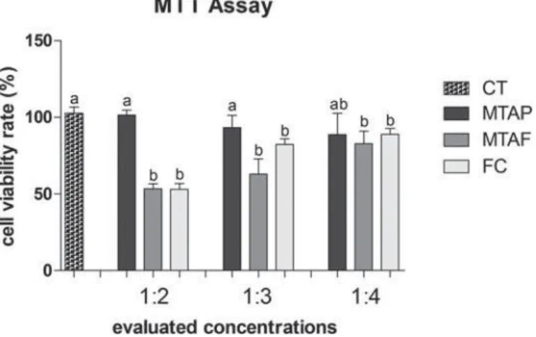
Results: MTT and NR results showed suitable cell viability rates for MTAP at all dilutions
( -13 ). Cells exposed to MTAF and FC (1:2 and 1:4 dilutions) showed signi cant low
viability rate when compared to CT in MTT. The NR results demonstrated cell viability for all materials tested. In MTAP group, the cells ALP activity was similar to CT in one and three days of exposure to the material. MTAF and FC groups demonstrated a decrease in ALP activity when compared to CT at both periods of cell exposure. Conclusions: The
hDPCs were suitable for the evaluation of new endodontic materials in vit ro. MTAP may be
considered a promising material for endodontic treatments.
Keyw ords: Dental pulp. Silicate cement. Cell viability. Alkaline phosphatase.
I N TRODUCTI ON
MTA is indicated as the material of choice for root-end- llings and root perforation treatment24.
The bioactivity of MTA led to the interest at the development of new dental materials based on calcium silicates, including root canal sealers26.
MTA Fillapex and MTA Plus are two endodontic calcium silicate-based sealers. MTA Fillapex (Angelus Industry Dental Products S/A, Londrina, PR, Brazil) composition consists of MTA associated with resins and pigments. MTA Plus (Avalon Biomed Inc.,
Sarasota, FL, USA) is composed of calcium silicates in smaller particles than MTA, according to the manufacturer. Moreover, MTA Plus presents a water-soluble salt-free gel as a vehicle. This gel might improve the consistency, solubility and adhesiveness to dentin walls18, allowing its use as a sealer.
Studies evaluating MTA Fillapex biocompatibility and bioactivity in osteoblastic cells showed cytotoxicity effect of this sealer in the rst periods of cells exposure20,27. However, for extended periods, cell
by Alizarin Red staining was observed as well, demonstrating the cement biocompatibility and bioactivity after the setting time20.
In other study, MTA Fillapex presented lower adhesion to dentin walls when compared to MTA Plus16. Furthermore, the authors suggest that MTA
Plus has a higher biomineralization potential than MTA Fillapex, being able to form apatite deposits in collagen brils inside the dentinal tubules, being similar to a tag between dentin-MTA Plus interface. When evaluating MTA Plus bioactive potential, a recent study showed that this cement is similar to ProRoot MTA (Dentsply Tulsa Dental Specialties, York, PA, USA), and releases calcium ions when in contact with water or HBSS (Hank’s saline based solution)6.
Although these sealers were launched in the market the last couple of years, literature still lacks information about their biological properties. The aim of this study was to evaluate the biocompatibility and bioactivity of MTA Plus and MTA Fillapex in primary culture of human dental pulp cells (hDPCs). The hDPCs are a suitable model to evaluate cytotoxicity of different dental materials11. This cell line is also
used to estimate the effects of MTA and several calcium silicate-based cements in cell culture, demonstrating its cytocompatibility and bioactivity
in vit ro17,26.
MATERI AL AN D METHODS
Dent al pulp cells explant s and cell cult ure condit ions
After approval from the Institutional Research Ethics Committee, patients with third molars at the stage of incomplete root formation, indicated to extraction for orthodontic reasons, were selected for this study. Dental pulps were collected via apical opening using a Hedstr en le (Maillefer, Dentsply, Chemin du Verger, Ballaigues, Switzerland) and stored in a culture dish (35x10 mm) containing Alpha Modification Minimum Essential Eagle’s Medium ( -MEM, Sigma-Aldrich, St. Louis, MO, USA) supplemented with 10% fetal bovine serum (FBS, Gibco, Life Technologies, Grand Island, NY, USA) and 1% penicillin and streptomycin (PenStrep, Gibco). The pulp tissues were sectioned into fragments of
approximately 1 mm3 and kept in supplemented
-MEM at 37 C, 5% humidity and 5% CO2. The culture medium was changed 24 h after the pulp collection and every 48 h to obtain explants of human dental pulp cells (hDPCs). Successive passages were prepared for cellular expansion. The experiments were performed with cells from the fourth passage, and were repeated three times independently (n=3/ group).
Preparat ion of endodont ic sealer ext ract s
The experimental groups and the preparation proportions are described in Figure 1. Endodontic sealers were prepared according to manufactures instructions. FillCanal (FC) was prepared according to Grossman technique9, and MTA Plus (MTAP)
was prepared with the vehicle for root canal lling consistency. Thereafter, 1 mL of each sealer was individually placed in wells of a 12-well plate (TPP, Techno Plastic Products, Zollstrasse, Trasadingen, Su a) and spread to ll the bottom completely. The plates with the samples were kept in an incubator at 37°C, 95% humidity and 5% CO2 during 24 h for the initial setting time of the materials. After this period, the specimens were exposed to ultraviolet light (UV) in a laminar ow for 30 min each side to disinfection13. The wells were lled with 5 mL
of -MEM supplemented with 1% PenStrep and incubated at 37°C, 95% humidity and 5% CO2 for additional 24 h, in order to obtain the sealer extracts.
Cyt ot oxicit y assays
C y t o t o x i c i t y w a s a s s e s s e d b y M T T (3-(4,5-dimethyl-thiazoyl)-2,5-diphenyl-tetrazolium bromide) and Neutral Red (NR) assays. hDPCs were plated at the concentration of 2x10⁵ cells per well
in 96-well plates (TPP) containing supplemented -MEM culture medium and cultured for 24 h at 37°C, 95% humidity and 5% CO2 to obtain adherent cells prior to the material exposure. hDPCs were exposed to the sealers extracts in dilutions using culture medium without FBS of 1:2, 1:3 and 1:4 for one day, according to ISO 10993-512.
MTT is based on the conversion of tetrazolium salt to insoluble violet formazan crystals, determining the cytotoxicity by measuring cell viability rate. For this experiment, sealers extracts were replaced by 100 μL
Group Material Reference Proportion
MTAP MTA Plus Avalon Biomed Inc.,
Bradenton, FL, USA
1 g : 350 μL
MTAF MTA Fillapex Angelus S/A, Londrina, PR,
Brasil
1 : 1
FC FillCanal Technew, Rio de Janeiro,
RJ, Brasil
1 g : 200 μL
of a 5 mg/mL MTT solution (Sigma-Aldrich), followed by incubation at 37°C, 95% humidity and 5% CO2 for 3 h. Thereafter, the content of the well was removed and the colorimetric product solubilized in 100 μL of acidi ed isopropanol 0.04 N (Sigma-Aldrich). The optical densities of the solutions were measured in a spectrophotometer (Elx800; Instrumentos Bio-Tek, Winooski, VT, USA) at 570 nm wavelength27.
The absorbance readouts were normalized with the absorbance of the group of cells exposed to -MEM (CT), and represented the activity of the viable cells.
NR is a vital dye that is incorporated through endocytosis and accumulated on the lysosome of cells that have their membranes intact. For this experiment, sealers extracts were replaced by 0.1 mL -MEM medium containing 50 μg NR/mL (Sigma-Aldrich) followed by incubation at 37°C, 95% humidity and 5% CO2 for 3 h. Then, the content of the well was removed and the colorimetric product solubilized in 100 μL of an ethanol solution (50% ethanol and 1% acetic acid) (Sigma-Aldrich). The optical densities of the solutions were measured in a spectrophotometer (Elx800) at 570 nm21.
ALP enzym e act ivit y
ALP is an enzyme present in the plasma membrane that is associated with the mineralization process, and it is one of the major markers of osteoblast activity in the cells3. The bioactivity assay
by ALP enzyme activity was performed with hDPCs at the concentration of 2x10⁴ cells per well in 12-well
plates (TPP), as previously described. The assay was performed in time intervals of one and three days. All assays were performed in triplicate and repeated three independent times (n=3/group). Cells exposed to culture medium were the control of group (CT).
ALP activity was evaluated in accordance to the release of tymolphtaleine from tymolphtaleine monophosphate using Alkaline Phosphatase kit (Labtest Diagnóstica, Lagoa Santa, MG, Brazil).
hDPCs were lysed for 30 min at ambient temperature using Sodium Lauryl Sulfate solution 1:1 (SDS, Sigma-Aldrich), and the assay was performed according to the manufacturer instructions. Absorbance was measured at the wavelength of 590 nm in a spectrophotometer. The data were expressed as ALP activity (μmol tymophtaleine/min/L)19.
St at ist ical analysis
The data were analyzed by Kolmogorov-Smirnov test, obtaining a normal distribution. Therefore, we performed ANOVA and Tukey tests post hoc, considering a level of signi cance of 5% (GraphPad Prism 5 software, GraphPad Software Inc., San Diego, CA, USA). All data is presented as mean and standard deviation of the mean.
RESULTS
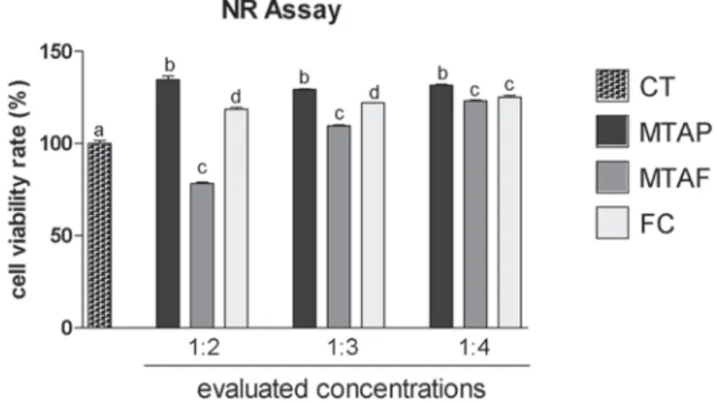
The MTT assay (Figure 2) showed 80-120% of viability to hDPCs exposed to MTAP at all concentrations evaluated, being similar to the negative control of cytotoxicity (p>0.05). The MTAP group viability rate was similar in the dilution of 1:4 to the other groups (p>0.05), although MTAF and FC groups viability rate showed signi cant difference in comparison to the CT. MTAF showed lower viability rate than CT at all concentrations evaluated. At NR assay (Figure 3), hDPCs exposed to MTAP showed signi cant higher viability rate (130-135%) than CT for all dilutions evaluated (p<0.05). MTAF showed the lowest viability of all groups in 1:2 dilution (p<0.05), however it increased in the dilutions of 1:3 and 1:4 (p<0.05). FC demonstrated good viability in all dilutions, being similar to MTAF in 1:4 (p>0.05).
In ALP bioactivity assay (Figure 4), MTAP showed similar activity to the control group at both periods of time evaluated (p>0.05), while the other groups demonstrated signi cant lower ALP activity when compared to CT (p<0.05). There was a tendency of
ALP activity increase in a time-dependent manner at all groups.
DI SCUSSI ON
hDPCs may represent a different population of differentiated and undifferentiated cells with great potential for pulp tissue and dentin regeneration. They can be successfully applied in studies to evaluate new biomaterials2, including endodontic
sealers. Studies in the literature reported that MTA is capable to stimulate odontogenic differentiation in dental pulp mesenchymal cells7,22. In this study,
human dental pulp was collected from third molars referred to extractions for orthodontic reasons, and obtained explants of hDPCs to investigate possible cytotoxic effects and the bioactivity of MTAP, MTAF and FC.
Furthermore, MTT and NR assays demonstrated higher viability rate to MTAP at all concentrations evaluated, and there was no evidence of cytotoxic
effect. All materials presented similar viability in the higher dilution (1:4) on MTT assay. Concentration-dependent results were also described by other authors when evaluating the viability of MTA ProRoot and MTAP in rat odontoblastic cells (MDPC-23), concluding that the cytotoxic risks become negligible after the dilution of the components8.
MTAF obtained similar results to FC. The viability of MTAF has been evaluated in several cell types, such as broblasts4,23, osteoblasts21 and human dental cell
cultures10, demonstrating cytotoxic results in less
than 7-day periods27. In this study, the cytotoxicity of
MTAF may be considered moderate to low (50-80%, approximately) and concentration-dependent, after 24 h setting-time. Zinc oxide and eugenol cements as FC are considered cytotoxic both in vit ro and in vivo, causing tissue in ammation and even bone resorption1. In addition, the literature demonstrated
that these cements are capable of inducing the expression of cyclooxygenase-2 and nuclear factor kappa B, an immune mediator involved in cellular
Figure 3- Cell viability rate (%) according to NR assay in human dental pulp cells (hDPCs) exposed to MTA Plus (MTAP), MTA Fillapex (MTAF), FillCanal (FC) and culture medium used as control (CT). Bars with different letters represent
responses to stress, cytokines, free radicals, UV radiation, oxidation of LDL and bacterial and viral antigens14. MTAF cytotoxicity can be associated with
its toxic components, such as salicylate resin, which can induce apoptosis5.
The bioactivity was analyzed in accordance to ALP enzymatic assay. The progressive increase in ALP activity is marked in bone cells15. Besides the
bioactivity, ALP expression indicates the potential of the cells for differentiation25. Therefore, the
evaluation of this enzyme allows the determination of the material bioactivity and its potential to promote repair with formation of mineralized tissue. The results showed an increase in ALP enzyme activity over time for cells exposed to all materials. MTAP presented results similar to the control group in both evaluated periods, and higher than the other cements groups. Other studies showed that after the initial period of cytotoxicity, MTAF sealer is able to promote bioactivity, stimulating the deposition of mineralized nodules and increasing ALP activity20,
corroborating the results of the present study.
CON CLUSI ON
T h i s s t u d y s h o w s t h a t M TA P p r e s e n t s biocompatibility and bioactivity in the primary culture of cells from human dental pulp. MTAP can be considered a suitable material for root canal lling. MTAF shows initial cytotoxicity.
ACKN OW LEDGEMEN TS
We would like to thank FAPESP - São Paulo Research Foundation (Processes 2011-18239-4 and 2012/13268-9).
We also thank CNPq - National Council for Scienti c and Technological Development (Process 475068/2010-6, MCT/CNPq 14/2010).
REFEREN CES
1- Al-Hiyasat AS, Tayyar M, Darmani H. Cytotoxicity evaluation of various resin based root canal sealers. Int Endod J. 2010;43:148-53. 2- Atari M, Barajas M, Hernández-Alfaro F, Gil C, Fabregat M, Ferrés Padró E, et al. Isolation of pluripotent stem cells from human third molar dental pulp. Histol Histopathol. 2011;26:1057-70.
3- Beck GR Jr. Inorganic phosphate as a signaling molecule in osteoblast differentiation. J Cell Biochem. 2003;90:234-43. 4- Bin CV, Valera MC, Camargo SE, Rabelo SB, Silva GO, Balducci I, et al. Cytotoxicity and genotoxicity of root canal sealers based on mineral trioxide aggregate. J Endod. 2012;38:495-500.
5- Braga JM, Oliveira RR, Castro Martins R, Vieira LQ, Sobrinho AP. Assessment of the cytotoxicity of a mineral trioxide aggregate-based sealer with respect to macrophage activity. Dent Traumatol. 2015. Epub ahead of print.
6- Camilleri J, Formosa L, Damidot D. The setting characteristics of MTA Plus in different environmental conditions. Int Endod J. 2013;46:831-40.
7- Chang SW, Lee SY, Kum KY, Kim EC. Effects of ProRoot MTA, Bioaggregate, and Micromega MTA on odontoblastic differentiation in human dental pulp cells. J Endod. 2014;40:113-8.
8- Eid AA, Gosier JL, Primus CM, Hammond BD, Susin LF, Pashley DH, et al. I n vit ro biocompatibility and oxidative stress pro les of different hydraulic calcium silicate cements. J Endod. 2014;40:255-60.
9- Grossman LI. Physical properties of root canal cements. J Endod. 1976;2:166-75.
10- G ven EP, Yalva ME, Kayahan MB, Sunay H, ah n F, Bayirli G. Human tooth germ stem cell response to calcium-silicate based endodontic cements. J Appl Oral Sci. 2013;21:351-7.
11- Guven EP, Yalvac ME, Sahin F, Yazici MM, Rizvanov AA, Bayirli G. Effect of dental materials calcium hydroxide-containing cement, mineral trioxide aggregate, and enamel matrix derivative on proliferation and differentiation of human tooth germ stem cells. J Endod. 2011;37:650-6.
12- International Organization for Standardization. ISO 10993-5: Biological evaluation of medical devices - Part 5: Tests for in vit ro
cytotoxicity. Geneva: ISO; 2009.
13- Katara G, Hemvani N, Chitnis S, Chitnis V, Chitnis DS. Surface disinfection by exposure to germicidal UV light. Indian J Med Microbiol. 2008;26:241-2.
14- Lee YY, Yang SF, Ho WH, Lee YH, Hung SL. Eugenol modulates cyclooxygenase-2 expression through the activation of nuclear factor kappa B in human osteoblasts. J Endod. 2007;33:1177-82. 15- Millán JL. Mammalian alkaline phosphatases: from biology to applications in medicine and biotechnology. 1st ed. Weinheim:
Wiley-VCH Verlag; 2006.
16- Neelakantan P, Grotra D, Sharma S. Retreatability of 2 mineral trioxide aggregate-based root canal sealers: a cone-beam computed tomography analysis. J Endod. 2013;39:893-6.
17- Paranjpe A, Smoot T, Zhang H, Johnson JD. Direct contact with mineral trioxide aggregate activates and differentiates human dental pulp cells. J Endod. 2011;37:1691-5.
18- Qi YP, Li N, Niu LN, Primus CM, Ling JQ, Pashley DH, et al. Remineralization of arti cial dentinal caries lesions by biomimetically modi ed mineral trioxide aggregate. Acta Biomater. 2012;8:836-42. 19- Rosa AL, Beloti MM. Development of the osteoblast phenotype of serial cell subcultures from human bone marrow. Braz Dent J. 2005;16:225-30.
20- Salles LP, Gomes-Cornélio AL, Guimarães FC, Herrera BS, Bao SN, Rossa-Junior C, et al. Mineral trioxide aggregate-based endodontic sealer stimulates hydroxyapatite nucleation in human osteoblast-like cell culture. J Endod. 2012;38:971-6.
21- Scelza MZ, Linhares AB, Silva LE, Granjeiro JM, Alves GG. A multiparametric assay to compare the cytotoxicity of endodontic sealers with primary human osteoblasts. Int Endod J. 2012;45:12-8. 22- Seo MS, Hwang KG, Lee J, Kim H, Baek SH. The effect of mineral trioxide aggregate on odontogenic differentiation in dental pulp stem cells. J Endod. 2013;39:242-8.
23- Silva EJ, Santos CC, Zaia AA. Long-term cytotoxic effects of contemporary root canal sealers. J Appl Oral Sci. 2013;21:43-7. 24- Torabinejad M, Parirokh M. Mineral trioxide aggregate: a comprehensive literature review – part II: leakage and biocompatibility investigations. J Endod. 2010;36:190-202. 25- Wang Y, Yan M, Wang Z, Wu J, Wang Z, Zheng Y, et al. Dental pulp stem cells from traumatically exposed pulps exhibited an enhanced osteogenic potential and weakened odontogenic capacity. Arch Oral Biol. 2013;58:1709-17.
26- Woo SM, Hwang YC, Lim HS, Choi NK, Kim SH, Kim WJ, et al. Effect of nifedipine on the differentiation of human dental pulp cells cultured with mineral trioxide aggregate. J Endod. 2013;39:801-5. 27- Yoshino P, Nishiyama CK, Modena KC, Santos CF, Sipert CR.
I n v it r o cytotoxicity of white MTA, MTA Fillapex® and Portland