THE METHODOLOGY FOR EVALUATING HALF-VALUE LAYER
AND ITS INFLUENCE ON THE DIAGNOSTIC RADIOLOGY*
Marco Aurélio de Sousa Lacerda1
, Teógenes Augusto da Silva2
, Arno Heeren de Oliveira3
OBJECTIVE: The critical analysis of the methodology for evaluating the x-ray beam half-value layer was based on technical standards for radiological protection and quality control in medical and dental diagnoses. MA-TERIALS AND METHODS: Exposure parameters, experimental set-up, scattering materials, instrumentation, size of radiation field and methodology of data analysis have been taken into consideration for measure-ments of half-value layer. RESULTS: The half-value layer, in good geometry, measured 2.44 ± 0.02 mmAl. A maximum deviation of 4.1% was found under other geometry conditions. A 98.8% deviation was ob-served with a different ionization chamber, and scattering material, and 29.5% with another type of detec-tor. CONCLUSION: Results showed that no relevant variation is observed in the half-value layer for different radiation field sizes. On the other hand, the influence of the radiation scattering on the half-value layer over-estimation was clearly observed, as well as the half-value layer reduction in the presence of lead used as scattering material. The practical methodology adopted for half-value layer evaluation has shown to be quite reliable, besides evidencing remarkable discrepancies resulting from the adoption of inappropriate method-ologies, emphasizing the necessity of establishing a standard procedure for measuring the half-value layer. Keywords: Half value layer; HVL; Quality control; Diagnostic radiology.
Influência da metodologia de avaliação da camada semi-redutora em radiologia diagnóstica.
OBJETIVO: A análise crítica da metodologia de medida da camada semi-redutora em feixes de raios X teve como base o regulamento técnico para proteção radiológica e controle de qualidade em radiodiagnóstico médico e odontológico. MATERIAIS E MÉTODOS: Na medida da camada semi-redutora, a técnica radiográ-fica, o arranjo experimental, os meios espalhadores, a instrumentação, o tamanho de campo de radiação e a metodologia de análise dos dados foram considerados. RESULTADOS: A camada semi-redutora obtida em condições de boa geometria, para a técnica escolhida, foi de 2,44 ± 0,02 mmAl. Em relação a este valor, observaram-se desvios máximos na camada semi-redutora de 4,1% na variação da geometria, de 98,8% na variação da câmara de ionização e do meio espalhador, e de até 29,5% com outro tipo de detector. CON-CLUSÃO: Os resultados mostraram que não ocorre variação significante na camada semi-redutora para di-ferentes tamanhos de campo de radiação, mas foram evidentes a influência do espalhamento na superesti-mação da camada semi-redutora e a redução desta na presença de blocos de chumbo como meio espalha-dor. O procedimento prático adotado mostrou-se bastante confiável e evidenciou a grande discrepância decorrente da adoção de metodologias impróprias, enfatizando a necessidade de estabelecer um procedi-mento padrão para a medida da camada semi-redutora.
Unitermos: Camada semi-redutora; CSR; Controle de qualidade; Radiologia diagnóstica. Abstract
Resumo
* Study developed at Centro de Desenvolvimento da Tecnolo-gia Nuclear/Comissão Nacional de EnerTecnolo-gia Nuclear (CDTN/ CNEN), Belo Horizonte, MG, Brazil.
1. PhD in Nuclear Sciences, Research Assistant at Centro de Desenvolvimento da Tecnologia Nuclear/Comissão Nacional de Energia Nuclear (CDTN/CNEN), Belo Horizonte, MG, Brazil.
2. PhD in Sciences – Nuclear Engineering, Titular Researcher and Head of Division at Centro de Desenvolvimento da Tecnolo-gia Nuclear/Comissão Nacional de EnerTecnolo-gia Nuclear (CDTN/ CNEN), Belo Horizonte, MG, Brazil.
3. Doctorate in “Genie Atomique”, Associate Professor at Department of Nuclear Energy – Universidade Federal de Minas Gerais (UFMG), Belo Horizonte, MG, Brazil.
Mailing address: Prof. Dr. Teógenes Augusto da Silva. Avenida Presidente Antonio Carlos, 6627, Pampulha. Belo Horizonte, MG, Brazil, 30123-970. E-mail: [email protected]
Received December 22, 2006. Accepted after revision Feb-ruary 13, 2007.
INTRODUCTION
The filtration of the radiation beam pro-duced by a x-ray tube reduces the patient
radiation exposure, since this procedure preferentially removes low energy photons unnecessary for the formation of the diag-nostic image of interest. The International Commission on Radiological Protection (ICRP) has established a minimum value for the total filtration thickness to be adopted with diagnostic x-ray beams; such value should not be lower than 2.5 mmAl (except for mammography equipment), and equal to 1.5 mmAl for odontological x-ray up to 70 kV(1). X-ray equipment
manufac-turers must ensure that the ICRP minimum filtration requirements are met.
Most frequently, the method employed to infer about the total filtration of an equipment associates the total filtration
with the quality of the x-ray beam, also de-nominated penetrability or penetrating en-ergy, numerically characterized by the half-value layer (HVL).
Several authors present correlations between HVL for a determined potential and the total filtration of the tube, also de-nominated quality diagrams(2,3).
Consider-ing the great discrepancies among these diagrams because of the several parameters influencing the correlation between HVL and total filtration, the Brazilian stan-dards(4), as well as the majority of
are obtained for HVL, it is assumed that the total beam filtration is in compliance with the ICRP standards.
The HVL may be more completely de-fined as the absorber material thickness necessary to reduce the x-ray beam inten-sity to half its incident magnitude, under good geometry conditions. This condition implies a configuration that minimizes the scattering influence which tends to increase the HVL value. Farr(5) and Trout et al.(6)
have studied the effects of variations in the radiation field size and focus-filter-detec-tor distances on HVL measurements. Trout et al.(6) have developed a method for
accu-rately determining the HVL for energies generated by 100–300 kV tensions, based on the extrapolation to size of the null field in the HVL curve as a function of size of the radiation field incident on the filter.
Besides geometry, other uncertainty sources, particularly those resulting from the dosimetry and analysis of beam attenu-ation data, affect the HVL measurement(3,6– 8). Technical, practical and economic
fac-tors should be always taken into consider-ation when a methodology is adopted; however, its reliability depends on the es-tablishment of all the relevant uncertainty components obtained by means of a de-tailed analysis of parameters directly influ-encing such measurements.
In the present study, a critical analysis is performed on the methodology for mea-surement of HVL in diagnostic x-ray beams, according to technical standards established by the Brazilian Ministry of Health(4). Relevant parameters for HVL
measurement and main uncertainty compo-nents are identified to assure compliance of the analyzed equipment with the perfor-mance standards.
MATERIALS AND METHODS
The experimental study was developed with a VMI, Pulsar 800 Plus model x-ray diagnostic unit with high frequency genera-tor, 16° angle rotating tungsten anode, double focus (FF = 1.0 mm and FG = 2.0 mm). This equipment presents a linear air-kerma rate with current and load, respec-tively, < 0.01 and < 0.02, accuracy and tension reproducibility, and exposure time < 2.5%, reproducibility of air-kerma
rate < 2%, and yielding = (3.86 ± 0.08) ×
10–² mGy/mA.s, at 80 kV and 1 m.
The choice of a methodology for HVL measurement involved the evaluation of the radiographic technique, experimental arrangement, attenuators, measurement instrumentation, radiation field size and procedure for analysis of beam attenuation data.
As regards the choice of the radio-graphic technique, the American Associa-tion of Physicists in Medicine(9,10)
recom-mends the utilization of a 80 kV tube po-tential, current between 200 mA and 400 mA, and exposure time > 50 ms. On the other hand, the Instituto de Eletrotécnica e Energia(11) recommends 80 kV tube
poten-tial and range of tube current x exposure time = 20 mA.s, suggesting a test with the air-kerma rate reproducibility to define which current provides the most reproduc-ible values for a same parameter. So, the present study considered a radiographic technique with 80 kV voltage, load of 20 mA.s, and current of 200 mA.
The attenuators consisted of commer-cial-type aluminium alloy 1200 H14, (mini-mum 99% aluminium) and maxi(mini-mum lim-its established by the NBR-7556(12). The
conclusions of the study developed by Piubelli(13), who had performed
compara-tive measurements of x-ray beam attenua-tion by standard high purity plates, and demonstrated a non-significant difference for the beam quality (< 0,1%), and the ex-pensiveness of commercial high purity plates were the main reason for the choice of commercial aluminium. The aluminium plate thickness with respective tolerance declared by the manufacturer (Alcan) was 0.50 ± 0.04 mm, confirmed by measure-ments with a Mitutoyo MFG digital mi-crometer, performed on three sites of each plate, with 0.01 mm maximum deviation. In order to minimize the uncertainties caused by the experimental arrangement (geometry) and by the dosimetry of mea-surements considered as a reference, a Radcal/MDH 10X5-6 model, low volume (6 cm³) ionization chamber with low en-ergy dependence (± 3% between 30 keV and 150 keV) attached to a Radcal/MDH 9010 model electrometer. Following the recommendation for a good geometry(6,9, 10,14,15), The chamber was placed at 100 cm
from the focus, and at 30 cm from the scat-tering means; the aluminium plates were placed at 50 cm from the focus, corre-sponding to half the focus-chamber dis-tance.
The beam attenuation measurements were performed for three ranges of field sizes incident on the ionization chamber: one smaller (8 × 8 cm²), one intermediate (11 × 11 cm²), and another, larger (14 ×
14 cm²), aiming at obtaining, by extrapo-lation, the value corresponding to the null field(6).
The HVL was determined by means of the exponential adjustment of the four points of beam attenuation, near the corre-spondent to half the initial intensity; so, by not utilizing the initial measurement with-out the beam attenuation, the uncertainty resulting from the data interpolation and exponential beam attenuation was mini-mized(16).
The geometry influence on the experi-mental arrangement for HVL determina-tion was evaluated by means of measure-ments with different focus-plate (FPD) and focus-chamber distances (FCD), with other parameters remaining unchanged. Addi-tionally, for evaluation of the dosimetry influence on the HVL, measurements were performed with different ionization cham-bers (both in shape and volume – 11 cm³, 60 cm³ and 180 cm³), attached to Radcal/ MDH 3036 and 9010 model electrometers; measurements with two other types of de-tectors — dosimetry pens 862L model, and fluoride lithium thermoluminescent TLD-100 dosimeters (Harshaw/Bicron Chemical Company) — also were performed. The different chambers and detectors were placed in air, over 8 cm-lead cubes and 30 cm-paper sheet layers for analysis of the scattering means influence.
The uncertainty components in the HVL determination were estimated according to the recommendations of Associação Brasileira de Normas Técnicas(17). For
calibra-tion and geometry, and aluminium plates thickness and purity. The quadratic combi-nation of this uncertainty components re-sulted in a combined standard uncertainty which, multiplied by the coverage factor k = 2, supplying the expanded uncertainty.
Aiming at evaluating the compliance of the equipment with the performance stan-dards established by the Brazilian regula-tory authorities(4), in a wider coverage range
in terms of peak tension, HVL at 60 kV, 100 kV and 110 kV were determined. The same 20 mA.s and 200 mA radiographic param-eters, Radcal/MDH 10X5-6 ionization chamber and geometry conditions of FCD = 100 cm and FPD = 50 cm employed for determining the optimum HVL value were utilized for this evaluation.
RESULTS
HVL values for the 8 × 8 cm², 11 × 11 cm² and 14 × 14 cm² radiation fields ob-tained with the 6 cm³ ionization chamber in good geometry, are shown on Table 1. The HVL values ranged between 2.44 mm and 2.45 mmAl, with uncertainties from 0.8% to 1.2% represented by the standard deviation.
Table 1 Mean HVL values for different x-ray beam field sizes in good geometry setup.
Field size (cm × cm)
8 × 8
11 × 11
14 × 14
HVL* (mmAl)
2.44 ± 0.02
2.44 ± 0.03
2.45 ± 0.03
* Uncertainty as regards a standard deviation.
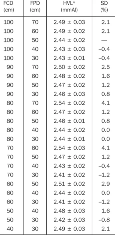
Table 2 HVL values for different focus-chamber distances and focus-plate distance and standard deviation in relation to the “optimum condition” with 80 kV, 200 mA, 20 mA.s.
FCD (cm) 100 100 100 100 100 90 90 90 90 80 80 80 80 80 70 70 70 70 60 60 60 50 50 40 FPD (cm) 70 60 50 40 30 70 60 50 30 70 60 50 40 30 60 50 40 30 50 40 30 40 30 30 HVL* (mmAl)
2.49 ± 0.03
2.49 ± 0.02
2.44 ± 0.02
2.43 ± 0.03
2.43 ± 0.01
2.50 ± 0.02
2.48 ± 0.02
2.47 ± 0.02
2.46 ± 0.03
2.54 ± 0.02
2.47 ± 0.02
2.46 ± 0.01
2.44 ± 0.02
2.44 ± 0.01
2.54 ± 0.03
2.47 ± 0.02
2.43 ± 0.02
2.41 ± 0.02
2.51 ± 0.02
2.44 ± 0.02
2.41 ± 0.02
2.48 ± 0.03
2.42 ± 0.03
2.49 ± 0.03 SD (%) 2.1 2.1 — –0.4 –0.4 2.5 1.6 1.2 0.8 4.1 1.2 0.8 0.0 0.0 4.1 1.2 –0.4 –1.2 2.9 0.0 –1.2 1.6 –0.8 2.1
* Uncertainty as regards a standard deviation. FCD, focus-chamber distance; FPD, focus-plate distance; SD, standard deviation.
Figure 2 illustrates the HVL x peak ten-sion relation for the minimum values estab-lished by the Brazilian Ministry of Health(4)
in comparison with the values obtained in the present study, considering the most rel-evant uncertainty components in the HVL measurements, as well as the estimated and propagated HVL value (see Table 5). There-fore, the expanded HVL value uncertainty, considering k = 2, for an interval with 95% confidence level corresponds to 3.7%.
DISCUSSION
HVL under good geometry conditions
HVL measurements under good geom-etry conditions (Table 1) did not show any variation in values with the radiation field size, not evidencing the presumed trend of decrease in HVL for the null field reported by Trout et al.(6). Therefore, there is no need
to perform measurements with several field sizes for determining HVL; an intermedi-ate field size may be adopted, covering, with a small deviation to spare, the whole ion-ization chamber sensible volume. As regards the number of measurements, for practical reasons, one may perform only one mea-surement considering for each site the mean value of three shots of the equipment.
Geometry influence on HVL
The results from the analysis of the ge-ometry influence on HVL (Table 2) have evidenced that, for a same FCD, HVL val-ues tend to decrease as the FPD decreases in relation to the plate-chamber distance (PCD), with 4.1% maximum deviation for a maximum relation (PCD = 10 cm). In this case, the scattering influence on the HVL overestimation becomes evident.
Table 2 also allows us to conclude that the HVL determination may be accurately achieved for a 50-100 cm FCD and a FPD/ PCD ratio between 50% and 30%. This assumption for distances < 70 cm is con-trary to Trout et al. recommendations(6) as
well as the recommendations of the major-ity of the protocols evaluated in the present study(9,11,14). A possible explanation for this
fact is that the classic study of Trout et al.(6),
upon which the mentioned protocols are based, was developed for 250 kV x-rays (for therapy purposes), a value about three times higher than the one considered in the The geometry influence on the HVL
was evaluated by means of different com-binations of FCD and FPD ranging be-tween 30 cm and 100 cm. The HVL values found for the different combinations with the respective standard deviation are com-pared with the values found for the mea-surements in good geometry (Table 2). The maximum deviation was 4.1%.
The influence of the dosimetry proce-dure on the HVL measurement was evalu-ated with ionization chambers of different volumes, attached to electrometers and placed in air and over different scattering means (air, lead, paper, table). The mean
values obtained for HVL, with respective standard deviation, are shown on Table 3. In the measurements with the 9010 elec-trometer, the HVL presented a deviation up to 7.8% in relation to the value in good geometry, in the presence of paper as a scat-tering mean. For the model 3036, the maxi-mum deviation was 98.8% under the same conditions.
present essays (80 kV). That is to say, for this voltage range, the scattering influence tends to be smaller, and does not signifi-cantly affect the results. Another explana-tion would be the different features of gen-erators and chambers utilized in the study of Trout et al.(6) and in the present study, a
result from the technological development of the last four decades.
Dosimetry influence on HVL
The results from the analysis of dosim-etry procedures influence on HVL values demonstrate that, for both electrometers, the HVL value increases proportionally to the ionization chambers volume, since sen-sitive scattering is shown to increase when higher volume ionization chambers are employed (Table 3). The significant differ-ence of HVL values in measurements with ionization chambers with the same and higher volumes but connected to different electrometers, is explained by the influence of the greater electronic sophistication present in the 9010 model as compared with the 3036 model. It may be concluded that the association electrometer-ionization chamber volume affects significantly the HVL value determination, so the sugges-tion is that low-volume ionizasugges-tion cham-bers (to decrease the scattering influence) are always utilized in conjunction with appropriate electrometers to reduce errors, particularly those resulting from noise.
The scattering material influence was analyzed with the results included on Table 3. it is observed that lead reduced the HVL value when placed below chambers 10X5-6, 10X5-60 and 30X6-11, demonstrating a decrease in the scattered radiation detected by the dosimeter. This may be explained by the high atomic number of lead which, for the studied voltage, favors the supremacy of the photoelectrical effect over the Comptom scattering, reducing the amount of scattered radiation on the detector.
The results also demonstrate an increase in the HVL values with the ionization chambers positioned on paper layers, which also demonstrates the scattering in-fluence on the HVL overestimation. Addi-tionally, it may be observed that this in-crease becomes more significant when higher volume ionization chambers are uti-lized, considering their higher sensitivity. Table 3 HVL values obtained with chambers of different volumes and shapes, coupled with different
electrometers, in air over different scattering means.
Chamber
10X5-6
10X5-60
10X5-180
10X5-6
30X6-11
10X5-60
10X5-180
Electrometer
9010
9010
9010
3036
3036
3036
3036
Scattering mean
Air Lead Paper Table
Air Lead Paper Table
Air Lead Table
Air Lead Paper Table
Air Lead Paper Table
Air Lead Paper Table
Air Lead Paper
HVL* (mmAl)
2.44 ± 0.02 2.43 ± 0.02 2.54 ± 0.03 2.46 ± 0.02
2.50 ± 0.03 2.47 ± 0.03 2.63 ± 0.03 2.54 ± 0.02
2.58 ± 0.04 2.59 ± 0.03 2.72 ± 0.04
2.43 ± 0.02 2.40 ± 0.03 2.55 ± 0.03 2.46 ± 0.03
2.52 ± 0.02 2.51 ± 0.02 2.58 ± 0.03 2.54 ± 0.02
2.69 ± 0.02 2.65 ± 0.03 2.87 ± 0.03 2.73 ± 0.02
4.15 ± 0.04 4.18 ± 0.05 4.83 ± 0.05
SD (%)
— –0.4
4.1 0.8
2.5 1.2 7.8 4.1
5.7 6.2 11.5
— 1.3 4.9 1.2
3.7 3.3 6.2 4.5
10.7 9.1 18.1 12.4
70.8 72.0 98.8
* Uncertainty as regards a standard deviation. SD, standard deviation.
Table 4 HVL values obtained with dosimetry pens and TLD-100 detectors in air over different scattering means, in comparison with the optimum condition.
Dosimeter
Ionization chamber
Dosimetry pen
TLD-100
Scattering mean
Air
Air Lead Paper Table
Air Lead Paper
HVL (mmAl)
2.44 ± 0.02
2.76 ± 0.25 2.71 ± 0.26 3.16 ± 0.29 3.07 ± 0.25
2.38 ± 0.15 2.52 ± 0.13 2.66 ± 0.16
SD (%)
—
13.1 11.1 29.5 25.8
–2.5 3.3 9.0
SD, standard deviation.
Table 5 Most relevant sources of uncertainty in the determination of HVL.
Source of uncertainty
Measurements repeatability (standard deviation)
Adjustment of points on the attenuation curve
Irradiation field size
Positioning of ionization chamber and attenuators
Dosimeters calibration
Instrument resolution
Ionization chamber energy dependence
Temperature and pressure correction
Attenuators thickness
Purity of attenuators
Uncertainty (%)
0.80
0.30
0.20
0.10
0.30
0.01
1.10
0.30
1.10
HVL values found with ionization cham-bers placed on examination tables demon-strated a small increase, sensibly lower than the one found with the same ionization chambers placed on paper layers. This dem-onstrates the scattering mean influence on the amount of scattered radiation detected. In summary, it may be concluded that: a) the utilization of lead cubes below the ionization chamber represents an excellent alternative to reduce the scattering influ-ence on the HVL determination; b) a low-volume ionization chamber (≤ 6 cm³) placed on the examination table or on the same lead cubes may yield reliable results for HVL determination.
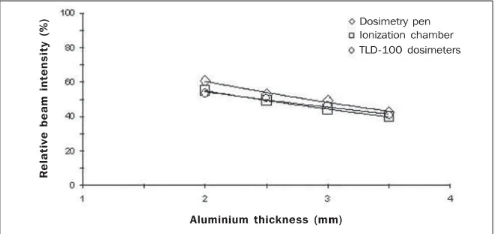
Table 4, also, shows that the values found for HVL utilizing dosimetry pens placed in air were higher than those found with the ionization chamber under the same conditions (optimum value). This is a re-sult from the difference in the inclination of the beam attenuation curves (Figure 1) caused by the higher dosimetry pen energy dependence as compared with the ioniza-tion chamber. Also, it is observed that the results with the dosimetry pen and TLD-100 detectors presented much higher un-certainties (standard deviation) than those found under “optimum conditions”, caused both by the higher energy dependence of these detectors and the low resolution of the dosimetry pen.
One may conclude that dosimetry pens should not be utilized for determining HVL because of the high uncertainty rates in measured values resulting particularly from the high energy dependence. Also, the uti-lization of TLD-100 detectors is not recom-mended for determining HVL, considering: a) the uncertainty magnitude resulting from the high sensitivity of these dosimeters to high temperatures, handling and chemical contamination; b) higher energy depen-dence; c) practical and economic difficul-ties.
Compliance of HVL values
The comparison between HVL values obtained in the present study and minimum value established by the Ministry of Health(4) (Figure 2) has shown that, for all
of the analyzed voltage ranges, HVL val-ues are about 7% higher than those, dem-onstrating that the x-ray diagnostic unit
utilized is not in compliance with the per-formance standards established by the regulatory authority, even if the uncertainty of about 4% found for the HVL value is considered. The present study has evi-denced that the dosimeters energy depen-dence, the attenuators thickness, and the repeatability of measurements were the uncertainty sources which have most con-tributed to the uncertainty in the HVL mea-surement.
CONCLUSIONS
The present study has demonstrated the feasibility of a practical procedure for de-termining HVL in diagnostic radiology, highlighting the utilization of a low-vol-ume, low energy dependent ionization chamber and an optimum geometry condi-tion to reduce the influence of scattered radiation.
The results of HVL measurements have testified to the metrological reliability of the practical procedure, showing, also, the remarkable discrepancies resulting from the adoption of inappropriate methodolo-gies and affirming the need for definition of the main influential parameters as well as the establishment of a standard proce-dure.
It is considered that the critical analy-sis of the HVL test as part of the program for radiodiagnosis quality control was ac-complished in detail, guaranteeing the re-sults reliability and confidence to certify the compliance or non-compliance of the x-ray unit with legal requirements.
Something similar should be done in re-lation to all the other tests in the quality pro-gram(4) aiming at the standardization of procedures as a contribution to the adop-tion of appropriate performance standards according to the Brazilian reality. Figure 1. X-ray beam attenuation during an essay with different dosimeter types.
R
e
la
ti
v
e
b
e
a
m
i
n
te
n
s
it
y
(
%
) Dosimetry pen
Ionization chamber TLD-100 dosimeters
Aluminium thickness (mm)
Figure 2. Relation between HVL and peak tension for the Health Ministry legal standards, as compared with the present study results.
Present study Ministry of Health
Peak tension (kV)
H
V
L
(
m
m
A
REFERENCES
1. International Commission on Radiological Pro-tection. Protection of the patient in diagnostic ra-diology. ICRP Publication 34. New York, NY: Pergamon Press, 1982.
2. Trout ED, Kelley JP, Furno EJ. A study of the in-herent filtration of diagnostic X-ray tubes. Radi-ology 1956;66:102–106.
3. Nagel HD. Limitations in the determination of to-tal filtration of x-ray tube assemblies. Phys Med Biol 1988;33:271–289.
4. Ministério da Saúde. Diretrizes de proteção radio-lógica em radiodiagnóstico médico e odontoló-gico. Portaria nº 453. Diário Oficial da União, 2 de junho de 1998.
5. Farr RF. The specification of roentgen ray output and quality. Acta Radiol 1955;43:152–160. 6. Trout ED, Kelley JP, Lucas AC. Determination of
half-value layer. Am J Roentgenol Radium Ther Nucl Med 1960;84:729–740.
7. Wagner LK, Archer BR., Cerra F. On the measure-ment of half-value layer in film-screen mammog-raphy. Med Phys 1990;17:989–997.
8. Sunde PB. Specification and selection of dosim-etry instrumentation for diagnostic radiology. Radiat Prot Dosim 1992;43:183–186. 9. American Association of Physicists in Medicine.
Performance acceptance testing for X-ray genera-tors and automatic exposure control devices. AAPM Report 14. New York, NY: American In-stitute of Physics, 1985.
10. American Association of Physicists in Medicine. Protocols for the radiation safety surveys of diag-nostic radiological equipment. AAPM Report 25. New York, NY: American Institute of Physics, 1988.
11. Instituto de Eletrotécnica e Energia. Procedimen-tos para testes de estado: equipamenProcedimen-tos de raios X. IEE. São Paulo: Universidade de São Paulo, 1999.
12. Associação Brasileira de Normas Técnicas. Cha-pas de alumínio e suas ligas – especificação. NBR 7556. Rio de Janeiro, RJ: Associação Brasileira de Normas Técnicas, 2000.
13. Piubelli SL. Influência da pureza de absorvedo-res comerciais na determinação da energia efetiva
e da camada semi-redutora para feixes de 24 a 50 kV, e propagação do erro para a taxa de exposi-ção. (Dissertação de Mestrado). Rio de Janeiro, RJ: Universidade Federal Fluminense, 1989. 14. Instituto de Radioproteção e Dosimetria. Controle
de qualidade em radiodiagnóstico. Protocolos IRD. Rio de Janeiro, RJ: Instituto de Radiopro-teção e Dosimetria, 2000.
15. International Commission on Radiation Units and Measurements. Recommendations of the Interna-tional Commission on Radiation Units and Mea-surements. Physical Aspects of Irradiation. Re-port 10b, 1962.
16. Lacerda MAS. Análise crítica da medida da ca-mada semi-redutora em radiologia diagnóstica. (Dissertação de Mestrado). Belo Horizonte, MG: Universidade Federal de Minas Gerais, 2002. 17. Associação Brasileira de Normas Técnicas. Guia