Revista Brasileira de Hematologia e Hemoterapia
Brazilian Journal of Hematology and Hemotherapy
www.rbhh.org
Original article
Influence of
β
S-globin haplotypes and hydroxyurea on tumor
necrosis factor-alpha levels in sickle cell anemia
Marília Rocha Laurentino
a, Pedro Aurio Maia Filho
a,
Maritza Cavalcante Barbosa
b, Izabel Cristina Justino Bandeira
a,
Lilianne Brito da Silva Rocha
a, Romelia Pinheiro Gonçalves
a,*
a Universidade Federal do Ceará (UFC), Fortaleza, CE, Brazil b Universidade Federal do Ceará (UFC), Maranguape, CE, Brazil
A R T I C L E I N F O
Article history:
Received 8 July 2013 Accepted 25 September 2013
Keywords:
Anemia, sickle cell Inflammation
Tumor necrosis factor-alpha Haplotypes
Hydroxyurea
A B S T R A C T
Background: Sickle cell anemia is a chronic inflammatory disease characterized by an increased production of proinflammatory cytokines including tumor necrosis factor-alpha. Hydroxyurea, by decreasing the polymerization of hemoglobin, reduces inflammatory states. The effect of the genetic polymorphisms of sickle cell patients on tumor necrosis factor-alpha levels remains unknown.
Objective: The aim of this study was to investigate the association of tumor necrosis factor-alpha levels with β-globin haplotypes and the use of hydroxyurea.
Methods: A cross-sectional study was performed of 67 patients with sickle cell anemia diagnosed at steady-state in a referral hospital in Fortaleza, Ceará, Brazil. A group of 26 healthy individuals was used as control. βS-haplotype analysis was performed by restriction fragment length polymorphism-polymerase chain reaction. The tumor necrosis factor-alpha levels were measured by the enzyme-linked immunosorbent assay test. Laboratory data (complete blood count and fetal hemoglobin) and information regarding the use of hydroxyurea were obtained from medical records. Statistical analysis was performed using R software with the Kruskal-Wallis and Mann-Whitney tests. Statistical significance was established for p-values < 0.05 for all analyses.
Results: The mean age of the participants was 35.48 years. Patients with sickle cell anemia had significantly higher tumor necrosis factor-alpha levels than controls (p-values < 0.0001). Tumor necrosis factor-alpha levels were lower in sickle cell anemia patients who were receiving hydroxyurea treatment than those who were not (p-value = 0.1249). Sickle cell anemia patients with Bantu/n genotype had significantly higher levels than patients with the Bantu/Benin genotype (p-value = 0.0021).
Conclusion: In summary, βS-globin haplotypes, but not hydroxyurea therapy, have a role in modulating tumor necrosis factor-alpha levels in sickle cell anemia adults at steady-state. Many previous studies have investigated prognosis and inflammatory states in sickle cell anemia patients, but the discovery that tumor necrosis factor-alpha levels vary according to the genetic polymorphism of the patient is a new finding.
© 2014 Associação Brasileira de Hematologia, Hemoterapia e Terapia Celular. All rights reserved.
*Corresponding author at: Rua Universidade Federal do Ceará, Faculdade de Farmácia, Odontologia e Enfermagem (FFOE), Rua Capitão Francisco Pedro, 1210, Rodolfo Teófilo, 60430-370, Fortaleza, CE, Brazil.
E-mail address: [email protected] (R.P. Gonçalves).
1516-8484/$ - see front matter © 2014 Associação Brasileira de Hematologia, Hemoterapia e Terapia Celular. Published by Elsevier Edi-tora Ltda. All rights reserved.
Introduction
Sickle cell anemia (SCA) is a genetic disorder characterized by a point mutation in the beta globin gene generating, when homozygous, an abnormal hemoglobin known as hemoglobin S (Hb S).1,2 Clinical manifestations result from
the tendency of Hb S to polymerize in the deoxygenated state, causing vascular obstructions and ischemia, and consequent painful crises associated with episodes of chronic hemolysis.1,3
The vaso-occlusive crisis is the most common symptom in SCA patients, manifesting in 50% of cases.4 This event is
mainly attributed to abnormal adhesion of sickled red blood cells, granulocytes and platelets to the vascular endothelium, with production of proinlammatory mediators that stimulate the production of endothelial adhesion molecules.5 SCA is
considered a chronic inlammatory disease characterized by an increased production of proinlammatory cytokines such as interleukin (IL)-6, IL-8 and tumor necrosis factor-alpha (TNF-a).6,7
TNF-a is a cytokine with a large variety of properties that is involved in the activation of endothelial cells and leukocytes, the stimulation of macrophages and the recruitment and chemotaxis of inflammatory cells;8 it
has been implicated in the pathogenesis of various acute and chronic states, including sepsis, chronic infections and inflammatory conditions. Additionally it plays an important role in the synthesis of acute phase proteins and in the expression of adhesion molecules by the vascular endothelium.9 In SCA, TNF-a has been linked as a risk factor
for the occurrence of painful crises, as well as participating in the occlusion of the microcirculation.10
Although the disease has a genetic cause common to all carriers, patients can manifest a variable clinical picture including severe alterations in the function of vital organs.5,9 Several factors may modulate the
clinical features of SCA, such as fetal hemoglobin (Hb F), the β-globin haplotypes and co-association with thalassemia.11 The different haplotypes may influence Hb
F levels and the clinical presentation of SCA patients. The Senegal haplotype, by producing the highest levels of fetal hemoglobin in the blood (> 15%), is associated with a less severe clinical evolution of SCA and a lower occurrence of organic damage. The clinical picture of the Benin haplotype is of intermediate severity, with Hb F levels between 5% and 15%. The Bantu or CAR haplotype is associated with greater clinical severity and the lowest Hb F levels (< 5%).12,13
Hydroxyurea (HU), used in the treatment of SCA, is a chemotherapeutic agent that increases the concentration of Hb F, thereby decreasing the polymerization of hemoglobin in deoxygenated states and reducing sickling.14 HU
contributes to reduce the plasmatic expression of adhesion molecules as well as clinical episodes of acute chest syndrome and painful crises.15 Despite the numerous
benefits that the use of HU can bring to users, not all patients have sufficient clinical and laboratory criteria to be included in the treatment protocol.16 In addition, some
patients refuse treatment because of the potential risks and side effects related to the use of HU.
In this context, the present study aimed to evaluate the profile of the proinflammatory cytokine TNF-a and its association with the haplotypes of the beta globin S and the use of HU.
Methods
A cross-sectional study was carried out with 67 volunteer patients of both genders, aged from 18-66 years with clinical and laboratory diagnosis of SCA, attended in the hematology service of a hospital in Fortaleza, Ceará, Brazil. Patients were at steady-state in accordance to criteria established by Ballas17 and were stratiied in groups according to genotype
and whether they received HU or not: Bantu/n [patients with Bantu/Bantu (n = 31) and Bantu/Atypical (n = 6) genotypes]; Bantu/Benin (n = 22) and Benin/Benin (n = 8). A group of 26 healthy individuals was used as a control group.
The analysis of the haplotype cluster βS was performed by restriction fragment length polymorphism-polymerase chain reaction (PCR-RFLP). The level of the cytokine TNF-α
was measured by the enzyme-linked immunosorbent assay (ELISA) test using cytokine kits (BD Biosciences ®). Laboratory data (complete blood count and Hb F concentration) and information regarding the use of HU were obtained from medical records.
The study was approved by the Research Ethics Committee of Hospital Universitário Walter Cantídio (HUWC) and was conducted in accordance with the Declaration of Helsinki as revised in 2008. Informed consent forms were signed by all participants.
Statistical analysis was performed with R software using the Kruskal-Wallis and Mann-Whitney tests. The results are expressed as means ± standard deviation (SD). The level of signiicance was set at 5% (p-value < 0.05).
Results
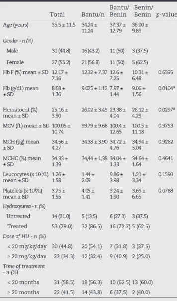
The clinical details of the steady-state SCA patients according to genotypes and the use of HU are shown in Tables 1 and 2, respectively. Of the 67 patients included in the study, 55.2% were female and 44.8% were male. The mean age was 35.48 (± 11.55) years. The patient group had a mean Hb concentration of 8.68 g/dL (± 1.36 g/dL), hematocrit (Ht) 25.16% (± 3.9%) mean corpuscular volume (MCV) 100.05 fL (± 10.74 fL), mean corpuscular hemoglobin (MCH) 34.56 pg (± 4.27 pg), mean corpuscular hemoglobin concentration (MCHC) 34.33% (± 1.39%), leukocytes 1.26 × 10⁹/L (± 1.58 ×
109/L), platelets 374.90 × 10⁹/L (± 154.59 × 10⁹/L), Hb F 12.17%
Total Bantu/n Bantu/Benin Benin/Benin p-value
Age (years) 35.5 ± 11.5 34.24 ±
11.24 37.37 ± 12.79 36.00 ± 9.89
Gender - n (%)
Male 30 (44.8) 16 (43.2) 11 (50) 3 (37.5)
Female 37 (55.2) 21 (56.8) 11 (50) 5 (62.5) Hb F (%) mean ± SD 12.17 ±
7.16 12.32 ± 7.37 12.6 ± 7.25 10.31 ± 6.48 0.6395 Hb (g/dL) mean
± SD 8.68 ± 1.36 9.025 ± 1.12 7.97 ± 1.44 9.06 ± 1.56 0.0104
a
Hematocrit (%)
mean ± SD 25.16 ± 3.90 26.02 ± 3.45 23.38 ± 4.04 26.12 ± 4.29 0.0297
a
MCV (fL) mean ± SD 100.05 ±
10.74 99.79 ± 9.68 100.4 ± 12.65 100.5 ± 11.18 0.9753 MCH (pg) mean
± SD 34.56 ± 4.27 34.38 ± 3.90 34.72 ± 4.76 34.94 ± 5.04 0.9262
MCHC (%) mean
± SD 34.33 ± 1.39 34,44 ± 1,38 34.04 ± 1.33 34.64 ± 1.64 0.4641 Leucocytes (x 109/L)
mean ± SD 1.26 ± 1.58 1.44 ± 2.09 9.86 ± 3.98 1.21 ± 3.34 0.1590
Platelets (x 109/L)
mean ± SD 3.75 ± 1.55 4.05 ± 1.41 3.24 ± 1.90 3.69 ± 6.65 0.0768
Hydroxyurea - n (%)
Untreated 14 (21.0) 5 (13.5) 6 (27.3) 3 (37.5) Treated 53 (79.0) 32 (86.5) 16 (72.7) 5 (62.5)
Dose of HU - n (%)
< 20 mg/kg/day 30 (44.8) 20 (54.1) 7 (31.8) 3 (37.5) ≥ 20 mg/kg/day 23 (34.3) 12 (32.4) 9 (40.9) 2 (25.0)
Time of treatment - n (%)
< 20 months 31 (58.5) 18 (56.3) 10 (62.5) 13 (60.0) ≥ 20 months 22 (41.5) 14 (43.8) 6 (37.5) 2 (40.0)
MCV: mean corpuscular volume; MCH: mean corpuscular hemoglobin; MCHC: mean corpuscular hemoglobin concentration.
a p-value < 0.05
Statistical analysis using Kruskal-Wallis test
Table 1 - Clinical details of steady-state sickle cell anemia patients (n = 67) according to genotypes.
Serum levels of TNF-a were signiicantly higher in SCA patients compared to healthy individuals (p-value < 0.0001; Figure 1).
Regarding the use of HU, TNF-a levels were decreased in patients who were receiving treatment compared to those who were not (p-value = 0.1249; Figure 2). Patients who were receiving HU at a dose of < 20 mg/kg/day did not have signiicantly different TNF-a levels compared to those who received doses ≥ 20 mg/kg/day (p-value = 0.3045). Regarding the duration of treatment with HU, patients receiving the drug for a period greater than or equal to 20 months showed no statistical difference in the TNF-a levels when compared to patients who were treated with HU for less than 20 months (p-value = 0.3034).
When comparing TNF-a levels between βS-globin
haplotypes, it was observed that the Bantu/n group had signiicantly higher TNF-a levels than the Bantu/Benin group (p-value = 0.0021; Figure 3).
Treated with
hydroxyurea Untreated p-value
Patients - n (%) 53 (79) 14 (21) Age (years) mean ± SD 35.35 ± 11.62 35.92 ± 11.67
Gender - n (%)
Male 23 (43.4) 7 (50) Female 30 (56.6) 7 (50)
Hb F (%) mean ± SD 13.28 ± 7.19 8.014 ± 5.470 0.0067a
Hb (g/dL) mean ± SD 8.93 ± 1.314 7.749 ± 1.168 0.0016a
Hematocrit (%) mean ± SD 25.85± 3.755 22.6 ± 3.458 0.0024a
MCV (fL) mean ± SD 100.9 ± 11.11 96.83 ± 8.846 0.1045 MCH (pg) mean ± SD 34.89 ± 4.334 33.30 ± 3.941 0.1082 MCHC (%) mean ± SD 34.35 ± 1.404 34.30 ± 1.419 0.4528 Leucocytes (× 109/L) mean
± SD 1.26 ± 1.76 1.28 ± 0.47 0.0421
a
Platelets (× 109/L) mean
± SD 3.70 ± 1.55 3.92 ± 1.57 0.3858
MCV: mean corpuscular volume; MCH: mean corpuscular hemoglobin; MCHC: mean corpuscular hemoglobin concentration.
ap-value < 0.05
Statistical analysis using Kruskal-Wallis test.
Table 2 - Clinical details of steady-state sickle cell anemia patients (n = 67) according to use of hydroxyurea.
Figure 1 – Box plot comparing TNF-a levels between Control Group and sickle cell anemia patients Mann-Whitney test (p < 0.0001).
Discussion
The recurrent vaso-occlusion-ischemic processes induce a chronic inlammatory response in SCA patients, which is characterized by high levels of proinlammatory cytokines that activate the endothelium.18 The activated endothelium
Regarding HU therapy, patients receiving treatment had lower TNF-a levels compared to patients who were not, but this difference was not signiicant. These results conirm those reported by Tavakkoli et al.19 who found no difference
in the TNF-a serum levels in SCA patients at steady-state in relation to receiving HU or not. Furthermore, the authors found no difference between the vaso-occlusive crises of these patients. On the other hand, Lanaro at al.,20 in a study with
50 SCA adult patients at steady-state on HU therapy, showed that they had signiicantly lower TNF-a levels compared to 26 untreated patients. So, the effect of HU therapy on the release of inlammatory mediators is not well understood.
In the current study, a higher prevalence of the Bantu haplotype was found compared to the Benin haplotype. This result is in agreement with the study by Silva et al.12 who used
the same population as ours and explained this result by the origins of the black population brought to the state of Ceará.
Studies of several different ethnic groups of SCA patients with distinct hematological characteristics suggested that the
βS-globin gene cluster haplotype may be useful as a predictor of disease severity.22-25 Furthermore, Bakanay et al.26 demonstrated
that the βS-globin gene cluster haplotype, independent of the Hb F level, is correlated to survival of SCA patients on HU therapy. In the present study, a signiicant increase of TNF-a
levels was observed in the Bantu/n group compared to the Bantu/Benin group. This result can be explained by the fact that patients with the Bantu haplotype have a tendency of having more severe clinical manifestations because of the increased concentration of Hb S and decreased concentration of Hb F.27 Sickled red blood cells exhibit abnormal adhesion to
the vascular endothelium, thus contributing to the expression of adhesion molecules and ampliication of inlammation by proinlammatory cytokines such as TNF-alpha.8,28
Conclusion
SCA expresses factors that inluence the clinical features of the patients. In this study one of these factors was evidenced, showing the modulatory role of the β-globin haplotypes on TNF-a levels in SCA adults at steady-state. Many previous studies have investigated the prognosis and inlammatory state of SCA patients, but the discovery that TNF-a levels vary according to the genetic polymorphism of the patient is a new inding.
Conflicts of interest
The authors declare no conflicts interest.
R E F E R E N C E S
1. Gualandro SF. A associação anemia falciforme e hemoglobina fetal. Rev Bras Hematol Hemoter. 2009;31:403-4.
the pancellular activation, which results in high circulating levels of the numerous inlammatory molecules found in SCA patients. Thus, there is a vicious cycle between production of inlammatory mediators and cell adhesion to the endothelium, leading to a chronic inlammation process.19
The results of the present study show that TNF-a levels were more elevated in patients with SCA compared to healthy individuals. Similar results were found by other studies in SCA patients at steady-state.20,21 This cytokine is fundamental for
inlammatory diseases because it up-regulates the cytokine cascade responsible for inlammation.
Figure 3 – Box plot comparing TNF-α levels between genotypes of sickle cell anemia patients. Kruskal-Wallis test. Statistical significance was observed between the Bantu/n and Bantu/Benin Groups (p = 0.0022).
Figure 2 – Box plot comparing TNF-α levels between sickle cell
2. Rees DC, Williams TN, Gladwin MT. Sickle-cell disease. Lancet. 2010;376:2018-31.
3. Di Nuzzo DV, Fonseca SF. Anemia falciforme e infecções. J Pediatr (Rio J). 2004;80:347-54.
4. Steinberg MH. Predicting clinical severity in sickle cell anaemia. Br J Haematol. 2005;129:465-81.
5. Zago MA, Pinto AC. Fisiopatologia das doenças falciformes: da mutação genética à insuficiência de múltiplos órgãos. Rev Bras Hematol Hemoter. 2007;29:207-14.
6. Santos JL, Chin CM. Anemia falciforme: desafios e avanços na busca de novos fármacos. Quím Nova. 2012;35:783-90. 7. Vargas AE. Expressão gênica e perfil imunogético de
pacientes com anemia falciforme. [thesis]. Porto Alegre: Universidade Federal do Rio Grande do Sul; 2009.
8. Cajado C, Cerqueira BA, Couto FD, Moura-Neto JP, Vilas-Boas W, Dorea MJ, et al. TNF-alpha and IL-8: Serum levels and gene polymorphisms (-308G>A and -251A>T) are associated with classical biomarkers and medical history in children with sickle cell anemia. Cytokine. 2011;56:312-7.
9. Raghupathy R, Haider MZ, Azizieh F, Souza TMD, Abdelsalam R, Adekile AD. Tumor necrosis factor-a is undetectable in the plasma of SS patients with elevated Hb F. Am J Hematol. 2000;64:91-4.
10. Cajado CS, Cerqueira BA, Barbosa CG, Lyra IM, Adorno EV, Gonçalves MS. IL-8 e TNF-alfa: Marcadores imunológicos no prognóstico da anemia falciforme. Gaz Med Bahia. 2010;80:56-61.
11. Figueiredo MS. Fatores moduladores da gravidade da evolução clínica da anemia falciforme. Rev Bras Hematol Hemoter. 2007;29:215-7.
12. Silva LB, Gonçalves RP, Rabenhorst SH. Análise dos haplótipos da anemia falciforme em Fortaleza revela as origens étnicas da população cearense. J Bras Patol Med Lab. 2009;45:115-8.
13. Fleury MK. Haplótipos do cluster da globina beta em pacientes com anemia falciforme no Rio de Janeiro: Aspectos clínicos e laboratoriais. RBAC. 2007;39:89-93.
14. Bandeira FM, Peres JC, Carvalho EJ, Bezerra I, Araújo AS, Mello MR, et al. Hidroxiuréia em pacientes com síndromes falciformes acompanhados no Hospital Hemope, Recife-PE. Rev Bras Hematol Hemoter. 2004;26:189-94.
15. Figueiredo MS. Agentes indutores da síntese de hemoglobina fetal. Rev Bras Hematol Hemoter. 2007;29:313-5.
16. Cançado RD, Lobo C, Ângulo IL, Araújo PI, Jesus JA. Protocolo clínico e diretrizes terapêuticas para uso de hidroxiureia na doença falciforme. Rev Bras Hematol. 2009;31:361-6.
17. Ballas SK. More definitions in sickle cell disease: Steady state v base line data. Am J Hematol. 2012;87:338.
18. Okapala I. Leukocyte adhesion cell disease. Curr Opin Hematol. 2006;13:40-4.
19. Tavakkoli F, Masoud N, Melville Q, Perlin E. Plasma levels of TNF-a in sickle cell patients receiving hydroxyurea. Hematology. 2004;9:61–4.
20. Lanaro C, Franco-Penteado SF, Albuqueque DM, Saad ST, Conran N, Costa FF. Altered levels of cytokines and inflammatory mediators in plasma and leukocytes of sickle cell anemia patients and effects of hydroxyurea therapy. J Leukoc Biol. 2009;85:235-42.
21. Saleh AW, Hillen HF, Duits AJ. Levels of endothelial, neutrophil and platelet-specific factors in sickle cell anemia patients during hydroxyurea therapy. Acta Haematol. 1999;102:31-7.
22. Ballas SK, Talacki CA, Adachi K, Schwartz E, Surrey S, Rappaport E. The XMN I site (-158, C fi T) 5¢ to the Gamma gene: Correlation with the Senegalese haplotype and Gc globin expression. Hemoglobin, 1991;15:393–405.
23. Dimovski AJ, Oner C, Agarwal S, Gu YC, Gu LH, Kutlar F, et al. Certain mutations observed in the 5’ sequences of the Gc-and Ac-globin genes of bS chromosomes are specific for chromosomes with major haplotypes. Acta Haematol. 1991;85:79-87.
24. Powars DR. Sickle cell anemia: bs-gene-cluster haplotypes as prognostic indicators of vital organ failure. Semin Hematol. 1991;28:202-8.
25. Zago MA, Figueiredo MS, Ogo SH. Bantu bs cluster haplotype predominates among Brazilian Blacks. Am J Phys Anthropol. 1992;88:295-8.
26. Bakanay SM, Dainer E, Clair B, Adekile A, Daitch L, Wells L, et al. Mortality in sickle cell patients on hydroxyurea therapy. Blood. 2005;105:545-7.
27. Galiza Neto GC, Pitombeira MS, Vieira HF, Vieira MLC, Farias, DAB. Análise dos haplótipos do gene da ßS-globina no Ceará. J Bras Patol Med Lab. 2005;41:315-21.