Artigo
Received 08/29/06. Accepted 05/26/07Brazilian Journal of Pharmacognosy 17(3): 349-355, Jul./Set. 2007
The infl uence of the essential oil of
Melaleuca alternifolia
on the
healing of infected dental alveoli: A histological study in rats
Maria Regina Orofi no Kreuger
1,2*, Carlos Eduardo Ternes
1, Leonardo Lamim Mello
1,
Alexandre Bella Cruz
2, Silvana Nair Leite
2, David Rivero Tames
11Laboratório de Pesquisas do Curso de Odontologia da Universidade do Vale do Itajaí, Rua Uruguai 458,
88303-202, Itajaí, SC, Brazil,
2Núcleo de Investigações Químico-Farmacêuticas (NIQFAR), Curso de Farmácia,
Universidade do Vale do Itajaí, 88303-202, Itajaí, SC, Brasil
RESUMO: “Infl uência do óleo essencial de Melaleuca alternifolia na cicatrização de alveolite dental infectada: um estudo histológico em ratos”. A planta Melaleuca alternifolia é nativa da Austrália e a destilação de suas folhas produz um óleo essencial conhecido por óleo de Melaleuca, ou Tea tree oil, usado como antimicrobiano. Nesse estudo, foi verifi cado a atividade deste óleo no processo de reparo de alvéolos dentais infectados. 48 ratos foram utilizados (Rattus novergicus albinus, Wistar) e após a extração do dente e posterior infecção do alvéolo com Staphylococcus aureus, os animais foram separados em três grupos: Grupo I: curetagem e irrigação do alvéolo com soro fi siológico; Grupo II: curetagem e irrigação com soro fi siológico e tratamento com Rifocina 25 mg; e Grupo III: curetagem e irrigação com soro fi siológico e aplicação tópica de óleo de Melaleuca 20%. Os animais foram sacrifi cados 24 horas, 7, 14 e 21 dias após o tratamento e o processo de reparo do alvéolo dental foi analisado por microscopia ótica. Os resultados foram submetidos à análise quantitativa e qualitativa e foi possível concluir que o óleo a 20% causou um retardo no processo de reparo dos alvéolos dentais infectados dos ratos, demonstrado por maior área de necrose e menor osteogênese.
Unitermos: Melaleuca alternifolia, Staphylococcus aureus, alvéolo dental.
ABSTRACT: The plant Melaleuca alternifolia is native to Australia. The distillation of its leaves produces an essential oil, commonly known as oil of Melaleuca, or Tea tree oil, which present antimicrobial activity. This study investigates the action of this oil on the repair process of infected dental alveoli. 48 rats were used (Rattus novergicus albinus, Wistar). After tooth extraction and posterior infection of the dental alveoli with Staphylococcus aureus, the animals were separated into three groups: Group I: curettage and irrigation with physiologic saline solution; Group II: curettage and irrigation with physiologic saline solution and topical application of rifamycin diethylamide B 25 mg; and Group III: curettage and irrigation with physiologic saline solution and topical application of oil of Melaleuca 20%. The animals were sacrifi ced 24 hours, 7, 14 and 21 days after the treatment with powder and the repair process of the dental alveoli was analyzed using an optical microscope. The results were submitted to qualitative and quantitative analysis and it was concluded that tea tree oil at 20% caused a delay in the repair process of infected dental alveoli in rats, as demonstrated by the presence of more necrosis area and less osteogenesis.
Keywords: Melaleuca alternifolia, Staphylococcus aureus, dental alveoli.
INTRODUCTION
The most common complication in the healing of extraction wounds is fi brinolytic alveolitis, also termed “dry sockets”. It is an infl ammatory disease that attacks the buccal cavity, specifi cally, the more superfi cial bony portions of the dental alveolus. It presents multiple etiology; however, the main cause is due to the penetration of bacteria, after extraction. This complication settles at around 48 or 72 hours after surgery (Gregory, 1996). Due to its high incidence rate among postoperative complications in surgery, alveolitis
has been studied in depth, in order to determine the best treatment and consequently, to lower its frequency (Shafer et al., 1987). A reduction in the incidence of this condition after the application of topical or systemic antibiotics has been reported. Rifocin M® or rifamycin diethylamide B, when applied topically, presents great effectiveness, and is used in dentistry for the treatment of infections, fi stulas or ulcer washes and in the irrigation of pulp chamber or root channels (Bazerke, 1978).
that oil of Melaleuca has antiseptic activities (Schulz, 2000). Oil of Melaleuca or Tea Tree Oil (TTO) has been reported to have anti-bacterial (Carson et al., 2006; Luz; Janaina, 2007), antifungic (Hammer et al., 1997, Oliva et al., 2003), antiviral (Bishop, 1995), anti-tumor activity (Calcabrini et al., 2004) and anti-infl ammatory properties in vitro (Brand et al., 2001; Hart et al., 2000). Oil of Melaleuca has a wide spectrum of activity that is mainly attributed to terpeno-4-ol. It possesses effective antimicrobial activity against Gram-positive bacteria, such as Staphylococcus aureus and Gram-negative bacteria, such as Escherichia coli (Cox et al., 2000) or
Salmonella typhimurium (Wilkinson; Cavanagh, 2005). Depending on the condition to be treated, the oil can be used either undiluted, or diluted in water (concentrations of 2-10% are common). The undiluted oil causes greater skin irritation or allergic reactions, and its internal use is not recommended (Schulz, 2000). TTO has been used for centuries as a botanical medicine, but it is only in recent decades that it has emerged in the scientifi c literature as a promising adjunctive wound treatment (Halcón et al., 2004). Due to this information, we decided to investigate the effect of TTO on the healing of dental sockets infected with Staphylococcus aureus.
MATERIAL AND METHODS
Animals
Forty-eight male Wistar rats (Rattus norvegicus albinus) weighing 280 to 320 g were used. The animals were kept in their cages and received water and food ad libitum during the experiment.
Bacteria
To investigate the effect of the infection, the bacterium Staphylococcus aureus (ATCC 6538P) was used. It is purchased from the tropical culture collection of “André Tosello Technology and Research Tropical Foundation”, Campinas, State of São Paulo, Brazil. The inoculum suspension was prepared from overnight broth culture (Mueller-Hinton) and adjusted to 0.5 McFarland turbidity equivalent (approximately 1.5 x 10⁸ cells/mL). A suspension with 50μL of inoculum of S. aureus was performed for each alveolus.
Gel preparation
A neutral gel of 75% Carbopol, 5% PVP and 20% oil of Melaleuca (Tea Tree Oil) was prepared.
Tooth extraction and treatment
For the tooth extraction, the animals were anesthetized intramuscularly with Ketamina 8 mg/kg weight and Xylazina 10 mg/kg weight (Teles et al., 1997).
The right maxillary incisor was extracted according to the technique previous described (Okamoto; Russo 1973). Next, the ischemia of the alveolus was provoked by the introduction of a paper cone soaked in adrenaline solution 1:1000 for 1 minute. After this, a suspension with 50 μL of 108 cells/mL of bacteria Staphylococcus aureus was injected into the dental alveolus. 48 hours after the induction of the infection, the dental sockets of the animals were curetted and irrigated with saline solution 0.9%. The animals were then divided into three groups, according to the treatment to be administered. Group I (n = 16), irrigation with 1.5 mL of saline solution, Group II (n = 16), topical application with 1.5 mL of rifamycin diethylamide B (Rifocin M®) 25 mg and Group III (n = 16), topical application of a neutral gel with oil of Melaleuca (Tea Tree Oil) in suffi cient quantities to cover the socket. Each alveolus received only one application of each chemical substance. Four rats from each group were sacrifi ced at 24 hours, 7, 14, and 21 days after treatment. The right maxilla was separated from the left one a median sagittal incision. Another incision was made, tangential to the distal surface of the molars, to obtain a block containing the right incisor alveolus. The blocks were fi xed in neutral 10% formalin, decalcifi ed in equal parts of 20% sodium citrate and 50% formic acid, and embedded in paraffi n. Sections were cut at 7 µM and stained with hematoxylin and eosin (HE) for histological examination.
Histological analysis
The middle third of the socket was analyzed. A single blind microscopic evaluation of three areas (anterior, medium and posterior) of serial sections from each lesion was carried out. A reticule eyepiece was used to measure the percentage of the necrosis areas and osteogenesis (objective 20x). The mononuclear cells and polymorphonuclear neutrophils were determined numerically using an optical microscope (objective 100 x). The averages for the three sets were determined.
Statistical analysis
Signifi cant differences in necrosis area, quantifi cation of cells and vessels were compared by using the analysis of variance (ANOVA). The differences between the groups were compared by Tukey’s test; P
values of < 0.05 were considered signifi cant.
RESULTS
24 Hours after treatment
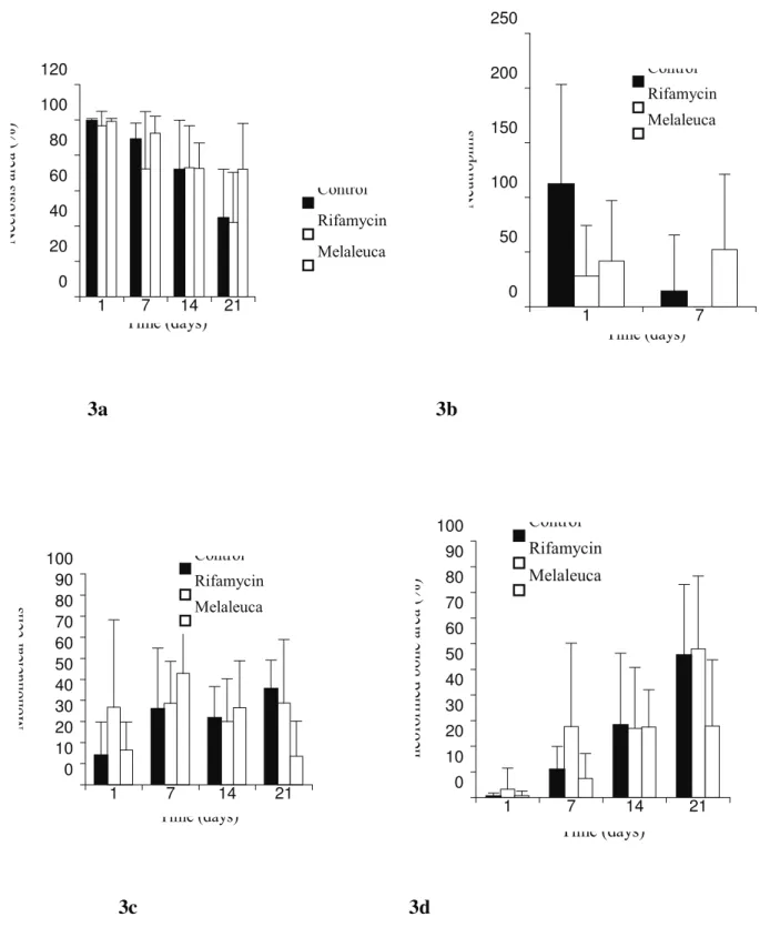
clotting and exudate. Figure 3a shows that the area of necrosis was similar in all the groups at this time. A large number of polymorphonuclear neutrophils were observed. The control group (treated with saline solution) demonstrated a higher number of this type of leukocyte. The value was not considered statistically signifi cant (P < 0.01) in relation to the group treated with TTO, but was in accordance with the group treated with rifamycin diethylamide B (Figure 3b). Moderate infi ltration of mononuclear leukocytes was present, which was higher in the rifamycin group (Figure 3c). For all the groups, it was impossible, at this time, to observe the proliferation of osteogenesis (Figure 3d).
7 Days after treatment
Necrosis area, exudate and clotting still persisted, but were less intense in the group treated with rifamycin (P < 0.01) than in the group treated with tea tree oil or the control group (Figure 3a). After 7 days, the sockets treated with oil of Melaleuca showed extensive infl ammation, infi ltrated with areas of necrosis and interspersed with areas of granulation tissue. Although in dental alveoli treated with rifamycin and alveoli irrigated with saline solution, blood clotting still persists, but neoformed bone trabeculae was observed, as well as the presence of granulation tissue, partially fi lling the alveolus. The dental alveolus treated with oil of Melaleuca presented little osteoneogenesis. Figure 3d represents the reading for bony neoformation after 7 days. The dental alveolus treated with rifamycin presented a greater area of neoformed bone (P < 0.05) when compared with the other two groups. In the analysis of leukocytes in the lesion area, a prevalence of polymorphonuclear neutrophils was obtained for the group treated with TTO. The other two groups presented small amount of these cells in the infl ammatory exudates, while they were almost non existent after treatment with rifamycin, and not very evident in the dental alveolus curetted and irrigated with physiological serum (Figure 3b). Figure 3c represents the average reading for the mononuclear leukocytes after 7 days of treatment, evidencing similar values in the number of these cells for all the groups studied.
14 Days after treatment
All the groups presented the dental alveolus fi lled with thin and regular bone trabeculae and granulation tissue. However, the characteristics of the trabecular bone were different in each group. Areas of remaining blood clotting still persisted in some analyzed lesions, which were less evident in the lesions treated with rifamycin. These animals showed thicker and matured bone compared to the others, particularly when compared with the dental alveolus treated with oil of Melaleuca, which presented immature and thinner
neoformed bone. Treatment with TTO unleashed a large area of necrosis in the sockets, compared with the two other groups but the average reading of the dental alveolus analyzed did not differ statistically among the three groups (Figure 3a). Some polymorphonuclear neutrophils were observed at this time (Figure 3b). Mononuclear cells were prevalent, and the group treated with TTO showed a higher number of this cell type, though this difference was not statistically signifi cant (Figure 3c). The mean bony neoformation did not differ among the all groups after 14 days, rather, the characteristics of the trabecular bone were different (Figure 3d).
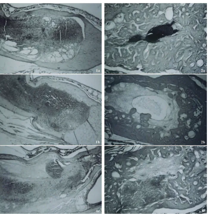
21 Days after treatment
The sockets treated with rifamycin diethylamide B or saline solution presented a dental alveolus fi lled with thick, mature trabecular bone in the peripheral area. The central portion of the alveolus showed portions of connective tissue without bone differentiation, and a few areas of blood clotting. The animals treated with oil of Melaleuca showed sockets with the same aspects at 14 days, and there still remained an extensive area of tissue necrosis. Many cells without contours and nuclear material were observed (Figures 2 a, b and c). The average percentage of necrosis area in the sockets treated with TTO was more extensive (P < 0.05) than in the sockets treated with rifamycin or saline solution (Figure 3a). Figure 3c shows the reduction in the number of mononuclear cells in the dental alveolus which received oil of Melaleuca, compared with the other two groups. The extensive necrosis in the alveoli treated with TTO prevented an adapted reading of the number of mononuclear cells in the lesion. This persistent area of necrosis also impaired the growth of new bone in the sockets with TTO (P < 0.01), compared with the other groups (Figure 3d).
DISCUSSION
Alveolitis still remains an unpleasant organic reaction following dental extraction, and is one of the most extensively studied infl ammatory diseases in dentistry. Oil of Melaleuca (Tea tree oil - TTO) has recently gained a reputation as a safe, natural and effective antiseptic. This has led to an increase in its popularity, and it has now become a major antimicrobial, or a natural preservative in many pharmaceutical and cosmetic products for external use (Halcón et al., 2004).
of trabecular bone, and at 21 days powder-treatment, an evident bony differentiation with bony trabeculae almost fi lling for total the alveolus. Treatment with rifamycin has proven to be effi cient against bacteria, especially Gram-positive bacteria such as S. aureus, and is used to treat infectious dental alveolitis (Ogawa; Mariano, 1997).
The results for group I results (irrigation with physiologic serum) were similar to those described in the literature, such as the presence of blood clotting fi lling the dental alveolus partially, and a high number of polymorphonuclear neutrophils in the fi rst few days, followed by the neoformation of granulation tissue and bony trabeculae partially fi lling the alveolus after 21 days (Carvalho et al., 1991; Poi et al., 2000). This group showed better development than the dental alveolus treated with oil of Melaleuca. The results of this study demonstrated a high level of cytotoxic activity in the lesions treated with TTO, impairing normal osteogenesis. The analysis showed that the treatment determined a larger area of necrosis and less osteogenesis in the sockets. This necrosis presented an acidophilic staining, resembling a coagulation necrosis of the cells. Some studies have demonstrated that this oil causes destructive effects in the cellular membrane of the bacteria. The analysis of Escherichia coli cells by electron microscopy after exposure to TTO showed a loss of cellular electrondense material and coagulation of cytoplasmic constituents (Gustafsson et al., 1998). The ability of TTO to disrupt the permeability barrier of cell membrane structures and the accompanying loss of chemiosmotic control, as the most likely source of its lethal action at minimum inhibitory level, was described (Brand et al., 2001). These benefi cial effects of TTO on the bacteria can prove toxic and lethal for the remaining healthy tissue in the alveolus, and for the granulation tissue in proliferation, justifying the smaller amount of osteogenesis and the persistent area of necrosis found in the lesions treated with this oil in the present study. In an in vitro study, concentrations of 300 mg/mL and 500 mg/mL of TTO caused 50% mortality of the human ephitelial cells and fi broblasts after 24 or 48 hours of incubation (Soderberg et al., 1996). In another study
in vitro, the tea tree oil emulsifi ed into culture medium containing 10% fetal calf serum was toxic to monocytes at a concentration of 0.016% v/v (Hart et al., 2000) and for neutrophils in the same concentration (Brand et al., 2001). The high concentration (20%) of neutral gel used in this experiment could be the reason for the toxicity of the cells.
In spite of the toxicity induced by TTO, it did not inhibit the infl ux of leukocytes to the area of the lesion. The presence of high levels of polymorphonuclear neutrophils in the group treated with TTO suggests high cytotoxic activity in the lesion. Leukocytes secrete free radicals from oxygen and enzymes that damage the tissue, but their ability to produce these nitrogen and oxygen
reactive species, enzymes and cytokines, is essential for antibacterial ability. The soluble components of oil of Melaleuca suppress the production of superoxide ions by human neutrophils in vitro, but not of monocytes (Bishop, 1995). It was demonstrated that terpeno-4-ol, the largest component of essential oil of Melaleuca, suppresses the synthesis of infl ammatory mediators for activated human monocytes. The composition inhibited the production of cytokines such as Tumor Necrosis Factor (TNF), Interleukin-1b (IL-1b), IL-8 and IL-10, as well as the lipidic mediator prostaglandin, when the monocytes are activated by bacterial lipopolysaccharide (Hart et al., 2000). This essential oil is a compound of approximately 100 components. Little is known of the toxicity of the individual components, or the oil as a whole. The lipophilic nature of tea tree oil and its ability to penetrate the skin may increase its toxicity (Carson; Riley, 1995).
Although there are many results demonstrating
in vitro the antibacterial activity of TTO, more animal studies in vivo need to be carried out. Given the widespread availability of an extensive range of tea tree oil products, the paucity of accurate toxicity data is a problem. There is a need for more research in this area to further demonstrate the effi cacy and animal or patient tolerance of tea tree oil as a topical treatment, in order to identify active components and the synergy or antagonism between the components.
ACKNOWLEDGMENTS
The authors are grateful to Maria de Lourdes Correa and Beatris Pacheco Correa for their technical support.
REFERENCES
Bazerke P 1978. Farmacologia Odontológica. Buenos Aires: Mundi.
Bishop CD 1995. Antiviral activity of essential oil of Melaleuca alternifolia (Maiden and Betche) Cheel (tea tree) against tobacco mosaic virus. J Essent Oil Res 7: 641-644.
Brand C, Ferrante A, Prager RH, Riley TV, Carson CF, Finlay-Jones JJ, Hart PH 2001. The water-soluble components of essential oil of Melaleuca alternifolia (tea tree oil) suppress the production of the superoxide by humam monocytes, but not neutrophils, activated in vitro. Infl amm Res 50: 213-219.
Calcabrini A, Stringaro A, Toccacieli L, Meschini S, Marra M, Colone M, Salvatore G, Mondello F, Arancia G, Molinari A 2004. Terpinen-4-ol, the main component of Melaleuca alternifolia (tea tree) oil inhibits the in vitro growth of human melanoma cells. J Invest Dermatol 122: 349-360.
Carson CF, Riley TV 1995. Toxicity of the essential oil of Melaleuca alternifolia or tea tree oil. J Toxicol Clin Toxic 33: 193-194.
Figure 1. After 24 h. Groups: Control (1a) rifamycin (1b) and TTO (1c). Middle third of the socket presents clot, exudate and leukocyte infi ltration (objective 20 x).
0 20 40 60 80 100 120
1 7 14 21
Time (days)
Necrosi
s area (%) Control
Rifamycin
Melaleuca
0 50 100 150 200 250
1 7
Time (days)
N
eutr
ophils
Control
Rifamycin
Melaleuca
3a 3b
Figure 3. 3a -Percentage of necrosis area in control, TTO and rifamycin treated dental alveoli. After 21 days, the TTO Group presented higher necrosis area (P > 0.005) compared with the other two groups (objective 20 x). 3b - Number of polymorphonuclear neutrophils in the dental alveolus that received saline or treatment (objective 100 x). 3c - Number of mononuclear cells in the dental alveolus (objective 100 x). 3d - Percentage of osteogenesis dental alveolus (objective 20 x). After 7 days, the rifamycin group presented higher osteogenesis (P > 0.05) compared with the others two groups. All the data correspond to the mean of three areas examined (n = 8).
0 10 20 30 40 50 60 70 80 90 100
1 7 14 21
Time (days)
Mononuclear
cells
Control
Rifamycin
Melaleuca
0 10 20 30 40 50 60 70 80 90 100
1 7 14 21
Time (days)
neoform
ed bone area (%
)
Control Rifamycin Melaleuca
alternifolia (Tea Tree) oil: a review of antimicrobial and other medicinal properties. Clin Microbiol Rev 19: 50-62.
Carvalho PSP, Okamoto T, Barbosa DZ 1991. Infl uência da limpeza cirúrgica e/ou aplicação de alveosan no processo de reparo em feridas de extração dental infectadas. Estudo histológico em ratos. Rev Odonto UNESP 20: 165-173.
Carvalho PSP, Mariano RC, Okamoto T 1997. Treatment of fi brinolytic alveolitis with rifamycin B diethylamide associated with gelfoam: a histological study. Braz Dent J 8: 3-8.
Cox SD, Mann CM, Markham JL, Bell HC, Gustafson JE, Warmington JR, Wyllie SG 2000. The mode of antimicrobial action of essential oil of Melaleuca alternifolia (tea tree oil). J Appl Microbiol 88: 170-175.
Gregori C 1996. Cirurgia Buco-dento-alveolar. São Paulo: Sarvier.
Gustafsson JE, Liew YC, Chews S, Markham J, Bell HC, Wyllie SG, Warmington JR 1998. Effects of tea tree oil on Escherichia coli. Lett Appl Microbiol 26: 194-198.
Halcón H, Milkus K 2004 Staphylococcus aureus and wounds: A review of tea tree oil as a promising antimicrobial. Am J Infection Control 32: 402-408.
Hammer KA, Carson CF, Riley TV 1997. In vitro susceptibility of Malassezia furfur to the essential oil of Melaleuca alternifolia. J Med Vet Mycol 35: 3705-3707. Hart PH, Brand C, Carson CF, Riley TV, Prager F,
Finlay-Jonnes JJ 2000. Terpinen-4-ol, the main component of Melaleuca alternifolia (tea tree) oil suppresses infl ammatory mediator production by activated human monocytes. Infl amm Res 49: 619-626.
Ogawa R, Mariano RC 1997. Alveolite e as complicações pós-extrações de terceiros molares inferiores retidos. Trabalho para obtenção do certifi cado de estágio na disciplina de Cirurgia Buco Maxilo-Facial. Escola de Farmácia e Odontologia de Alfenas, MG.
Okamoto T, Russo MC 1973. Wound healing following tooth extraction. Histological study in rats. Rev Fac Odontol Araçatuba 2: 153-164.
Oliva P, Piccirili E, Ceddia T, Pontieri T, Aureli P, Ferrini AM 2003. Antimycotic activity of Melaleuca alternifolia essential oil and its major components. Lett Appl Microbiol 37: 85-87.
Packer JF, Luz MMS 2007. Método para avaliação e pesquisa da atividade antimicrobiana de produtos de origem natural. Rev Bras Farmacogn 17: 102-107.
Poi WR, Carvalho ACP, Okamoto T 2000. Infl uência da pasta sultan sobre o processo de reparo em alvéolo dental infectado. Análise histológica em ratos. Robrac 9: 9. Schulz V 2000. Rational phytoterapy: A physician’s guide to
herbal Medicine 2000. West Lafayette: Springer. Shafer WG, Hine MK, Levy BM 1987. Tratado de patologia
bucal. Rio de Janeiro: Guanabara-Koogan.
Soderberg TA, Johansson A, Gref R 1996. Toxic effects of some conifer resin acids and tea tree oil on human epithelial and fi broblast cells. Toxicol 107: 99-109. Teles R, Wang CY, Stashenko P 1997. Increased susceptibility
of RAG-2 mice to dissemination of endodontic infections. Infect Immun 6: 3781-3787.
Wilkinson JM, Cavanagh HM 2005. Antibacterial activity of