Article
J. Braz. Chem. Soc., Vol. 27, No. 3, 599-604, 2016. Printed in Brazil - ©2016 Sociedade Brasileira de Química 0103 - 5053 $6.00+0.00
A
*e-mail: [email protected]
Chemotaxonomy of the Amazonian
Unonopsis
Species Based on Leaf Alkaloid
Fingerprint Direct Infusion ESI-MS and Chemometric Analysis
Felipe M. A. Silva,*,a Francinaldo A. Silva Filho,a Bruna R. Lima,a
Richardson A. Almeida,a Elzalina R. Soares,a Hector H. F. Koolen,a,b
Afonso D. L. Souzaa and Maria L. B. Pinheiroa
aDepartamento de Química,Universidade Federal do Amazonas, 69077-000 Manaus-AM, Brazil
bGrupo DeMpSter de Espectrometria de Massas, Universidade do Estado do Amazonas, 69050-010 Manaus-AM, Brazil
Unonopsis (Annonaceae) is a neotropical genus constituted by nearly fifty species, with fifteen described in Brazil. In the state of Amazonas seven species are found, including U. guatterioides, which displays problems from the botanical viewpoint. Previous studies showed this genus as a promising source of aporphinoid alkaloids. In order to investigate the potential of the leaf alkaloid fingerprint for chemotaxonomic approaches, twelve Unonopsis specimens, representing five species commonly found in the state of Amazonas were subjected to acid-base partitioning to yield the respective alkaloidal fractions. These fractions were analysed by direct infusion electrospray ionization multiple stage mass spectrometry (ESI-MSn). The obtained data were treated through
chemometric tools [principal components analysis (PCA) and hierarchical cluster analysis (HCA)]. Multivariate analysis pointed to aporphine, proaporphine and tetrahydroprotoberberine alkaloids as the responsible compounds for segregation of the investigated species, being these alkaloids tentatively identificated by multiple stage mass spectrometry. The alkaloid fingerprint along with multivariate analysis provided a simple and effective approach to differentiate Unonopsis species commonly found in the state of Amazonas.
Keywords: alkaloid fingerprint, aporphinoid alkaloids, chemotaxonomy, Unonopsis
Introduction
Unonopsis (Annonacaeae) is a Neotropical genus (Central and tropical South American regions) constituted by approximately fifty species, being twenty five relatively new.1 This genus presents taxonomic problems clearly
observed through the numerous attempts to position it within the Annonaceae family, as well as the difficulty of classify some species such as U. guatterioides, which recently had 13 species incorporated as synonyms.1
In Brazil, fifteen Unonopsis species are described, some of them presenting restricted geographical distribution such as U. duckei. In the state of Amazonas besides U. duckei, the species U. floribunda, U. guatterioides, U. rufescens, U. stipitata, U. spectabilis and U. veneficiorum are also cataloged.1 The leaves of some species are used for
medicinal purposes, as example, leaves of U. stipitata and
U. veneficiorum are added in the food of indigenous people presenting brain disorders.2
Chemical studies point the Unonopsis genus as a promising source of aporphinoid alkaloids, being aporphine sensu stricto, oxoaporphines, azafluerenones, phenanthrenes and proaporphines recurrent in the literature.3,4 In addition,
the triterpene polycarpol, considered a chemotaxonomic marker in Annonaceae family, has been identified in all Unonopsis species subjected to phytochemical approaches.5
Chemosystematics, the science by which chemical characters are applied in the formal systematics of plants, fungi, etc., is considered a useful tool, along with the morphology, anatomy and cytogenetics, in the systematic organization.6 However, it is observed that
the success of this approach depends on the maximum accumulation of information about the distribution of secondary metabolites in a particular taxon.7 Considering
consequently a large amount of chemical data is obtained, making necessary the use of statistical tools for maximum exploitation of the information.9,10
Electrospray ionization mass spectrometry (ESI-MS) fingerprinting has been demonstrated to be a useful technique for the screening of organic compounds of plant origin.11 For example, the technique has been applied to
the screening of alkaloids in Unonopsis4 and Bocageopsis
species.12 This technique provides immediate compositional
information about ESI ionizable compounds through direct infusion, such as: products of acid-base, complexation and redox reactions,11,13 allowing the identification of
characteristic compounds in extracts.14,15 Although direct
infusion ESI-MS analyses present problems related to matrix effects, which results in reduced sensitivity and capabilities for metabolite identification,16 it has been
successfully applied as a fast fingerprinting method.4,11,12,14
Some applications presented classification and prediction results comparable to LC-MS analysis.16
In this work, the alkaloid fractions from the leaves of the twelve Unonopsis specimens, representing five species commonly found in the state of Amazonas, were analyzed using direct infusion ESI-MS and multiple stage mass spectrometry (MSn). The data were treated through
chemometric tools to investigate the potential of the leaf alkaloid fingerprint for chemotaxonomic approaches.
Experimental
Plant material
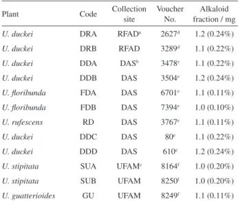
For this study, twelve Unonopsis specimens were used. The leaves from the cited species were collected from the three main sites in the state of Amazonas: Distrito Agropecuário da SUFRAMA (DAS), Reserva Florestal Adolpho Ducke (RFAD) and Universidade Federal do Amazonas (UFAM) campus. From DAS, 4 specimens of U. duckei, 2 specimens of U. floribunda, and 1 specimen of U. rufescens were collected. From RFAD, 2 specimens of U. duckei were collected. From UFAM, 2 specimens of U. stipitata and 1 specimen of U. guatterioides were collected. All the species were sampled in September 2012. A voucher specimen of each individual is deposited according to Table 1.
Alkaloid extraction
The alkaloid fractions were obtained according to a previously reported method.12 The leaves were immediately
dried over ambient temperature (ca. 20 °C) during 20 days, then powdered. The pulverized (500 mg each, except
FDA, FDB, RD and GU, which were used 1,000 mg each) was vigorously stirred using a vortex mixer for 1 min with a mixture of 10% ammonium hydroxide (NH4OH)
aqueous solution (pH 11) (5 mL) and dichloromethane (CH2Cl2) (5 mL) in a glass container. The organic phase
was transferred to another glass container and vigorously stirred (1 min) with a 10% acetic acid (CH3CO2H) aqueous
solution (pH 0.5) (5 mL). The aqueous phase was removed, treated with NH4OH to achieve pH 10 and extracted under
vigorous stirring (1 min) with CH2Cl2 (5 mL). The organic
phase was dried over anhydrous sodium sulfate, transferred to a new container and the solvent evaporated to dryness under a nitrogen gas stream to yield the crude alkaloid fraction. The obtained masses for the alkaloidal fractions are presented in Table 1.
Mass spectrometry analysis
Stock solutions (1 mg mL−1) of the leaves alkaloidal
fractions, were prepared with methanol. Aliquots (5 µL) of the stock solutions were further diluted to 5 µg mL−1
and analyzed by direct infusion into the mass spectrometer. All mass spectra were acquired in a continuous monitoring mode (Thermo LCQ Fleet Tune application) using a LCQ Fleet ion-trap mass spectrometer (Thermo LCQ Fleet, San Jose, CA, USA) with an electrospray ionization (ESI) interface and running in the positive ion mode to perform ESI-MS and ESI-MSn analyses. Spectra
were obtained from the mean of at least 10 scans per Table 1. Unonopsis collected in Amazonas state, Brazil
Plant Code Collection
site
Voucher No.
Alkaloid fraction / mg
U. duckei DRA RFADa 2627d 1.2 (0.24%)
U. duckei DRB RFAD 3289d 1.1 (0.22%)
U. duckei DDA DASb 3478e 1.1 (0.22%)
U. duckei DDB DAS 3504e 1.2 (0.24%)
U. floribunda FDA DAS 6701e 1.1 (0.11%)
U. floribunda FDB DAS 7394e 1.0 (0.10%)
U. rufescens RD DAS 3767e 1.1 (0.11%)
U. duckei DDC DAS 80e 1.1 (0.22%)
U. duckei DDD DAS 610e 1.2 (0.24%)
U. stipitata SUA UFAMc 8164f 1.0 (0.20%)
U. stipitata SUB UFAM 8250f 1.0 (0.20%)
U. guatterioides GU UFAM 8249f 1.1 (0.11%) aReserva Florestal Adolpho Ducke; bDistrito Agropecuário da
SUFRAMA; cUniversidade Federal do Amazonas; dherbarium of Instituto
Nacional de Pesquisas da Amazônia (INPA); ebotany collection of
Projeto Dinâmica Biológica de Fragmentos Florestais (PDBFF)/INPA;
spectrum. Samples were directly infusedinto the ion source through the instrument syringe pump (10 µL min−1). The
MS analytical conditions were: spray voltage, 5 kV; sheath gas, 10 arb; auxiliary gas, 5 arb; sweep gas, 0 arb; capillary temperature, 200 °C; capillary voltage, 40 V; tube lens, 115 V; mass range, m/z 200 to 400. Helium was used as collision gas, and the ESI-MSn spectra were obtained using
collision energies ranging from 20 to 30%.
Chemometric analysis
The multivariate analysis was performed through the software Chemoface,17 version 1.5. Ions with intensity
below 5% relative to the most abundant ion were neglected during data analysis.11 Principal components analysis
(PCA) was calculated through the variation of 54 variables, corresponding to the 54 registered ions. Hierarchical cluster analysis (HCA) was calculated through the Euclidian distances and average linkage of the first four principal components, whose cumulative variance represents 99.18%.
Results and Discussion
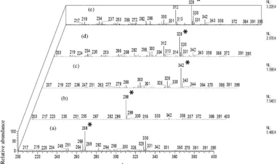
The fingerprint direct infusion of the leaves alkaloidal fractions from m/z 200 to 400 revealed several ions with even m/z values (Figures S1 to S12 in the Supplementary Information (SI) section), suggesting the potential presence of alkaloids. This proposal was based on previous studies with different isoquinoline-derived alkaloids with an odd number of nitrogens such as: aporphines,
tetrahydroprotoberberines,benzylisoquinolines and proaporphines, where protonation process occurs in higher proportions when compared to other reactions.3,4,12 The
base peaks corresponding to ions at m/z 268, 298 and 342 [M + H]+ were observed in U. guatterioides, U. duckei and
U. stipitata mass spectra, respectively, while the base peak at m/z 328 [M + H]+ was observed in U. floribunda and
U. rufescens mass spectra (Figure 1). Several others ions with even m/z values were observed pointing to complex samples with high diversity of alkaloids.
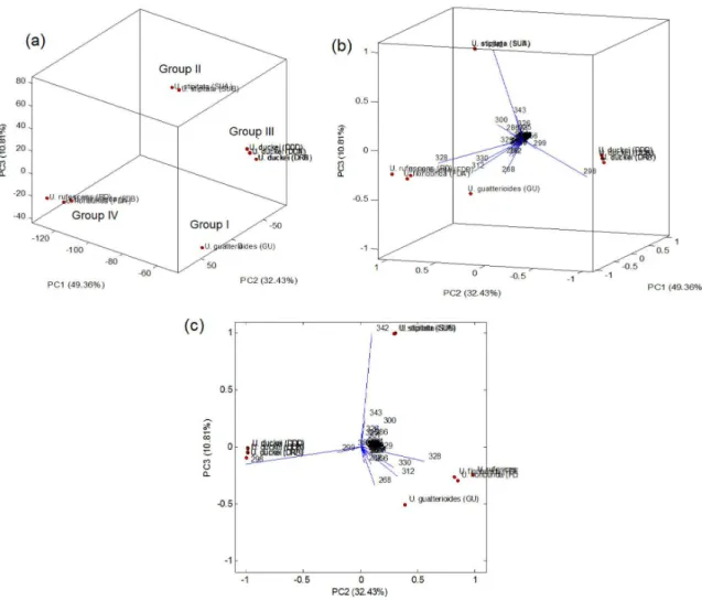
Analysis of the MS spectra revealed that 54 ions remained after elimination of the 5% least intense ions (Table S1 in the SI section). These 54 ions and their relative intensities were then subjected to the PCA and HCA analysis. In the PCA score plot (Figure 2a) were observed four main groups, being the specimens of U. guatterioides (group I), U. stipitata (group II) and U. duckei (group III) clearly separated. Group IV was constituted by U. rufescens and U. floribunda specimens, which is not surprising since these botanically close1 species were recently suggested to
be chemically close.18
According to the PCA biplot (Figures 2b and 2c), the ions at m/z 268, 298, 328 and 342 [M + H]+ were the
main responsible for the segregation of the groups I-IV. Through the MSn spectra of these ions (Figures S13 to
S17 in the SI section) were observed key fragmentations previously described for aporphine, proaporhine and tetrahydroprotoberberine alkaloids.3,4,12,19
The MSn spectra of the ions at m/z 268 and 298 [M + H]+
presented strong evidence of the aporphine skeleton, being observed initial losses of 17 (−NH
3) (m/z 268 → 251) and
31 u (−NH
2CH3) (m/z 298 → 267), which is in accordance
with the absence and presence, respectively, of a methyl at the heterocyclic nitrogen.19 In addition, subsequent losses
of 32 u (−CH3OH) (m/z 251 → 219 and m/z 267 → 235)
and 28 u (−CO) (m/z 219 → 191 and m/z 235 → 207) were
observed. These last fragmentations point to the presence of vicinal hydroxyl and methoxyl groups on aporphine skeletons.19 Fragmentation pathway observed to the ion at
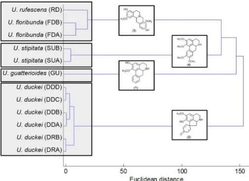
m/z 268 is consistent with the structure of the aporphine alkaloid asimilobine (1),3,4,12 previously reported in leaves
of U. guatterioides,3 while the fragmentation observed
to the ion at m/z 298 is in accordance with the structure of the proaporphine alkaloid glaziovine (2), previously reported in leaves of U. duckei.4 In the MS2 spectra of
the ion at m/z 328 were observed several fragment ions highlighting an intense product ion at m/z 178 and a minor ion at m/z 151. High mass losses are unusual in aporphine skeleton but are commonly observed in benzylisoquinolines and tetrahydroprotoberberines alkaloids.12,20,21 The major
ion at m/z 178 and the minor at m/z 151 were previously described as key fragments for tetrahydroprotoberberine
compounds containing methoxyl and hydroxyl groups at the ring A.12 Although it is possible to predict the substitution
patterns of this compound (scoulerine type) (3), it is not possible to guarantee the exact substitution positions once for this skeleton different positions with the same substituents will provide the same fragment ion.12 In the
MSn spectra of the ions at m/z 342, besides the initial loss
of 17 u (−NH3) (m/z 342 → 325), MS3 losses of 15 u (·CH3)
(m/z 325 → 310) and 31 u (·OCH
3) (m/z 325 → 294), and
subsequent MS4 loss of 15 u (·CH
3) (m/z 310 → 295) were
observed. These key fragmentations are in accordance with the presence of aporphines skeleton containing a methyl group at the heterocyclic nitrogen and adjacent methoxyl groups.19 The fragmentation pathway observed
to the ion at m/z 342 is consistent with the structure of the aporphine alkaloid norglaucine (4) previously reported in leaves of U. duckei.4 All theobserved fragmentations were
summarized in the Table 2.
and norglaucine (aporphine) play a fundamental role in the segregation of U. guatterioides, U. duckei and U. stipitata species, respectively. The unknown tetrahydroprotoberberine alkaloid was responsible for the formation of group IV, constituted by U. floribunda and U. rufescens species.
In the HCA dendrogram (Figure 3) were observed two subgroups in the U. duckei group, corresponding to different collection sites (RFAD and DAS). The chemical variability observed among these specimens was close to the observed between U. stipitata specimens (group II), while for the U. floribunda and U. rufescens (group IV) highest chemical variability was observed. The highest chemical similarity was observed between group II and IV, being this major group chemically close to the U. guatterioides (group I). Group III presented lower chemical similarity than the other groups.
In recent published work, the leaves essential oils of the same Amazonian Unonopsis species were analyzed by gas chromatography coupled to mass spectrometry (GC-MS) and the data treated by chemometric tools (PCA and HCA).18
Despite multivariate analysis showed significant differences between the species and their collection sites, the chemical variability among studied species was larger than presented in this work, suggesting that the non-volatile constituents (alkaloids) present better potential for chemotaxonomic purposes than volatile constituents (terpenoids). Another point that supports the use of the alkaloid fingerprint in chemotaxonomic approaches is the recurrent presence of these substances in Unonopsis. For example, glaziovine, norglaucine and glaucine were reported in the leaves of U. duckei,4 while asimilobine, anonaine, nornuciferine,
lisicamine and liriodenine were reported in the leaves of U. guatterioides.2 The presence of tetrahydroprotoberberine
Table 2. Precursor ions and key fragments observed by ESI-MSn experiments
Plant [M + H]+ MS2 MS3 MS4 Alkaloid type
U. guatterioides 268a 251a (−17 u)b 219a (−32 u) 191 (−28 u) aporphine
U. duckei 298a 267a (−17 u) 235a (−32 u) 207 (−28 u) proaporhine
U. stipitata 342a 325a (−17 u) 310a (−15 u)
294 (−31 u)
295 (−15 u) aporphine
U. floribunda 328a 178 (−150 u)
151 (−177 u)
− − tetrahydroprotoberberine
U. rufescens 328a 178 (−150 u)
151 (−177 u)
− − tetrahydroprotoberberine
aPrecursor ion; bneutral loss observed.
alkaloids in U. rufescens and U. floribunda species is an important fact, once this type of alkaloid was not reported in the Unonopsis genus. This observation along with previous chemical evidences18 could support a future approximation
of these species. Morphologically, the difference between U. floribunda and U. rufescens is the glaucous monocarps and the thicker monocarp wall.1
Conclusions
Multivariate analysis pointed to aporphine, proaporphine and tetrahydroprotoberberine alkaloids as responsibles for segregation of five Amazonian Unonopsis species, being these compounds tentatively identificated. New chemical evidences that support the botanical approximation between the close-related species U. rufescens and U. floribunda were found. The ESI-MS and ESI-MSn data along with
multivariate analysis provided a simple and effective approach to differentiate Unonopsis species commonly found in the state of Amazonas.
Supplementary Information
Supplementary information is available free of charge at http://jbcs.sbq.org.br as PDF file.
Acknowledgements
The authors are grateful to CAPES, CNPq, FINEP, and FAPEAM for financial support. We thank PDBFF for providing logistical and field support. This is publication number 680 in the PDBFF Technical Series.
References
1. Maas, P. J. M.; Westra, L. Y. T.; Vermeer, M.; Blumea2008, 52, 413.
2. Adams, M.; Gmunder, F.; Hamburger, M.; J. Ethnopharmacol.
2007, 113, 363.
3. Silva, F. M. A.; Koolen, H. H. F.; Almeida, R. A.; Souza, A. D. L.; Pinheiro, M. L. B.; Costa, E. V.; Quim. Nova2012, 35, 944.
4. Silva, F. M. A.; Souza, A. D. L.; Koolen, H. H. F.; Barison, A.; Vendramin, M. E.; Costa, E. V.; Ferreira, A. G.; Pinheiro, M. L. B.; Phytochem. Anal.2014, 25, 45.
5. Silva, F. M. A.; Lima, B. R.; Soares, E. R.; Almeida, R. A.; Silva-Filho, F. A.; Corrêa, W. R.; Salvador, M. J.; Souza, A. Q. L.; Koolen, H. H. F.; Souza, A. D. L.; Pinheiro, M. L. B.; Rev. Bras. Farmacogn.2015, 25, 11.
6. Waterman, P. G.; Gray, A. I.; Nat. Prod. Rep. 1987, 4, 175. 7. Waterman, P. G.; Phytochemistry 2007, 68, 2896.
8. Chiaradia, M. C.; Collins, C. H.; Jardim, I. C. S. F.; Quim. Nova 2008, 31, 623.
9. Moita Neto, J. M.; Moita, G. C.; Quim. Nova1998, 21, 467. 10. Barros Neto, B.; Scarminio, I. S.; Bruns, R. E.; Quim. Nova
2006, 29, 1401.
11. Schiozer, A. L.; Cabral, E. C.; Godoy, L. A. F.; Chaves, F. C. M.; Poppi, R. J.; Riveros, J. M.; Eberlin, M. N.; Barata, L. E. S.; J. Braz. Chem. Soc.2012, 23, 409.
12. Soares, E. R.; Silva, F. M. A. S.; Almeida, R. A.; Lima, B. R.; Silva-Filho, F. A.; Barison, A.; Koolen, H. H. F.; Pinheiro, M. L. B.; Souza, A. D. L.; Phytochem. Anal.2015, 26, 339. 13. Crotti, A. E. M.; Vessecchi, R.; Lopes, J. L. C.; Lopes, N. P.;
Quim. Nova2006, 29, 287.
14. Abreu, I. N.; Mazzafera, P.; Eberlin, M. N.; Zullo, M. A. T.; Sawaya, A. C. H. F.; Rapid Commun. Mass Spectrom.2007, 21, 1205.
15. Bataglion, G. A.; Silva, F. M. A.; Santos, J. M.; Santos, F. N.; Barcia, M. T.; Lourenço, C. C.; Salvador, M. J.; Godoy, H. T.; Eberlin, M. N.; Koolen, H. H. F.; Food Res. Int. 2014, 64, 472. 16. Lin, L.; Yu, Q.; Yan, X.; Hang, W.; Zheng, J.; Xing, J.;
Huang, B.; Analyst2010, 135, 2970.
17. Nunes, C. A.; Freitas, M. P.; Pinheiro, A. C. M.; Bastos, S. C.; J. Braz. Chem. Soc. 2012, 23, 2003.
18. Silva, F. M. A.; Koolen, H. H. F.; Pereira, J. L. S.; Flach, A.; Costa, L. A. M. A.; Souza, A. D. L.; Pinheiro, M. L. B.; Rec. Nat. Prod.2015, 9, 530.
19. Stévigny, C.; Jiwan, J. L. H.; Rozenberg, R.; Hoffmann, E.; Leclercq, J. Q.; Rapid Commun. Mass Spectrom.2004, 18, 523. 20. Schmidt, J.; Raith, K.; Boettcher, C.; Zenk, M. H.; Eur. J. Mass
Spectrom.2005, 11, 325.
21. Shim, H. J.; Lee, J. Y.; Kim, B.; Hong, J.; Mass Spectrom. Lett. 2013, 4, 79.
Submitted: August 5, 2015