Quim. Nova, Vol. 33, No. 4, 871-874, 2010
Artigo
*e-mail: olivia@butantan.gov.br
#Current address: Laboratório de Bioquímica e Biofísica, Instituto Butantan, Av. Vital Brasil, 1500, 05503-900 São Paulo - SP, Brasil
ANTIMYCOBACTERIAL AND CYTOTOXICITY ACTIVITIES OF FREE AND LIPOSOME-ENCAPSULATED 3-(4’-BROMO[1,1’-BIPHENYL-4-YL)-3-(4-BROMO-PHENYL)-N,N-DIMETHYL-2-PROPEN-1-AMINE
Ana O. de Souza*# e Célio L. Silva
Faculdade de Medicina de Ribeirão Preto, Universidade de São Paulo, Av. dos Bandeirantes, 3900, 14049-900 Ribeirão Preto - SP, Brasil Nelson Durán
Instituto de Química, Universidade Estadual de Campinas, CP 6154, 13083-970 Campinas - SP, Brasil Maria H. Andrade-Santana
Faculdade de Engenharia Química, Universidade Estadual de Campinas, CP 6066, 13083-970 Campinas - SP, Brasil
Recebido em 17/6/09; aceito em 2/12/09; publicado na web em 26/3/10
The antimycobacterial activity of 3-(4’-bromo[1,1’-biphenyl-4-yl)-3-(4-bromo-phenyl)-N,N-dimethyl-2-propen-1-amine (BBAP), free or incorporated in preformed liposomes, on extracellular M. tuberculosis H37Rv was 8 and 25 µM (MIC),respectively. Extracellular antimycobacterial activity was not signiicantly improved by entrapment of BBAP in liposomes, but there was a 6.1-fold reduction of BBAP cytotoxicity on J774 macrophages. Liposomal BBAP or its free form showed IC50 values of 165 and 27 µM, resulting in a selectivity index (SI=IC50/MIC) of 3.4 and 6.6, respectively. Free BBAP in concentrations from 10 to 80 µM were quite effective in eliminating intracellular M. tuberculosis while liposomal formulation was less effective at these concentrations.
Keywords: liposome; tuberculosis; 3-(4’-bromo[1,1’-biphenyl-4-yl)-3-(4-bromo-phenyl)-N,N-dimethyl-2-propen-1-amine.
INTRODUCTION
Tuberculosis still ranks among the world’s most deadly infectious diseases, killing around 2-3 million people per year.1 A third of the
world’s population may carry a latent tuberculosis infection, and the lifetime risk of developing tuberculosis ranges between 10 and 20%.2 Although tuberculosis is a preventable and treatable disease,
chemotherapy fails to reach its aim due to several reasons that include daily multiple drug administrations for several months, poor patient compliance, drug toxicity and emergence of drug resistance. Thus, one of the current strategies to enhance therapeutic activity, while minimizing toxicity, is to entrap drugs in a delivery system, assuring slow release over extended time periods. Liposome-encapsulated drugs often exhibit reduced toxicity and have also been shown to enhance retention of drugs in tissues, resulting in an improved overall therapeutic eficacy.3 Macrophage-speciic delivery systems are
cur-rently a subject of much interest, since macrophages act as host cells for many parasites and bacteria, which give rise to the outbreak of many human deadly diseases, such as leishmaniasis and tuberculosis.
It has been previously shown that 3-(4’-bromo[1,1’-biphenyl-4-yl)-3-(4-bromo-phenyl)-N,N-dimethyl-2-propen-1-amine (BBAP) is a drug with good antimycobacterial activity in vitro4-8 but also
cytotoxic to mammalian cells. Our group recently prepared inclusion complexes of BBAP in β-cyclodextrin8,9 in order to reduce
cytotoxi-city and to improve the antimycobacterial effect. Several combined spectroscopy techniques indicated the formation of a complex of BBAP/β-CD in the molar proportion of 1:1 and 1:2.
Biological assays regarding cytotoxicity towards mammalian cells (J774) and towards a permanent lung ibroblast cell line (V79) indicated no signiicant toxic effects with the BBAP/β-CD complexes. The complexes were more effective than the free compound on several mycobacteria species. Similar behavior of the inclusion complexes BBAP/β-CD (1:1 and 1:2) and rifampicin, a front-line antitubercular
drug, was observed for M. tuberculosis H37Rv growing inside J774 macrophages.
These previous results indicate that the entrapment of BBAP is a promising strategy for reduction of cytotoxicity and enhancement of its effectiveness against mycobacteria. In this study, the eficacy of a formulation containing liposome -entrapped BBAP was evalu-ated in vitro regarding cytotoxicity, extracellular and intracellular antimycobacterial activities.
EXPERIMENTAL
Preparation of liposomes
BBAP was synthesized as previously described.10 Liposomes
were composed by phospholipid:cholesterol (60:40 molar), a known composition which provides stability of the liposomal membrane for drug encapsulation. The phospholipid used in this study was the hydrogenated soybean phosphatidylcholine Epikuron 200SH (Lipoid), a commercial soy lecithin useful for liposome preparations due to high percentage of the phosphatidylcholine structural lipids (distearoylphosphatidylcholine (DSPC:0-15%) and dipalmitoylphos-phatidylcholine (DPPC:85-100%).
Small unilamellar vesicles containing BBAP were prepared by the classical Bangham method as described previously.11 Briely, the
lipids (phospholipid: cholesterol 60:40%) and BBAP were dissolved in chloroform:methanol (9:1 v/v) in BBAP/phospholipid ratios of 0 (empty liposomes); 0.1; 0.2; 0.3; 0.4; 0.5; and 0.6 and a dried ilm was formed by rotatory evaporation at 55 °C. The drug-lipid ilm was hydrated with 5 mL of Hepes buffer (10 mM, pH=7.4 containing NaCl 120 mM), forming a 7.5 mM lipid concentration and the suspension sonicated for 30 min (Ultrasonic Cleaner Unique) to produce small liposomes and maintained in repose during 2 h and 30 min.
Preformed liposomes were sized by multiple extrusions (15 times) through a drain disk (Millipore C32WP02500) and two stacked polycarbonate membranes (pore sizes 100 nm - Osmomics K01CP02500) by a high-pressure extruder (10 kgf/cm2) (Lipex
de Souza et al.
872 Quim. Nova
from the liposome suspension by ultrailtration through a membrane (YM 10 NMWL 10.000 - Amicon 13622) in an Amicon cell (Model 850) under nitrogen pressure (2.5 kgf/cm2).
Characterization of liposomes
Phospholipid content in liposomes was characterized by quanti-ication of total phosphate in samples, according to the methodology developed by Chen et al..12
To measure BBAP concentration, the liposomes were lysed with ethanol and the absorbance of the solution measured spectrophoto-metrically at 266 nm (Spectrophotometer, Hitachi U-200). BBAP concentrations in the liposomes were determined by comparison with a standard curve prepared with BBAP from 0 to 20 µg/mL and the trapping eficiency was calculated as follows:
% E = 100*(mols BBAP/mols lipids)inal/(mols BBAP/mols lipids)initial % E =Percent of encapsulation
Liposome mean diameter and particle size distribution were de-termined by quasi elastic light scattering at 90º with a He-Ne laser, 633 nm and at 25 °C (QLS - Light Scatter - Malvern Instruments Autosizer model 4700, UK).
Biological assays
Bacteria
M. tuberculosis H37Rv ATCC 27294, was grown in Loweinstein-Jensen medium at 37 °C for 3 weeks and subcultured in Middlebrook 7H9 broth medium, supplemented with OADC at 37 °C for 10 d. Cell concentration in the mycobacterial suspension was adjusted by optical absorbance in comparison with a standard curve and diluted in Middlebrook 7H9 broth medium to 4.0x10⁵ mycobacteria/mL to
determine the minimal inhibitory concentration (MIC) or in RPMI 1640 (5.0x10⁶ mycobacteria/mL) to detect macrophage infection in
intracellular antimycobacterial activity.13
J774 Macrophages
J774 cells were grown as monolayers in RPMI 1640 medium supplemented with 10% heat inactivated fetal calf serum (FCS), 100 IU/mL penicillin and 100 µg/mL streptomycin in a humidiied incubator with a 5% CO2 atmosphere at 37 °C.
Extracellular antimicrobial susceptibility testing – minimal inhibitory concentration (MIC)
Free BBAP dissolved in dimethylsulphoxide (DMSO), empty and BBAP-containing liposomes were sterilized by passage through a 0.22 µm PFTE ilter and diluted to the range of 10 to 160 µM in Middlebrook 7H9. Rifampicin was dissolved in DMSO, diluted in Middlebrook 7H9 broth and used as a reference drug at 12 µM.
Tests were performed by the microplate Alamar Blue assay, as previously described.14M. tuberculosis H37Rv (100 µL) were seeded
in a 96-well microplate (4.0x10⁵ mycobacteria/mL) containing 100 µL
of serial dilutions of BBAP - entrapped liposomes, empty liposomes or free BBAP. Plates were incubated at 37 °C for 6 d, followed by the addition of 25 µL mixed(1/1) Alamar Blue reagent and 10% Tween 80 (v/v) to each well. Plates were reincubated at 37 °C and after 24 h, a change in color from blue to pink, indicated mycobacterial growth.
Control wells consisted of either bacterium only, drugs with me-dium or only meme-dium. The visual MICs were deined as the lowest drug concentration that prevented a color change from blue to pink.
Cytotoxicity to mammalian cells – J774 macrophages
The BBAP cytotoxic effect was assayed on J774 macrophages by measuring the reduction of 3-(4,5-dimethylthiazole-2-yl)-2,5-diphenyl tetrazolium bromide (MTT) according to Denizot and Lang15 and expressed as IC
50 values (concentration in which at least
50% of the cells are viable). Stock solutions of BBAP were prepared in DMSO and diluted in RPMI 1640 without phenol red. The inal solvent concentrations in the assay were less than 0.3% and each sample concentration was tested in six replicates, and repeated three times in separate experiments. Cells (2.0x10⁶ cells/mL) added to
96-well plates had the medium removed after 12 h and replaced by a medium containing free or liposomal BBAP in concentrations ranging from 10 to 160 µM. As controls, macrophages were trea-ted with empty liposomes and with RPMI 1640, and considered as 100% viable. Cells were exposed for 24 h to test medium with or without drugs (controls).
After exposure to the test compounds, the medium in the wells was removed and replaced by 0.1 mL of serum-free medium contai-ning MTT (0.5 mg/mL). Following 4 h incubation, the supernatant was removed and the blue formazan product obtained was dissolved in 0.1 mL of isopropanol in 0.1 M HCl. Plates were stirred for 15 min on a microtiter plate shaker and absorbance was read at 570 nm. Intracellular antimicrobial susceptibility testing
J774 macrophages were harvested and plated at a concentra-tion of 5.0x10⁵ cells per well in 24-well tissue culture plates.
After overnight incubation, the cells were overlaid with 1 mL of a suspension of M. tuberculosis H37Rv (5.0x10⁶ mycobacteria/mL)
adjusted to yield a multiplicity of infection of 10 mycobacteria per macrophage. Cells were infected for 3-4 h and washed with phosphate-buffered saline (PBS), pH=7.4, to eliminate unbound mycobacteria. M. tuberculosis H37Rv-infected cells were fed again with culture medium containing free or liposomal BBAP in concentrations ranging from 10 to 160 µM.8,13
Control experiments included, RPMI 1640 medium, empty liposomes and rifampicin at 12 µM. Rifampicin and the empty liposomes were administered diluted in Middlebrook 7H9 broth. After 72 h, cells were washed with PBS to eliminate extracellular bacteria and lysed by addition of 500 µL of 0.25% (wt/vol) sodium dodecyl-sulphate in phosphate-buffered saline (PBS - pH=7.4). The lysates were serially diluted and 100 µL aliquots were dis-persed onto 7H10 agar plates. The colony formation units (CFU) of M. tuberculosis H37Rv were counted 2-4 weeks after incubation at 37 °C with 5% CO2.
Selectivity index
Selectivity index (SI) was calculated for free and liposomal BBAP formulations taking into account the MIC against M. tuberculosis H37Rv and the IC50 on J774 cells (SI=IC50/MIC) by the MTT assay.8
Statistical analysis
Results of cytotoxicity assays were expressed as cellular viabi-lity and displayed graphically using the computer software package Origin-Data Analysis and Technical Graphics, version 6.0 (Copyri-ght Software, Inc.). The IC50 values, concentrations that produced a 50% inhibitory effect on the evaluated parameter, were obtained by data interpolation.
Antimycobacterial and cytotoxicity activities 873 Vol. 33, No. 4
RESULTS AND DISCUSSION
Liposomes characterization
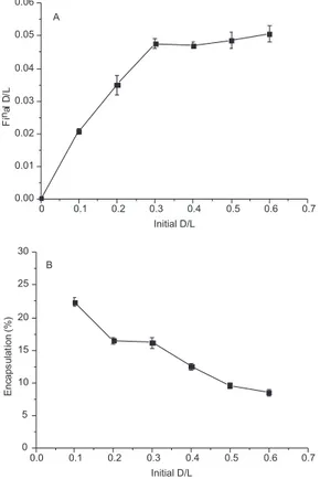
Figure 1A shows the proile relating the initial and inal drug/lipid ratios. The BBAP encapsulation increased from 0.1 to 0.3, remaining constant at higher drug/lipid ratios. These results indicate the satu-ration of the lipid bilayer, from which the encapsulation eficiencies decreased strongly (Figure 1B). The inal drug/lipid ratios were 10 times lesser than the initial ones, characterizing a low capability of the lipid matrix for BBAP entrapment. Further optimization of the formulation in terms of phospholipids and cholesterol concentration could enhance the packing of lipids in the matrix, providing higher entrapment eficiencies.
The average diameter of liposomes sizes was 130 nm and the polydispersity index was 0.3. The distributions were monodisperse in the range of 100 to 150 nm.
Biological assays
Antimycobacterial activity of free BBAP and of liposomes car-rying BBAP was assayed on extracellular M. tuberculosis H37Rv and the MIC values obtained were, respectively, 8 and 25 µM (Table 1).
As shown in Figure 2 liposomes entrapment of BBAP succes-sfully decreased the cytotoxicity of BBAP. The IC50 of liposomes with free and entrapped BBAP were 27 and 165 µM, respective-ly. The cytotoxicity of the compound was reduced by 6.1 fold, showing that this pharmaceutical formulation was important in reducing the toxicity of the drug, although the increase of the extracellular antimycobacterial activity was modest. Empty liposomes were not toxic to the cells (Figure 2). Table 1 shows
the selectivity indexes (SI=IC50/MIC) of free BBAP and BBAP entrapped in liposomes and the values are 3.4 and 6.6, respectively. For a drug to be considered a good candidate against tuberculosis its SI should be higher than 10.
Although lower eficacy of liposomal BBAP compared to free BBAP, these results indicate that BBAP liposomal formulation is promising for antimycobacterial activity as demonstrated by decre-asing of BBAP cytotoxicity and the selectivity index. However, an optimization of liposome composition, drug encapsulation and release as well as in vitro and in vivo studies are required for a complete evaluation of antimycobacterial eficacy of liposomal BBAP.
Free BBAP in concentrations ranging from 10 to 80 µM was signiicantly effective in eliminating intracellular M. tuberculosis H37Rv while BBAP liposomal formulation was not so eficient at the same concentrations (Figure 3) in comparison to control cells, which were infected and treated only with culture medium or empty liposomes (data not shown). The effects of the BBAP liposomal formulation on the biological results could be related to the slow release of drug and liposome phagocytosis. Enhancements on these Figure 1. Proile relating the initial and inal drug/lipid ratios (A) and
en-capsulation eficiencies of BBAP in liposomes (B). D/L = drug/lipid ratios. Bars represent mean ± SEM of two independent experiments
Figure 2. Cytotoxicity to J774 macrophages of free BBAP (), empty liposo-mes () and liposome-entrapped BBAP () determined by the MTT assay. Bars represent mean ± SEM of three independent experiments
Table 1. Antimycobacterial parameters of free and liposome-entrapped BBAP Samples MIC* (µM) IC50
** (µM) SI=IC 50/MIC
Free BBAP 8 27 3.4
Liposomal BBAP 25 165 6.6
*MIC = corresponds to the MIC against M. tuberculosis H37Rv; **IC50 = cor-responds to the IC50 on J774 macrophages by the MTT assay; SI=IC50/MIC.
de Souza et al.
874 Quim. Nova
aspects could be provided by adding lipids of low phase transition temperature and cationic lipids to the formulation.
As expected rifampicin at 12 µM was effective and eliminated more than 2 log in the bacillus number.
CONCLUSION
The encapsulation of BBAP in liposomes and the in vitro evalu-ations of the cytotoxicity, extracellular and intracellular antimyco-bacterial activities were characterized. The liposomal formulation is promising for the treatment of M. tuberculosis infections due to its reduced citotoxicity and higher selectivity index, compared to free BBAP.
ACKNOWLEDGMENTS
The authors thank Conselho Nacional de Desenvolvimento Cientíico e Tecnológico (CNPq) for inancial support to A. O. de Souza (Proix-CNPq) and Brazilian Tuberculosis Research Network (REDE TB - Instituto do Milênio).
REFERENCES
1. Corbett, E. L.; Watt, C. J.; Walker, N.; Mayer, D. B. M.; Willians, B. G.; Raviglione, M. C.; Dye, C.; Arch. Intern. Med.2003, 163, 1009. 2. Vynnycky, E.; Fine, P. E.; Epidemiol. Infec.1997, 119, 183. 3. Pandey, R.; Khuller, G. K.; J. Antimicrob. Chemother.2005, 55, 430.
4. De Souza, A. O.; Aily, D. C. G.; Sato, D. N.; Durán, N.; J. Antimicrob. Chemother. 1998, 42, 407.
5. De Souza, A. O.; Junior, R. R. S.; Ferreira-Julio, J. F.; Rodriguez, J. A.; Melo, P. S.; Haun, M.; Sato, D. N.; Durán, N.; Eur. J. Med. Chem. 2001, 36, 843.
6. De Souza, A. O.; Pereira, D. G.; Durán, N.; Ann. Rev. Biomed. Sci. 2002, 4, 53.
7. De Souza, A. O.; Hemerly, F. P.; Busollo, A. C.; Melo, P. S.; Machado, G. M. C.; Miranda, C. C.; Santa-Rita, R. M.; Haun, M.; Leon, L. L.; Sato, D. N.; De Castro, S. L.; Durán, N.; J. Antimicrob. Chemother. 2002, 50, 629.
8. De Souza, A. O.; Santos, R. R.; Sato, D. N.; De Azevedo, M. M. M.; Ferreira, D. A.; Melo, P. S.; Haun, M.; Silva, C. L.; Durán, N.; J. Braz. Chem. Soc. 2004, 15, 682.
9. De Souza, A. O.; Alderete, J. B.; Faljoni-Alario, A.; Silva, C. L.; Durán, N.; J. Chil. Chem. Soc.2005, 50, 591.
10. De Conti, R.; Gimenez, S. M. M.; Haun, M.; Pilli, R. A.; De Castro, S. L.; Durán, N.; Eur. J. Med. Chem.1996, 31, 915.
11. Bangham, A. D.; Standish, M. M.; Watkins, J. C.; J. Mol. Biol. 1965, 13, 238. 12. Chen, P. S.; Toribara, T. Y.; Warner, H.; Anal. Chem.1956, 28, 1756.
13. Oh, Y. K.; Nix, D. E.; Straubinger, R. M.; Antimicrob. Agents Chemother. 1995, 39, 2104.
14. Collins, L. A.; Franzblau, S. G.; Antimicrob. Agents Chemother. 1997, 41, 1004.