Quim. Nova, Vol. 33, No. 4, 846-849, 2010
Ar
ti
go
*e-mail: [email protected]
STEROIDAL AND PHENOLIC COMPOUNDS FROM Sidastrum paniculatum (L.) FRYXELL AND EVALUATION OF CYTOTOXIC AND ANTI-INFLAMMATORY ACTIVITIES
José Marcílio Sobral Cavalcante, Tiago Bezerra de Sá de Souza Nogueira, Anna Cláudia de Andrade Tomaz, Davi Antas e Silva, Maria de Fátima Agra e Maria de Fátima Vanderlei de Souza*
Laboratório de Tecnologia Farmacêutica “Prof. Delby Fernandes de Medeiros”, Centro de Ciências da Saúde, Universidade Federal da Paraíba, CP 5009, 58051-970 João Pessoa - PB, Brasil
Paulo Roberto Cavalcanti Carvalho, Sílvia Rafaelli Ramos, Silene Carneiro do Nascimento e Teresinha Gonçalves-Silva Departamento de Antibióticos, Universidade Federal de Pernambuco, 50670-901 Recife - PE, Brasil
Recebido em 28/5/09; aceito em 17/11/09; publicado na web em 2/3/10
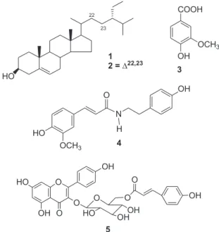
Sidastrum paniculatum (L.) Fryxell belongs to the family Malvaceae and is popularly known as “malva roxa” or “malvavisco”. The phytochemical study of the hexane, CHCl3 and EtOAc phases from the crude ethanol extract of S. paniculatum led to the isolation of six compounds: a mixture of b-sitosterol and stigmasterol, 4-methoxy-3-hydroxybenzoic acid, 4-methoxy-3-hydroxybenzaldehyde, N-trans-feruloyltyramine and kaempferol-3-O-b-D-(6’’-E-p-coumaroyl) glucoside. The structural identiication of the compounds was made on the basis of spectroscopic methods such as IR, 1H and 13C NMR with the aid of including two-dimensional techniques, besides comparison with literature data. The b-sitosterol and stigmasterol mixture showed a signiicant anti-inlammatory activity.
Keywords: Sidastrum paniculatum; Malvaceae; anti-inlammatory activity.
INTRODUCTION
Sidastrum paniculatum (L.) Fryxell is a shrub belonging to the family Malvaceae with wide distribution in the neotropics.1 In Northeast of Brazil this species ispopularly known as “malva-roxa” or “malvavisco”. For long time, this species was belonging to the genus Sida, as Sida paniculata L., and was transferred to Sidastrum by Frixell.2
Sidastrum Baker f. is one of 70 genera informally included by Bayer and Kubitzki as a member of the tribe Malveae, subfamily Malvoideae (Malvaceae).3 The genus comprises about eight spe-cies with neotropical distribution, occurring from Mexico to West Indies to Argentina.1,3 In Brazil, the northeast region is probably the diversity core of the genus Sidastrum since a great number of species can be found there.1
Recent molecular studies of the Sida generic alliance4 strongly support the phylogenetic relationships between Sidastrum and the genus Meximalva as well as an Australian group of Sida species, which form a well-supported clade as suggested before by Fryxell1 based on morphological characters.
Previous phytochemical studies on species from Malva-ceae, specially of the genus Sida have reported the presence of steroids,5-8 phenols,6,7,9 lavonoids,6-8,10,11 triterpenes,5,9 essential oils,12 alkaloids,13 sesquiterpene lactone,14 fatty acids,15 and pha-eophorbide.6
Aiming at contributing to the chemotaxonomic study of the family Malvaceae, subfamily Malvoideae, and considering the absence of data in literature concerning the chemical constitution of the genus Sidastrum, the species Sidastrum paniculatum was submitted to a phytochemical study to isolate and identify its chemical constituents, through usual chromatographic and spectroscopic methods, besides comparison with literature data. In addition to that, an evaluation of the cytotoxic and anti-inlammatory activities with some of the isolated compounds will be herein described.
EXPERIMENTAL General procedures
NMR spectra (HOMOCOSY, HETCOR, HMQC, HMBC and NOESY) were registered in CDCl3, CD3OD and recorded on a Mer-cury Varian instrument operating at 200 MHz and 50 MHz for 1H and 13C, respectively. The solvent signal was used as internal standard. IR spectra were measured on a Perkin-Elmer, FT-IR-1750 spectrometer in KBr pellets. Chromatography columns were carried out on silica gel (Merck) and Sephadex LH-20 (Merck). TLC were performed on silica gel PF254 plates and the spots were visualized under UV light (254 and 366 nm) and by exposure to the iodine vapor.
Plant material
The whole plant of S. paniculatum was collected in Pedra da Boca, in the municipality of Araruna, State of Paraíba, on February 2004, and a voucher specimen (M. F. Agra et al. 6051) was deposi-ted at the Herbarium Prof. Lauro Pires Xavier (JPB), Universidade Federal da Paraíba.
Extraction and isolation
Steroidal and phenolic compounds from Sidastrum paniculatum 847 Vol. 33, No. 4
silica gel column eluted with hexane, CHCl3 and methanol providing 15 sub-fractions which were analysed and joined through analytical TLC. The sub-fraction 11/15 showed itself as crystals, yielding 0.093 g of a b-sitosterol (1) and stigmasterol (2) mixture. The CHCl3 phase provided a precipitate (5.0 g) and a supernatant (13.0 g). 6.0 g of the latter were submitted to chromatography on a column packed with silica gel and eluted with hexane, EtOAc and methanol, giving 200 fractions that were analysed and joined through analytical TLC. Fraction 5/15 (0.81 g) was also chromatographed on silica gel column using the above chromato-graphic procedures, yielding 120 sub-fractions.
The sub-fraction 26/57 (0.05 g) showed itself as an amorphous solid, deined as 4-hydroxy-3-methoxybenzoic acid (3). Fraction 26/102 (0.45 g), on the other hand, was chromatographed follow-ing the same methodology, providfollow-ing 96 sub-fractions of which the sub-fraction 43/56 was recrystallized in chloroform and methanol, yielding 0.025 g of the 4-hydroxy-3-methoxybenzaldehyde (3). 0.15 g of the precipitate was submitted to preparative TLC, using a mixture of hexane:ethyl acetate (1:1) as eluent, resulting on the isolation and puriication of 0.021 g of N-trans-feruloyltyramine (4). The ethyl acetate phase (3.0 g) was subjected to chromatography column packed with Sephadex LH-20 and eluted with methanol, providing 23 frac-tions that were analysed and combined through analytical TLC. The sub-fraction 12/16 (0.087 g) was also chromatographed on Sephadex LH-20, yielding 14 fractions. The sub-fraction 5/8 (0.032 g) was deined as kaempferol-3-O-b-D-(6’’-E-p-coumaroyl) glucoside (5).
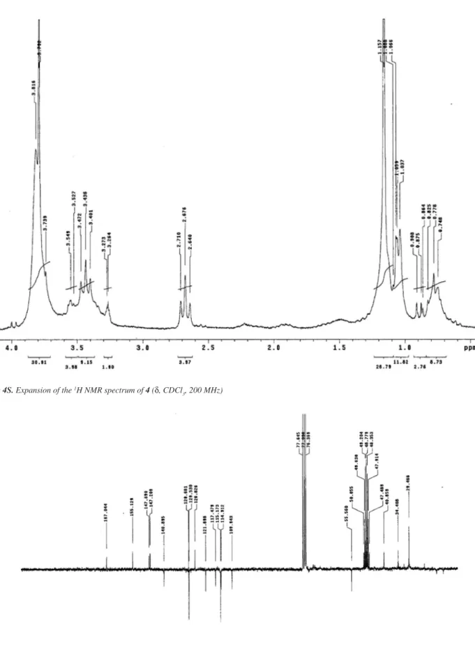
4 (N-trans-feruloyltyramine)1H NMR (d, CDCl
3, 200 MHz): 7.37 (d, J=15.8 Hz, H-7), 6.96 (d, J=8.4 Hz, H-2’/6’), 6.93 (brd, J=8.4 Hz, H-6), 6.90 (brs, H-2), 6.74 (d, J=8.4 Hz, H-5), 6.68 (d, J=8.4 Hz, H-3’/5’), 6.16 (d, J=15.8 Hz, H-8), 3.79 (s, OCH3-3), 3.43 (t, J=6.9 Hz, H-8’), 2.67 (t, J=6.9 Hz, H-7’). 13C NMR (50 MHz): 167.04 (C-9), 155.17 (C-4´), 147.69 (C-3), 147.20 (C-4), 155.17 (C-4´), 140.89 (C-7), 129.68 (C-1´), 129.53 (C-2´/6´), 126.82 (C-1), 121.89 (C-6), 117.47 (C-8), 115.17 (C-3’/5’), 114.91 (C-5), 109.70 (C-2), 55.56 (OCH3-3), 40.89 (C-8´), 34.41 (C-7’).
Cells
The cytotoxicity of b-sitosterol and stigmasterol mixture was tested against NCI-H292 (carcinoma of human lungs), HEp-2 (human
larynx epidermoid carcinoma) and KB (mouth carcinoma) cells all obtained from the Bank of Cells, Rio de Janeiro, Brazil. The cells were grown in DMEM – Minimum Essential Medium Eagle modiied Dulbecco’s- medium supplemented with 10% fetal bovine serum, 2 mM glutamine, 100 μg/mL streptomycin, 100 UmL−1 penicillin at 37 °C with a 5% CO2 atmosphere.
Animals
Swiss albino femalemice weighing 25-30 g, obtained from the Animal House of Departamento de Antibióticos from Universidade Federal de Pernambuco, Brazil, were used. The animals were housed in cages with free access to food and water. All animals were kept under 12h light/dark cycle (lights on at 6:00 a.m.). The animals were treated according to the ethical principles of animal experimenta-tion of COBEA (Brazilian College of Animal Experiments), Brazil, and the rules of the National Institute of Health Guide for Care and Use of Laboratory Animals. The Animal Studies Committee of the Universidade Federal de Pernambuco approved the experimental protocols (number 116/07).
Cell proliferation assays
The tumor cell growth was quantiied by the ability of living cells to reduce the yellow dye 3-(4,5-dimethyl-2-thiazolyl)-2,5-diphenyl-2H-tetrazolium bromide (MTT) to a purple formazan product. A cellular suspension (105 cells/mL) was prepared and distributed in 96-well (225 mL/well) and incubated during 24 h at 37 °C. After that, the b-sitosterol and stigmasterol mixture was added. At the end of incubation (72 h), the plate was centrifuged and the medium was then replaced by fresh medium (200 μL) containing 25 μL of MTT. Three hours later, the MTT formazan product was dissolved in 100 μL DMSO, and the absorbance was measured using a multiplate reader.16,17 The drugs effects were quantiied as the percentage of control absorbance of reduced dye at 595 nm. The tested concentra-tions were 10.0; 5.0; 2.5 and 1.25 μg/mL and the experiments were done in triplicate.
Statistical analysis
Results are presented as means ± s.e.m. for groups of six animals for in vivo experiments, and they are representative of two independent experiments. The differences between the experimental groups were compared by analysis of variance (ANOVA) complemented with Student’s t-tests. P < 0.05 was considered as indicative of signiicance. Anti-inlammatory activity
Air pouches were produced on the dorsal cervical region of mice by subcutaneous injection of 2.5 mL on day 0, followed by a second injection of 2.5 mL of sterile air 3 days later. At day 6, 1 mL of a carrageenan solution 1% (w/v) was injected into the cavity. The B-sitosterol and stigmasterol mixture (10, 30 and 90 mg/kg) and dexamethasone (3 mg/kg) were administered (p.o.) 1 h before injection of carrageenan. After killing the animals (6 h after inlam-mation induction), pouches were washed with 3 mL of saline solution containing 3 mM EDTA. Samples from the air pouch exudates were collected for determinations of total and differential leukocyte con-tents. Total leukocyte counts were performed in a Neubauer chamber after diluting the exudates with Turk’s solution (1:20) and cytospin preparations of exudates were stained with May-Grunwald Giemsa for the differential leukocyte count, which was performed under an oil immersion objective.16
Cavalcante et al.
848 Quim. Nova
RESULTS AND DISCUSSION
The structural assignments of compounds 1-3 and 5 were made based on the spectral analysis and are in good agreement with those reported in the literature. Thus, their structures were identiied as the mixture of the steroids b-sitosterol and stigmasterol (1),18 4-hydroxy-3-methoxybenzoic acid (2),7,19 4-hydroxy-3-methoxybenzaldehyde (3)20 and kaempferol 3-O-b-D-(6’’-E-p-coumaroyl) glucopyranoside (5) (tiliroside).6,7,10 All compounds are being reported here for the irst time in the genus Sidastrum. The compound 5 (tiliroside) was previously isolated from the Malvaceae species Sida galheirensis,6 Bakeridesia pickelli,7Herissantia crispa,8 and Herissantia tiubae.10 However, the presence of the compound 5 (tiliroside) in species of Malvaceae does not represent it as a chemical marker, because it was reported also to be present in other species of angiosperms like Solanum21 belonging to the Solanaceae family, as an example.
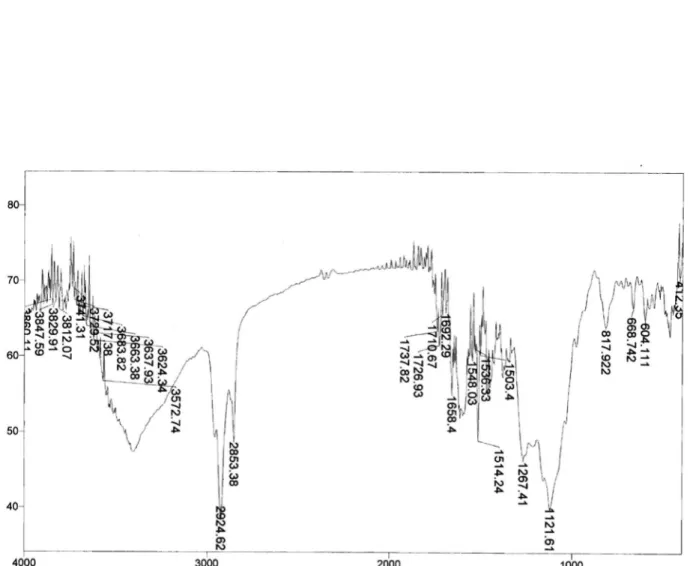
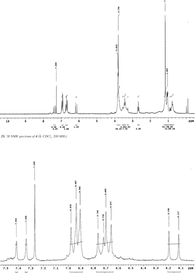
The IR spectrum of compound 4 showed a large absorption band at 3300-3350 cm-1 (N-H bending) and at 1658 cm-1 (C=O stretch) suggesting an amide function. The aromatic skeleton was evidenced by the absorptions between 1600 and 1450 cm-1. The 1H NMR spectra showed a pair of doublets at d 6.68 and 6.96 (2H, J = 8.4 Hz), which suggests an AA’BB’-type system of hydrogens bonded to aromatic carbon. It showed yet a doublet at d 6.74 (1H, J = 8.4 Hz) together with a broad doublet at d 6.93 (1H, J = 8.4 Hz) and a broad singlet at d 6.90 (1H), characterizing AMX system.
Those IR and 1H NMR data together with a pair of doublets at d 6.16 (H-8, J = 15.8 Hz) and 7.37 (H-7, J = 15.8 Hz) referring to trans oleinic hydrogens and a pair of triplets at d 2.67 and 3.43 (H-7’and H-8’), suggested the presence of the coumaroyl and tyramine units, respectively. A methoxyl group in one of the aromatic nuclei was inferred by the signal at d 3.79.
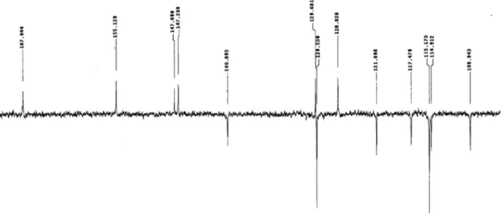
The 13C NMR spectral data strengthened the information provided by the IR and 1H NMR spectra, emphasizing the presence of the am-ide function due to the signal at d 167.04 (α,b-unsaturated carbonyl) thus reinforcing the coumaroyl unit because of the α,b-unsaturated carbons (C-8 and C-7) at d 117.47 and 140.89, respectively, as well as the tyramine unit whose methylene carbons were shown to be at d 34.41 (C-7’) and 40.89 (C-8’). The absorptions at d 115.17 and 129.53 (2C, C-3’/5’ and C-2’/6’) corroborated the AA’BB’ system, while a methoxyl group was suggested by the signal at d 55.56. Non-hydrogenated carbons bound to oxygen groups were inferred by the signals at d 147.20, 147.60 and 155.12.
The heteronuclear correlation spectrum HMBC conirmed the suggestion of the α,b-unsaturated amide strengthening the presence of the coumaroyl and tyramine units since it showed, respectively, two- and three-bond correlations (2,3J
CH) between H-7’ and H-8’ with C-1’, and 2J
CH and 3J
CH correlations between H-8 and H-7 with the carbonyl carbon, respectively (Figure 2).
The homonuclear correlation spectrum NOESY revealed the space interactions between the methoxyl hydrogens with H-2, thus determining the meta position for the methoxyl group in the couma-royl portion of the molecule (Figure 2).
The other assignments of carbons and hydrogens were determined based on all spectral data and in comparison with the literature ones,22 which allowed to unambiguously identify substance 4 as being N-trans-feruloyltyramine (Figure 3), substance already isolated from other vegetal species23 with studies demonstrating its action against weeds and improvement of seed germination,24 as well as its activity as a melanin biosynthesis inhibitor,22 being described for the irst time in the family Malvaceae.
In vitro activity of b-sitosterol and stigmasterol mixture against three human tumor cell lines was determined. However, the mixture assayed did not show cytotoxicity at the tested concentrations.
According to the protocol of National Cancer Institute (NCI-USA), natural products from plants are considered cytotoxic if IC50 30 μg/mL. The mixture tested exhibited IC50 values greater than 10 μg mL−1 for all tumor cell lines tested. The results suggested that the compounds tested showed low cell toxicity, which is desirable in case of anti-inlammatory activity. Literature shows that some compounds isolated from Hisbiscus taiwanensis (Malvaceae) also did not show cytotoxic activity against human cells H.549 (lung carcinoma) and NC5-7 (breast carcinoma) up dose of 20 mg/mL.25
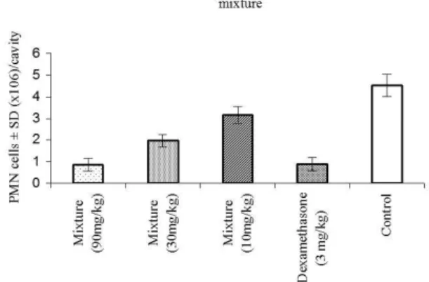
The b-sitosterol and stigmasterol mixture showed a signiicant anti-inlammatory activity when compared to control group, inhibiting the carrageenan-induced cell migration by at rates of 30, 56.8 and 81.2% of doses 10, 30 and 90 mg/kg, respectively (Figure 3). The number of polymorphonuclear leucocytes which migrated to the peri-toneal cavity in the control group was 4.53 ± 0.8 and in dexamethasone pattern was 0.90 ± 0.3 x 10⁶ cells per cavity (p<0.05 for all doses).
The anti-inlammatory activity of b-sitosterol26 in the carrageenan paw edema model has been reported. It has been demonstrated that the stigmasterol was active in reducing the edema by 41% inhibi-tion at doses of 0.5 mg/ear.27 Most articles on anti-inlammatory activity describe the most common type of evaluation which are the carrageenan-induced paw edema26 and the TPA-induced ear edema.27 However, as several authors describe in the literature, the paw edema test is not speciic for anti-inlammatory drugs, as anti-cholinergic drugs also reduce the edema. Furthermore, we found only one study that evaluates the activity of MPO;27 however, no studies evaluate the cell migration of the mixture b-sitosterol and stigmasterol, and it is known that cell migration is an important step in the developing of acute inlammation. Besides, we also aimed at verifying whether the mixture of those steroids possess synergistic action, and our data suggest that they do, since anti-inlammatory effects of b-sitosterol isolated in the model of carrageenan-induced paw edema were not Figure 2. Main correlations observed at the HMBC (nJ) and NOESY (NOE)
spectra of 4
Steroidal and phenolic compounds from Sidastrum paniculatum 849 Vol. 33, No. 4
observed.28 The b-sitosterol and stigmasterol mixture showed a prom-ising anti-inlammatory activity, when compared to dexamethasone, being greater than that of the isolated products, suggesting synergistic action between b-sitosterol and stigmasterol. However, this was only one preliminary study with the mixture; further studies are necessary to provide the investigation of its mechanism of action.
SUPPLEMENTARY MATERIAL
Available in http://quimicanova.sbq.org.br, in format .PDF, with free access.
REFERENCES
1. Fryxell, P. A.; Brittonia 1997, 49, 204. 2. Fryxell, P. A.; Brittonia1978, 30, 447.
3. Bayer, C.; Kubitzki, K. In Flowering plants, dicotyledons: Malvales, Capparales, and nonbetalain Caryophyllales; Kubitzki, K.; Bayer, C., eds.; Springer-Verlag: Berlin, 2003, 225.
4. Aguilar, J. F.; Fryxell, P. A.; Jansen, R. K.; Syst. Bot.2003, 28, 352; Tate, J. A.; Aguilar, J. F.; Wagstaff, S. J.; La Duke, J. C.; Slotta, T. B.; Simpson, B. B.; Am. J. Bot.2005, 92, 584.
5. Ahmed, Z.; Kazmi, S. N. H.; Walik, A.; J. Nat. Prod. 1990, 53, 1342. 6. Silva, D. A.; Silva, T. M. S.; Lins, A. C. S.; Costa, D. A.; Cavalcante, J.
M. S.; Matias, W. N.; Souza, M. F. V.; Quim. Nova2006, 29, 1250. 7. Costa, D. A.; Silva, D. A.; Cavalcanti, A. C.; Medeiros, M. A. A.; Lima,
J. T.; Cavalcante, J. M. S.; Silva, B. A.; Agra, M. F.; Souza, M. F. V.; Quim. Nova2007, 30, 901.
8. Costa, D. A.; Matias, W. N.; Lima, I. O.; Xavier, A. L.; Costa, V. B. M.; Diniz, M. F. F. M.; Agra, M. F.; Batista, L. M.; Souza, M. F. V.; Silva, D. A.; Quim. Nova2009, 32, 48.
9. Silva; D. A.; Falcão-Silva, V. S.; Gomes, A. Y. S.; Costa, D. A.; Lemos, V. S.; Agra, M. F.; Braz-Filho, R.; Siqueira-Junior, J. P.; Souza, M. F. V.; Pharmaceut. Biol.2009, 29, 279.
10. Silva D. A.; Chaves M. C. O.; Costa, D. A.; Moraes, M. R. R.; Nóbrega, F. B. P.; Souza, M. F. V.; Pharmaceut. Biol.2005, 43, 197.
11. Silva, D. A.; Costa, D. A.; Silva, D. F.; Souza, M. F. V.; Agra, M. F.; Medeiros, I. A.; Barbosa-Filho, J. M.; Braz-Filho, R.; Rev. Bras. Farmacogn.2005, 15, 23.
12. Ames, J. M.; Macleod, G.; Phytochemistry1990, 29, 1201. 13. Ghosal, S.; Chauhan, R.; Mehta, R.; Phytochemistry1975, 14, 830. 14. Sharma, P. V.; Ahmad, Z. A.; Phytochemistry1989, 28, 3525. 15. Carmody, D. R.; Dejong, W.; Smith, T. R.; Oil & Soap1945, October,
263; Vickery, J. R.; J. Am. Oil Chem. Soc.1980,February, 87; Schimid, K. M.; Patterson, G. W.; Phytochemistry1988, 27, 2831; Nakatani, M.; Fukunaga, Y.; Hase, T.; Phytochemistry1986, 25, 449.
16. Klemm, P.; Harris, H. J.; Perretti, M.; Eur. J. Pharmacol.1995,281, 69. 17. Alley, M. C.; Scudiero, D. A.; Monks, A.; Hursey, M. L.; Czerwinski,
M. J.; Fine, D. L.; Abbott, B. J.; Mayo, J. G.; Shoemaker, R. H.; Boyd, M. R.; Cancer Res. 1998, 48, 589.
18. Kojima, H.; Sato, N.; Hatano, A.; Ogura, H.; Phytochemistry1990, 29, 2351.
19. Tomaz, A. C. C.; Nogueira, R. B. S. S.; Pinto, D. S.; Agra, M. F.; Souza, M. F. V.; Da-Cunha, E. V. L.; Rev. Bras. Farmacogn.2008, 18, 47. 20. França, V. C.; Tese de Doutorado, Universidade Federal da Paraíba,
Brasil, 2002.
21. Esteves-Souza, A.; Silva, T. M. S.; Alves, C. C. F.; Carvalho, M. G.; Braz-Filho, R.; Echevarria, A.; J. Braz. Chem. Soc.2002, 13, 838. 22. Efdi, M.;Ohguchi, K.;Akao, Y.;Nozawa, Y.;Koketsu, M.; Ishihara, H.;
Biol. Pharm. Bull. 2007, 30, 1972.
23. Chen, C. Y.; Chang, P. R.; Wu, Y. C.; J. Chin. Chem. Soc.1997, 44, 313; Kim, H. R.; Min, H. Y.; Jeong, Y. H.; Lee, S. K.; Lee, N. S.; Seo, E. K.; Arch. Pharmacal Res.2005, 28, 1224.
24. Miyaichi, Y.;Nunomura, N.;Kawata, Y.; Kizu, H.; Tomimori, T.; Watanabe, T.; Takano, A.; Malla, K. J.; Chem. Pharmaceut. Bull.2006, 54, 136; Oliveira, P. E. S.; Conserva, L. M.; Lemos, R. P. L.; Biochem. System. Ecol.2008, 36, 134.
25. Lajide, D.; Escoubas, P.; Mizutani, J.; Phytochemistry1995, 40, 1105. 26. Gupta, M. B.; Nath, R.; Srivastava, N.; Shanker, K.; Kishor, K.;
Bhargava, K. P.; Planta Med. 1980, 39, 157.
27. García, M. D.; Sáenz, M. T.; Gómez, M. A.; Fernández, M. A.; Phytother. Res.1999, 13, 78.
Quim. Nova, Vol. 33, No. 4, S1-S10, 2010
M
ate
ri
al
S
up
le
m
enta
r
*e-mail: [email protected]
STEROIDAL AND PHENOLIC COMPOUNDS FROM Sidastrum paniculatum (L.) FRYXELL AND EVALUATION OF CYTOTOXIC AND ANTI-INFLAMMATORY ACTIVITIES
José Marcílio Sobral Cavalcante, Tiago Bezerra de Sá de Souza Nogueira, Anna Cláudia de Andrade Tomaz, Davi Antas e Silva, Maria de Fátima Agra e Maria de Fátima Vanderlei de Souza*
Laboratório de Tecnologia Farmacêutica “Prof. Delby Fernandes de Medeiros”, Centro de Ciências da Saúde, Universidade Federal da Paraíba, CP 5009, 58051-970 João Pessoa - PB, Brasil
Paulo Roberto Cavalcanti Carvalho, Sílvia Rafaelli Ramos, Silene Carneiro do Nascimento e Teresinha Gonçalves-Silva Departamento de Antibióticos, Universidade Federal de Pernambuco, 50670-901 Recife - PE, Brasil
Cavalcante et al.
2 Quim. Nova
Figure 2S.1H NMR spectrum of 4 (δ, CDCl
3, 200 MHz)
Figure 3S. Expansion of the 1H NRM spectrum of 4(δ, CDCl
Steroidal and phenolic compounds from Sidastrum paniculatum 3
Vol. 33, No. 4
Figure 4S. Expansion of the 1H NMR spectrum of 4 (δ, CDCl
3, 200 MHz)
Figure 5S.13C-APT NMR spectrum of 4 (δ, CDCl
Cavalcante et al.
4 Quim. Nova
Figure 6S. Expansion of the 13C-APT NMR spectrum of 4 (δ, CDCl
3, 50 MHz)
Figure 7S. Expansion of the 13C-APT NMR spectrum of 4 (δ, CDCl
Steroidal and phenolic compounds from Sidastrum paniculatum 5
Vol. 33, No. 4
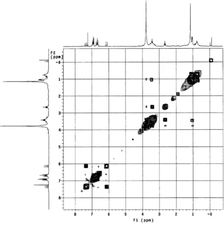
Figure 8S. 1H x 1H-COSY NMR spectrum of 4 (δ, CDCl
3, 200 MHz)
Figure 9S. Expansion of the 1H x 1H-COSY NMR spectrum of 4 (δ, CDCl
Cavalcante et al.
6 Quim. Nova
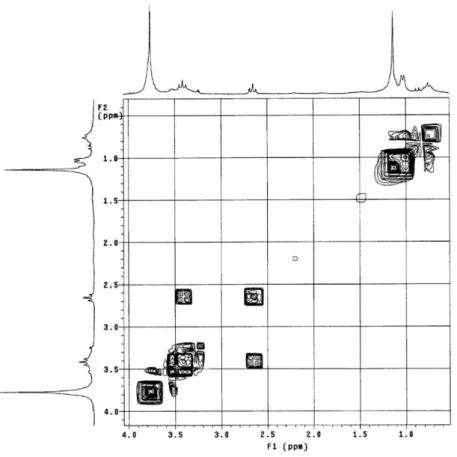
Figure 10S. Expansion of the 1H x 1H-COSY NMR spectrum of 4 (δ, CDCl
3, 200 MHz)
Figure 11S.1H x 13C-HETCOR NMR spectrum of 4 (δ, CDCl
Steroidal and phenolic compounds from Sidastrum paniculatum 7
Vol. 33, No. 4
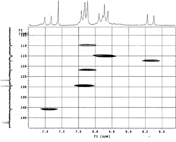
Figure 12S. Expansion of the 1H x 13C-HETCOR NMR spectrum of 4 (δ, CDCl
3, 200 MHz)
Figure 13S. Expansion of the 1H x 13C-HETCOR NMR spectrum of 4 (δ, CDCl
Cavalcante et al.
8 Quim. Nova
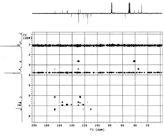
Figure 14S.1H x 13C-HMBC NMR spectrum of 4 (δ, CDCl
3, 200 MHz)
Figure 15S. Expansion of the 1H x 13C-HMBC NMR spectrum of 4 (δ, CDCl
Steroidal and phenolic compounds from Sidastrum paniculatum 9
Vol. 33, No. 4
Figure 16S. Expansion of the 1H x 13C-HMBC NMR spectrum of 4 (δ, CDCl
3, 200 MHz)
Figure 17S.1H x 1H-NOESY NMR spectrum of 4 (δ, CDCl
Cavalcante et al.
10 Quim. Nova
Figure 18S. Expansion of the 1H x 1H-NOESY NMR spectrum of 4 (δ, CDCl