http://dx.doi.org/10.1590/s2175-97902017000116136
A
r
*Correspondence: B. T. Tung. School of Medicine and Pharmacy. Vietnam National University. Hanoi, Oice 506, Building Y1, 144, Xuan Thuy, Cau Giay, Ha Noi, Vietnam. E-mail: [email protected]
Hepatoprotective effect of Phytosome Curcumin against
paracetamol-induced liver toxicity in mice
Bui Thanh Tung
*, Nguyen Thanh Hai, Phan Ke Son
School of Medicine and Pharmacy, Vietnam National University, Hanoi, Vietnam
Curcuma longa, which contains curcumin as a major constituent, has been shown many pharmacological
efects, but it is limited using in clinical due to low bioavailability. In this study, we developed a phytosome curcumin formulation and evaluated the hepatoprotective efect of phytosome curcumin on paracetamol induced liver damage in mice. Phytosome curcumin (equivalent to curcumin 100 and 200 mg/kg body weight) and curcumin (200 mg/kg body weight) were given by gastrically and toxicity was induced by paracetamol (500 mg/kg) during 7 days. On the inal day animals were sacriiced and liver function markers (ALT, AST), hepatic antioxidants (SOD, CAT and GPx) and lipid peroxidation in liver homogenate were estimated. Our data showed that phytosome has stronger hepatoprotective efect compared to curcumin-free. Administration of phytosome curcumin efectively suppressed paracetamol-induced liver injury evidenced by a reduction of lipid peroxidation level, and elevated enzymatic antioxidant activities of superoxide dismutase, catalase, glutathione peroxidase in mice liver tissue. Our study suggests that phytosome curcumin has strong antioxidant activity and potential hepatoprotective efects.
Uniterms: Curcumin/effects. Curcumin/antioxidant activity. Curcuma longa. Phytosome. Hepatoprotective.
INTRODUCTION
Curcumin is a polyphenol extracted from the rhizomes of Curcuma longa. It has a yellow color, and
traditional used in many foods. Curcumin has been
shown many beneficial biological activities including
antioxidant, anti-inlammatory, anticoagulant, antitumor
and hepatoprotective activities (Anand et al., 2008) .
However, curcumin has a limited to use as a drug to treat the disease because of its poor solubility in water and its low oral bioavailability (Prasad, Tyagi, Aggarwal, 2014; Siviero et al., 2015). Therefore, it is needed to develop a
new preparation of curcumin, to enhance the absorption and pharmacological activity.
Some intents to improve the bioavailability of curcumin, such as the using of nanoparticles, liposomes,
structural analogues and phospholipid complexes
(Gupta, Patchva, Aggarwal, 2013). Phytosome is a
technology, which incorporate natural product compound
or extract into phospholipids to produce lipid compatible molecular complex and then increase their absorption and bioavailability. Marczylo have prepared a formulation
of curcumin with phosphatidylcholine (phytosome curcumin) to increase its oral bioavailability. The author
showed that the maximum plasma concentration and area
under the plasma concentration time curve values for
phytosome curcumin after administration were ivefold
higher than the equivalent values seen after curcumin free
in rats plasma (Marczylo et al., 2007). In other study, the
bioavailability of phytosome curcumin was investigated in human clincal trial. Phytosome curcumin showed the total curcuminoid absorption was 29-fold higher than for
its corresponding curcuminoid free. Interestingly, the main
plasma curcuminoid after administration of phytosome
curcumin was demethoxycurcumin, but not curcumin (Cuomo et al., 2011).
Many study has demonstrated that the main causes
of the damage liver is linked to reactive oxygen species (ROS) (Jaeschke, Ramachandran 2011). ROS can attack
the proteins groups and DNA bases. The cells had to develop eiciency defense systems to prevent damages with their own antioxidant enzyme system. Paracetamol has been widely used in model of hepatotoxicity in
mice (McGill et al., 2012). The biochemical changed
with paracetamol toxicity seems to be a significantly
increasing in serum alanine aminotransferase (ALT) and asparatate aminotransferase (AST) levels and decreasing
the antioxidant enzyme endogenous in liver (Hinson,
Roberts, James, 2010).
In our recent publication, we have prepared saponin-phospholipids complex to increase the pharmacological
effect of saponin having poor oral absorption (Kim et al., 2016). Continue studying to improve bioavailability
of natural product, in this paper we aimed to prepare phytosome curcumin and determine its physicochemical
characteristics and evaluate its hepatoprotective efect in
paracetamol-induced mice, compare with curcumin free.
MATERIAL AND METHODS
Preparation of phytosome curcumin
Phytosome curcumin was prepared by reaction
between curcumin and phosphatidylcholine at diferent molar ratios: 1:1; 1:2; 1:4, each ratio was repeated three times. Weigh exactly 2.04 g curcumin powder and 4.35 g phosphatidylcholine and put to 100 mL round bottom lask, then added 30 mL of dichloromethane. The mixture was reluxed at 40 oC with magnetic stirring for 2 hours. Then solution was evaporated to remove dichloromethane
and added 50 mL n-hexane. The obtained complex was
precipitated, iltered and dried under vacuum to obtain the phytosome complex.
Morphology and structure of phytosome curcumin
Using the method of negative staining transmission electron microscopy (TEM) scanning electron microscope (SEM).
Determination of curcumin content in the phytosome curcumin
Standard curve of curcumin concentration
Approximately 100 mg curcumin powder was dissolved in methanol in 25 mL volumetric lask (4 mg/ mL). This solution was diluted 1000 – 10000x (4 µg/mL- 0.4 µg/mL) and iltered through membrane 0.45 µm and prepared a standard curve of curcumin by using a HPLC
method. The mobile phase used was water and acetonitrile gradient as following:
Mobil phase
Using water and acetonitrile gradient as following:
Determination of curcumin content in the complex
Approximately 50 mg of phytosome was dissolved in methanol in 25 mLvolumetric lask (2 mg/mL). These solutions were diluted 1000x (2 µg/mL). This solution was iltered through membrane 0.45 µm and percentage of curcumin was determined by using the HPLC method
as described above.
Yield of process
Weight exactly amount of phytosome, curcumin, disperse in water with 1:100 (w/v). Centrifugate at 8000 rpm during 30 min. Filter, take the solid and disolve in methanol. Curcumin’s was concentration was determined
by using a HPLC method.
Yield of process was calculated by:
Infrared (IR) spectroscopy
Infrared spectrum of curcumin, phosphatidylcholine and phytosome was measured by Fourier transform infrared spectroscopy (FTIR).
Differential scanning calorimetry (DSC)
The samples were sealed in the aluminum crimp
cell and heated at the speed of 10 oC/min from 0 to 800 oC
in nitrogen atmosphere (60 mL/min). The peak transition onset temperature of the obtained complexes were determined and compared with the help of a Mettler DSC 30S (Mettler Toledo, US) (Shyam, Kumar, 2012).
Time (min) % water % acetonitrile
0.00 70.0 30.0
20.00 50.0 50.0
23.00 70.0 30.0
Detector: UV –VIS, 425 nm. Flow rate: 1,0 mL/ min Injection volume: 10 µL
Column:Agilent ZORBAX Eclipse Plus 95Å C18, 4.6 x
1H-NMR Spectroscopy
1H-NMR (500 MH
Z CDCl3, δ in ppm) spectroscopy of curcumin, phosphatidylcholine, and phytosome curcumin was carried out by using Brucker Advance
DRX-500, BruckerBioSciences Corporation, Billerica, MA, USA) at 500 MHz (Maiti et al., 2007).
Particle Size and Zeta Potential analysis
The particle size and zeta potential of phytosome curcumin were determined at 25°C using photon correlation spectroscopy (ZetaSizer Nano-ZS90, Malvern, UK). The analysis consisted of 100 mg of phytosome curcumin powders were dispersed in about 15 mL of
double-distilled water before analysis.
Solubility studies
Solubility of curcumin, phytosome curcumin and
physical mixture of curcumin and phosphatidylcholine were evaluated by adding excess of the samples to 5 mL of water
in glass container at room temperature. The liquids were
shaken for 24 h and centrifuged at 5000 rpm for 10 min. The supernatant was iltered by membrane 0,45μm. Dilute 1 mL with methanol to 10 mL and their curcumin’s concentration were measured by HPLC method (Maiti et al., 2007).
Animals
Fifty (25–30 g) Swiss mice were used to study the
hepatoprotective activity of the phytosome curcumin.
The animals were kept at 27 ± 2 °C, relative humidity 44–56% and light and dark cycles of 12 h, for 5 days before and during the experiments. Animals were
provided with standard diet and water ad libitum. Mice were randomly divided into five groups, each group consisting of ten mice.
Group I received a single daily dose of 1 mL/kg of saline orally (NC group).
Group II was given a single daily dosing of paracetamol (500 mg/kg) orally (PAR group).
Group III was given orally a single daily dose of both 500 mg/kg paracetamol and 200 mg/kg of Curcumin (CUR
group).
Group IV was given orally a single daily dose of both 500 mg/kg paracetamol and amount phytosome curcumin equivalent to curcumin 100 mg/kg b.w (Phyt 100 group). Group V was given orally a single daily dose of both 500 mg/kg paracetamol and amount phytosome curcumin equivalent to curcumin 200 mg/kg b.w (Phyt 200 group).
Phytosome curcumin was administered three hours after the administration of paracetamol. The treatments were continued for seven days and on the final day of
the experiment blood were taken from carotid artery of
all animals and then mice were sacrificed by cervical dislocation. All liver tissues were dissected, washed in
0.9% NaCl and frozen rapidly at -80 °C. Frozen tissues defrosted, weighted and homogenized in ice-cold lysis
buffer, containing 50 mM Tris-HCl (pH 7.5), 8 mM
MgCl2, 5 mM ethylene glycol bis (2-aminoethyl
ether)-N,N,N’,N’-tetraacetic acid (EGTA), 0.5 mM EDTA, 0.01 mg/mL leupeptin, 0.01 mg/mL pepstatin, 0.01 mg/mL aprotinin, 1 mM phenylmethylsulfonyl luoride (PMSF) and 250 mM NaCl. Homogenates were then centrifuged at 12000 × g for 10 min at 4 °C. The supernatants was collected and stored until use at -80oC. Protein
concentration was determined by Bradford’s method (Noble, Bailey, 2009).
Hepatotoxicity
Serum levels of ALT and AST as markers of
hepatic function were measured by using a ALT Activity Assay Kit and AST Activity Assay Kit (Sigma-Aldrich,
Singapore) according to the manufacturer’s instructions.
Lipid peroxidation assay
Lipid peroxidation assay was performed by
determining the reaction of malonaldehyde with two
molecules of 1-methyl-2-phenylindole at 45°C as
described previously (Thanh et al., 2016; Thanh et al.,
2015). The reaction mixture consisted of 0.64 mL of 10.3 mM 1-methyl-2-phenylindole, 0.2 mL of sample and 10 µL of 2 µg/mL butylated hydroxytoluene. After vigorously mixing, 0.15 mL of 37% v/v HCl was added. The mixture was incubated at 45°C for 45 min and centrifuged at 10,000 g for 10 min. Cleared supernatant absorbance was recorded at 586 nm. A calibration curve prepared from 1,1,3,3- tetramethoxypropane
(Sigma-Aldrich, Singapore) was used for calculation.
Peroxidized lipids were expressed as nmol MDA
equivalents/mg protein.
Superoxide dismutase (SOD) activity determination
SOD activity was determined as described previously
(Thanh et al., 2015). This method is based on the capacity
of SOD to inhibit the autoxidation of pyrogallol. Each
Catalase (CAT) activity determination
CAT activity was measured in triplicate by
monitoring the disappearance of H2O2 at 240 nm as described previously (Thanh et al., 2015). Each assay was
measured in triplicate.
Glutathione peroxidase (GPx) activity determination
Glutathione peroxidase (GPx) activity was measured with a coupled enzyme assay described previously (Thanh
et al., 2015). Each assay was measured in triplicate.
Statistical analysis
All data are expressed as the mean ± standard deviation (SD). One-way analysis of variance (ANOVA)
was used to determine significance among groups.
Statistical signiicance was set at p<0.05.
RESULTS
Physical properties of phytosome curcumin
The resultant complexes after dried under vacuum were kept in petri disk (Figure 1). All phytosomes have orange–yellow powders, but the phytosome
with the ratio curcumin and phosphatidylcholine 1:1 were softer and smoother while phytosome with ratio
curcumin and phosphatidylcholine 1:2 and 1:4 seemed to be more sticky and clammy. That may be explained
by the more presence of phosphatidylcholine in the samples.
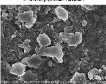
Morphology of phytosome curcumin
SEM and TEM images demonstrate the formation
of particles. As shown in Figure 2 (A, B) the particles
observed under SEM showed spherical-shaped particles. The TEM showed that particles phytosome curcumin are uniforms and homogeneous.
Yield of phytosome preparation process
Yield of phytosome preparation process was presented in Table I.
FIGURE 1 - Physical properties of curcumin phytosome at diferent ratios.
Table I showed that total yield of the preparation
process increased following the rise of phosphatidylcholine
amount added in the complexes. With the curcumin: phosphatidylcholine ratio is 1:2 and 1:4, the yield can be about 94%. However, this yield is only about 81% when the ratio is 1:1. This can be explained by adding the large
amount of phosphatidylcholine, the ability of interaction between curcumin and phosphatidylcholine was higher,
lead to form more phytosome curcumin complex.
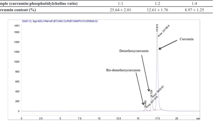
Determination of curcumin content in the complex
The curcumin content in the phytosome was shown
in Table II.
The curcumin content in the complex was determined by HPLC method. A HPLC chromatographical of
phytosome curcumin with curcumin: phosphatidylcholine ratio (mol:mol) 1:1 is shown in Figure 3. The percentages of curcumin in the phytosome in sample at ratio 1:1 are higher than curcumin in other ratio. The amount
of curcumin reached 25.64% in the complexes with
curcumin: phospholipis ratio 1:1. However, curcumin
content reached 12.61 % and 8.97 % in the complexes
with 1:2 and 1:3 ratios, respectively.
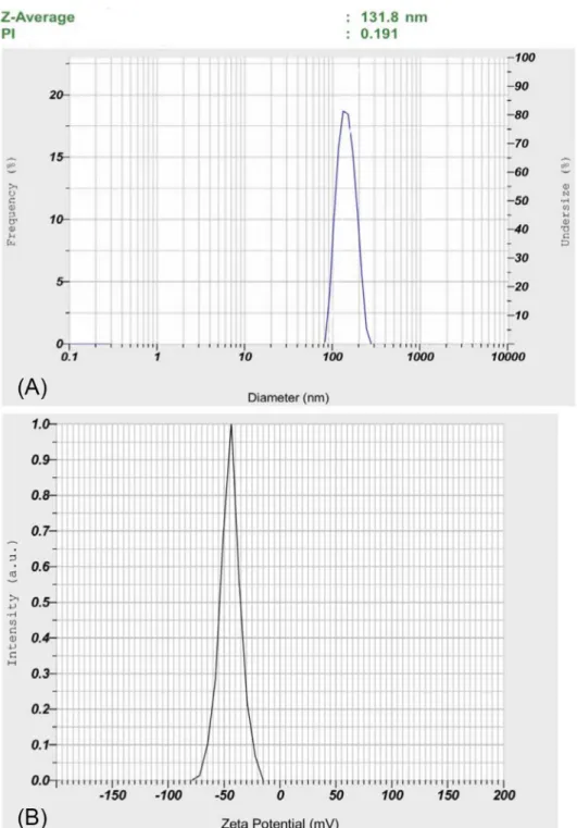
Particles size and zeta potential of phytosome curcumin
The distribution of the size of phytosome curcumin was shown in Figure 4. The average size of phytosome curcumin was 131.8 nm and PDI was 0.191. The zeta potential of PEG-CUR was -44.5 mV. One important parameter of nanoparticles is the polydispersity index (PDI), which is measured the particle size distribution. If PDI is smaller than 0.1, the particles are typically referred
to as “monodisperse” (Moreira, Gaspar, Allen, 2001; Pereira-Lachataignerais et al., 2006; Pham et al., 2015).
Our particles of phytosome curcumin are shown to be
TABLE I - Total yield of phytosome preparation process
Curcumin:
phosphatidylcholine ratio Total yield (w/w,%)
1:1 87.74 ± 1.73
1:2 91.61 ± 2.24
1:4 94.64 ± 2.64
TABLE II - Curcumin content in the phytosome
Sample (curcumin:phosphatidylcholine ratio) 1:1 1:2 1:4
Curcumin content (%) 25.64 ± 2.01 12.61 ± 1.76 8.97 ± 1.25
quite uniforms with PDI was 0.191 and have an average size of 131.8 nm.
The index used to evaluate the stability of particle system is zeta potential value. If the particles possess the absolute value of zeta potential greater than 30 mV, then
the particle system is highly stable and able to prevent the
aggregation of particles. If zeta potential values are in the range of 20-30 mV, the particle system is relatively stable
(Eloy et al., 2014; Pham et al., 2015). Our PEG-CUR has
zeta potential values of -44.5 mV, it can be considered they
are a relatively stable system.
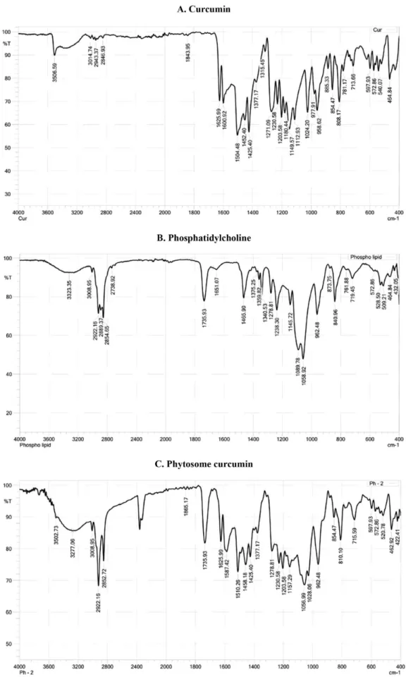
Infrared (IR) spectroscopy
IR spectra of curcumin, phosphatidylcholine and phytosome are shown in Figure 5. The IR spectra of curcumin showed two peaks at λ 1625.99 and 1600.92 cm-1
which represent for C=C and C=O bonds, but disappear
in phytosome’s IR spectra. The peak at λ 1734.01 cm-1
presents in both IR spectra of phosphatidylcholine and phytosome curcumin. Moreover, the peak at λ 3506.59
cm-1 of OH group in curcumin structure and two peaks
at λ 2916.37 and 2848.86 cm-1 in phosphatidylcholine
structure are also presented in phytosome curcumin’s IR spectrum. Interestingly, there is a new peak at λ 3745.76
cm-1 and some fluctuations appearing from 3200 to
4000 cm-1 in phytosome’s IR spectrum. This confirms the presence of hydrogen bonds between curcumin and phosphatidylcholine. Therefore, it is suggested that in the phytosome, curcumin bounds to the polar head
of phosphatidylcholine while the non–polar part of
phosphatidylcholine still are freely and envelops the polar part containing curcumin molecules.
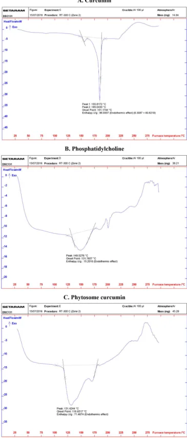
Differential scanning calorimetry (DSC)
Figure 6 reveals the DSC thermographs of curcumin (A), phosphatidylcholine (B) and phytosome curcumin (C). DSC spectrum of curcumin showed the onset temperature of the melting processes is 155.8172oC corresponds to
peak at 185.0430 oC which is the curcumin‘s melting
temperature. Phosphatidylcholine has a melting peak at 149.5276 oC corresponds to the temperature when hydrocarbon tails of phosphatidylcholine transform from
gel state to liquid crystalline. DSC of phytosome curcumin do not present the endothermic peak of both curcumin and phosphatidylcholine. The melting peak of curcumin and phosphatidylcholine were completely disappeared. Instead, it showed new melting peaks with lower endothermic efect of phosphatidylcholine (onset temperature 131.4244 oC
and meilting peak 116.6517 oC). This proves that curcumin reacts with phosphatidylcholine to form the chemical bonds between the OH in phenol group of curcumin structure with
phosphatidylcholine‘s polar head.
1H NMR spectroscopy
T h e p r o t o n N M R s p e c t r u m o f c u r c u m i n , phosphatidylcholine and phytosome curcumin was
represented in Figure 7. In 1H-NMR of phytosome curcumin signals at δ 0.876, 1.296 and 2.764 ppm showed that the signals of protons of methyl, methylene group
of aliphatic side chain and methylene protons linked to–C(=O)–C group, respectively. They are characteristics
of nonpolar portion of phosphatidylcholine molecule. The signal of protons at δ 5.334 ppm is due to signals of
second methylene group proton near N atom (-CH2CH2N)
of choline and signals at δ 3.362 ppm is due to protons
of methyl group attached to N–atom of choline which
represented a signal broadening due to their involvement
in phytosome curcumin (Sikarwar et al., 2008). The
protons signals at 5.779-5.983; 6.459-6.491; 6.926-6.942; 7.049-7.052; 7.112-7.132 and 7.572-7.604 [m, ArH] are
due to aromatic ring of curcumin. These data suggested
that phenyl group of curcumin was complexed with
choline part of phosphatidylcholine.
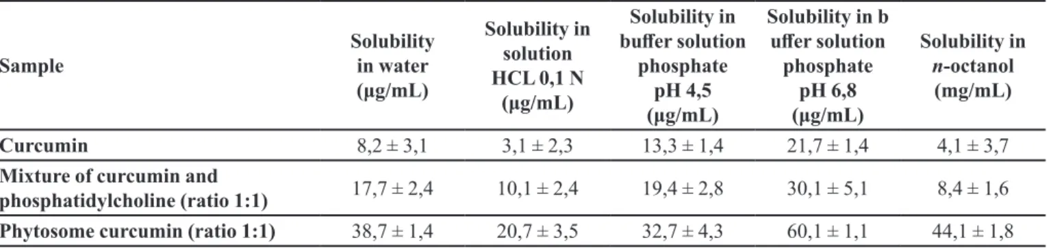
Solubility studies
Solubility of curcumin, phytosome and physical
mixture of curcumin and phosphatidylcholine in water0 was shown in Table III.
Curcumin, a hydrophobic molecule, is practically
insoluble in water, especially at pH 1.2. Therefore,
curcumin has very low bioavailability; this is a barrier
of using curcumin in clinical application. When pH was
increased, curcumin’s solubility tends to increase but still
is very low. Solubility of curcumin in n-octanol is also very low. Therefore, bioavailability of curcumin by oral via
or derma via is extremely low. Phytosome curcumin can signiicantly improve solubility of curcumin in solution at diferent pH and also in n-octanol. Thereby phytosome curcumin can increase the partition coeicient O/W, lead curcumin easily to difuse into the cell membrane, easily
transferred from the aqueous phase to the lipid phase, then increasing its bioavaibility.
Biological activity
Enzyme such as AST and ALT are main liver
transaminases have been used for the assessment of liver damage (Howell et al., 2014).
Serum ALT and AST activities were signiicantly
increased in PAR group as compared with control group
(Table IV). When mice were treated with phytosome,the
ALT and AST activities were signiicantly decreased as compared to the PAR group. These enzymes activities tended to decrease in Cur 200 group compared with PAR
group.
Lipid peroxidation
The levels of lipid peroxidation product (MDA)
from the liver tissues in the studied groups were shown
in Table V and Figure 8. An increased in MDA level was observed signiicantly in the PAR group when compared with control group (p<0.05). A decreased in MDA levels were observed signiicantly in group of Phyt 100 and Phyt 200 (p<0.05). The level of MDA tended to decrease in Cur 200 group compared with PAR group.
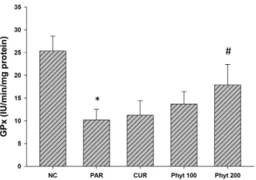
Antioxidant enzyme analysis
We also determined the activities of several
antioxidant enzymes including CAT, SOD and GPx in the liver tissue of control and experimental animals. The results were reported in Table V and Figures 9, 10 and 11. Mice in PAR group showed a signiicant lower in the activities of these antioxidants compared with control group (p<0.05). Mice in groups Phyt 100 and Phyt 200 exhibited significant increasing in the levels of CAT, FIGURE 7 - Proton NMR Spectrum of Curcumin (A),
TABLE III - Solubility of curcumin, phytosome and mixture of curcumin and phosphatidylcholine in diferent medium
Sample
Solubility in water
(μg/mL)
Solubility in solution
HCL 0,1 N (μg/mL)
Solubility in
bufer solution
phosphate pH 4,5
(μg/mL)
Solubility in b
ufer solution
phosphate pH 6,8
(μg/mL)
Solubility in n-octanol
(mg/mL)
Curcumin 8,2 ± 3,1 3,1 ± 2,3 13,3 ± 1,4 21,7 ± 1,4 4,1 ± 3,7
Mixture of curcumin and
phosphatidylcholine (ratio 1:1) 17,7 ± 2,4 10,1 ± 2,4 19,4 ± 2,8 30,1 ± 5,1 8,4 ± 1,6
Phytosome curcumin (ratio 1:1) 38,7 ± 1,4 20,7 ± 3,5 32,7 ± 4,3 60,1 ± 1,1 44,1 ± 1,8
TABLE IV - Efect of phytosome curcumin on liver marker enzymes
Parameters Control PAR Cur 200 Phyt 100 Phyt 200
AST (IU/L) 38.24 ± 4.23 101.28 ± 11.25* 81.28 ± 12.27 47.25 ± 5.24# 41.25 ± 5. 32#
ALT (IU/L) 31.12 ± 5.21 97.67 ± 11.84* 68.58 ± 14.45 42.15 ± 6.38# 37.17 ± 3.25#
TABLE V - The efect of administration of phytosome curcumin on lipid peroxidation and antioxidant enzymes in mice liver tissue
Parameters Control PAR Cur 200 Phyt 100 Phyt 200
MDA 0.52 ± 0.14 1.89 ± 0.15* 1.61 ± 0.13 1.42± 0.12# 1.28 ± 0.11#
SOD 0.573 ± 0.09 0.187 ± 0.08* 0.210 ± 0.07 0.282 ± 0.10# 0.362 ± 0.12#
CAT 298.87 ± 39.15 89.8 ± 13.19* 129.8 ± 14.21 165.15 ± 21.12# 196.12 ± 23.15#
GPx 25.35 ± 3.27 10.19 ± 2.32* 11.23 ± 3.18 13.68 ± 2.72 17.86 ± 4.54#
Value represents as mean ± S.D. *p< 0.05, signiicant diference compared with control group, #p< 0.05, signiicant diference
compared with paracetamol group (n = 10).
SOD and GPx compared with PAR group (p<0.05). These enzymes activities tended to increase in Cur 200 group
compared with PAR group.
DISCUSSION
Preparing new formulation for curcumin delivery is
important because of many beneicial efect of curcumin. Curcumin has poor absorption because of its low solubility
(Maiti et al., 2007). Phospholipids are now using in many
application of drug delivery technology. The advantage
of phospholipids is increasing solubilizing property of many natural products. In this study, we have developed
successfully phytosome curcumin which can improve the bioavailability of curcumin. The physicochemical
data showed that curcumin formed a complex with
phosphatidylcholine by hydrogen bonds. We also showed that the phytosome has increased the solubility
of curcumin in diferent medium.
Paracetamol have been used in many study for
induce liver damage (Farghaly, Hussein 2010). Liver is
the main organ in body, which metabolizes chemicals and drug. It has been well known that enzyme AST and ALT are markers of hepatocyte damage and the high level of AST and ALT is an important marker for liver injury (McGill, Jaeschke, 2013). In our study, paracetamol signiicant induced hepatic damage in mice by increasing
the level of AST and ALT. Our data are agreed with Nithianantham et al. (2011) they have showed that
parcetamol significant increased the ALT, AST, and bilirubin levels in mice.
Phytosome curcumin protects the mice from PAR-induced acute liver injury in vivo. After administration of paracetamol, serum ALT and AST levels in mice were significantly greater than those in control group, and phytosome curcumin could reduce those levels. Our results indicate that phytosome curcumin protects hepatocytes in vivo from damage induced by paracetamol administration.
Our data are in line with previous report that curcumin can protect damages in liver caused by paracetamol
(Kheradpezhouh et al., 2010).
FIGURE 8 - Efect of phytosome curcumin on lipid peroxidation of control and treated animals in paracetamol-induced liver injury in mice. NC: Control group; PAR: mice received 1g/ kg b.w of Paracetamol; CUR: mice received 200 mg/kg b.w curcumin and 1g/kg b.w of Paracetamol; Phyt 100: mice received amount phytosome curcumin equivalent to curcumin 100 mg/kg b.w and 1g/kg b.w of Paracetamol; Phyt 200: mice received amount phytosome curcumin equivalent to curcumin 200 mg/kg b.w and 1g/kg b.w of Paracetamol; Value represents as mean ± S.D. *p<0.05, signiicant diference compared with
control group, #p< 0.05, signiicant diference compared with
paracetamol group (n = 10).
FIGURE 9 - Efect of phytosome curcumin on CAT activity of control and treated animals in paracetamol-induced liver injury in mice. Value represents as mean ± S.D. *p<0.05, signiicant diference compared with control group, #p<0.05, signiicant diference compared with paracetamol group (n = 10).
FIGURE 10 - Efect of phytosome curcumin on SOD activity of control and treated animals in paracetamol-induced liver injury in mice. Value represents as mean ± S.D. *p<0.05, signiicant diference compared with control group, #p<0.05, signiicant diference compared with paracetamol group (n = 10).
FIGURE 11 - Efect of phytosome curcumin on GPx activity of control and treated animals in paracetamol-induced liver injury in mice. Value represents as mean ± S.D. *p<0.05, signiicant diference compared with control group, #p<0.05, signiicant diference compared with paracetamol group (n = 10).
fatty acids and disrupt the cell membrane. It leads to oxidative lipid and forms MDA, a product of lipid peroxidation. Increasing production of liver MDA observed in our experiments by PAR are in agreement with previous study which reported that PAR increased extracellular
MDA level (Boonruamkaew, Chonpathompikunlert et al.,
2016). In addition, we have shown phytosome curcumin may diminish the level of MDA in liver tissues. Our data are
in line with study of Hatem et al. which showed that hepatic
lipid peroxidation level was suppressed by administration of curcumin to paracetamol-treated rats (Farghaly, Hussein 2010).
The organism can develop a mechanism, such as
endogenous enzymatic against ROS. SOD is antioxidant enzyme that converts superoxide anion O2
•- to H 2O2.
CAT converts H2O2 to water and O2. GPx catalyzes the reduction of H2O2 and other peroxides by coupling reduced glutathione (Madrigal-Santillan et al., 2014; Thanh et al., 2015). Our data have showed the activities of these
enzymes in PAR group were declined. Interestingly
when mice were treated with phytosome curcumin, these
enzymes can be reversed signiicantly.
The hepatoprotective effect of phytosome was
signiicantly higher than curcumin free at the same dose. Free curcumin at the dose of 200 mg/kg only slightly
reduced the damage conditions in mice induced by
paracetamol. The phytosome at dose equivalent to 100 mg/kg of curcumin showed higher restored the damage
of mice liver compared with curcumin free at double dose
(200 mg/kg). The increasing hepatoprotective eicacy of phytosome curcumin may be explained by increasing
bioavailability of the curcumin.
In summary, this study demonstrates that phytosome curcumin had a strong protective efect against
paracetamol-induced acute hepatic damage in mice. The
hepatoprotective efect of phytosome curcumin may be explained by increasing levels of antioxidant enzymes and decreasing the lipid peroxidation and liver enzyme
on paracetamol-induced damage in mice.
CONFLICTS OF INTERESTS
The authors declare no conlict of interest.
ACKNOWLEDGEMENTS
The authors would like to thank the inancial support of project (Grant number QG.16.25) from the Vietnam
National University Ha Noi, Vietnam.
REFERENCES
ANAND, P.; THOMAS, S.G.; KUNNUMAKKARA, A.B.; SUNDARAM, C.; HARIKUMAR, K.B.; SUNG, B.; THARAKAN, S.T.; MISRA, K.; PRIYADARSINI, I.K.; RAJASEKHARAN, K.N. Biological activities of curcumin and its analogues (Congeners) made by man and Mother Nature. Biochem. Pharmacol., v.76, n.11, p.1590-1611,
2008.
BOONRUAMKAEW, P.; CHONPATHOMPIKUNLERT, P.; NAGASAKI, Y. Redox nanoparticle therapeutics for acetaminophen-induced hepatotoxicity in mice. Oxid. Med. Cell. Longev., v.2016, p.1-10, 2016.
CUOMO, J.; APPENDINO, G.; DERN, A.S.; SCHNEIDER, E.; MCKINNON, T.P.; BROWN, M.J.; TOGNI, S.; DIXON, B.M. Comparative absorption of a standardized curcuminoid mixture and its lecithin formulation. J. Nat. Prod., v.74, n.4, p.664-669, 2011.
ELOY, J.O.; SOUZA, M.C.; PETRILLI, R.; BARCELLOS, J.P.A.; LEE, R.J.; MARCHETTI, J.M. Liposomes as carriers of hydrophilic small molecule drugs: Strategies to enhance encapsulation and delivery. Colloids Surf., B, v.123, p.345-363, 2014.
FARGHALY, H.S.; HUSSEIN, M.A. Protective effect of curcumin against paracetamol-induced liver damage. Aust. J. Basic Appl. Sci., v.4, n.9, p.4266-4274, 2010.
GUPTA, S.C.; PATCHVA, S.; AGGARWAL, B.B. Therapeutic roles of curcumin: lessons learned from clinical trials. AAPS J., v.15, n.1, p.195-218, 2013.
HINSON, J.A.; ROBERTS, D.W.; JAMES, L.P. Mechanisms of acetaminophen-induced liver necrosis. Handb. Exp. Pharmacol., v.2010, n.196, p.369-405, 2010.
HOWELL, B.; SILER, S.; SHODA, L.; YANG, Y.; WOODHEAD, J.; WATKINS, P.B. A mechanistic model of drug‐induced liver injury aids the interpretation of elevated liver transaminase levels in a phase I clinical trial. CPT: Pharmacometrics Syst. Pharmacol., v.3, n.2, p.1-8, 2014.
JAESCHKE, H.; RAMACHANDRAN, A. Reactive oxygen species in the normal and acutely injured liver. J. Hepatol., v.55, n.1, p.227-228, 2011.
KHERADPEZHOUH, E.; PANJEHSHAHIN, M.-R.; MIRI, R.; JAVIDNIA, K.; NOORAFSHAN, A.; MONABATI, A.; DEHPOUR, A.-R. Curcumin protects rats against acetaminophen-induced hepatorenal damages and shows synergistic activity with N-acetyl cysteine. Eur. J. Pharmacol., v.628, n.1, p.274-281, 2010.
MADRIGAL-SANTILLAN, E.; MADRIGAL-BUJAIDAR, E.; ALVAREZ-GONZALEZ, I.; SUMAYA-MARTINEZ, M.T.; GUTIERREZ-SALINAS, J.; BAUTISTA, M.; MORALES-GONZALEZ, A.; GARCIA-LUNA, M.; GONZALEZ-RUBIO, Y.; AGUILAR-FAISAL, J.L.; MORALES-GONZALEZ, J.A. Review of natural products with hepatoprotective efects. World J. Gastroenterol., v.20,
n.40, p.14787-14804, 2014.
MAITI, K.; MUKHERJEE, K.; GANTAIT, A.; SAHA, B.P.; MUKHERJEE, P.K. Curcumin–phospholipid complex: Preparation, therapeutic evaluation and pharmacokinetic study in rats. Int. J. Pharm., v.330, n.1/2, p.155-163, 2007.
MARCZYLO, T.H.; VERSCHOYLE, R.D.; COOKE, D.N.; MORAZZONI, P.; STEWARD, W.P.; GESCHER, A.J. Comparison of systemic availability of curcumin with that of curcumin formulated with phosphatidylcholine. Cancer Chemother. Pharmacol., v.60, n.2, p.171-177, 2007.
MCGILL, M.R.; JAESCHKE, H. Metabolism and disposition of acetaminophen: recent advances in relation to hepatotoxicity and diagnosis. Pharm. Res., v.30, n.9, p.2174-2187, 2013.
M C G I L L , M . R . ; W I L L I A M S , C . D . ; X I E , Y. ; RAMACHANDRAN, A.; JAESCHKE, H. Acetaminophen-induced liver injury in rats and mice: comparison of protein adducts, mitochondrial dysfunction, and oxidative stress in the mechanism of toxicity. Toxicol. Appl. Pharmacol., v.264, n.3, p.387-394, 2012.
MOREIRA, J.N.; GASPAR, R.; ALLEN, T.M. Targeting stealth liposomes in a murine model of human small cell lung cancer. Biochim. Biophys. Acta, v.1515, n.2, p.167-176,
2001.
NITHIANANTHAM, K.; SHYAMALA, M.; CHEN, Y.; LATHA, L.Y.; JOTHY, S.L.; SASIDHARAN, S. Hepatoprotective potential of clitoria ternatea leaf extract against paracetamol induced damage in mice. Molecules, v.16, n.12, p.10134, 2011.
NOBLE, J.E.; BAILEY, M.J. Quantitation of protein. Methods Enzymol., v.463, p.73-95, 2009.
PEREIRA-LACHATAIGNERAIS, J.; PONS, R.; PANIZZA, P.; COURBIN, L.; ROUCH, J.; LOPEZ, O. Study and formation of vesicle systems with low polydispersity index by ultrasound method. Chem. Phys. Lipids, v.140, n.1/2,
p.88-97, 2006.
PHAM, T.M.H.; NGUYEN, L.T.; NGUYEN, V.L.; LE, P.L.; HO, A.S.; NGUYEN, T.H.; BUI, T.T. Developing and evaluating in vitro effect of pegylated liposomal doxorubicin on human cancer cells. J. Chem. Pharm. Bull., v.7, n.3, p.2239-2243, 2015.
PRASAD, S.; TYAGI, A.K.; AGGARWAL, B.B. Recent developments in delivery, bioavailability, absorption and metabolism of curcumin: the golden pigment from golden spice. Cancer Res. Treat., v.46, n.1, p.2-18, 2014.
SHYAM, K.R.; KUMAR, G.M. Prepration, charactaerization and antioxidant activities of gallic acid-phospholipids complex. Int. J. Res. Pharm. Sci., v.2, n.1, p.138-148, 2012.
SIKARWAR, M.S.; SHARMA, S.; JAIN, A.K.; PARIAL, S.D. Preparation, characterization and evaluation of Marsupsin-phospholipid complex. AAPS PharmSciTech, v.9, n.1, p.129-137, 2008.
SIVIERO, A.; GALLO, E.; MAGGINI, V.; GORI, L.; MUGELLI, A.; FIRENZUOLI, F.; VANNACCI, A. Curcumin, a golden spice with a low bioavailability. J. Herbal Med., v.5, n.2, p.57-70, 2015.
THANH, T.B.; THANH, H.N.; MINH, H.P.T.; LE-THI-THU, H.; LY, H.D.T.; DUC, L.V. Protective efect of Tetracera scandens L. leaf extract against CCl4-induced acute liver injury in rats. Asian Pacific J. Trop. Biomed., v.5, n.3,
p.221-227, 2015.
THANH, H.N.; MINH, H.P.T.; DUC, L.V.; THANH, T.B. Protective efect of coenzyme Q10 on methamphetamine-induced neurotoxicity in the mouse brain. Trends Med. Res., v.11, n.1, p.1-10, 2016.