http://dx.doi.org/10.1590/s2175-97902017000115210
*Correspondence: M. C. Valadares. Faculdade de Farmácia. Universidade Federal de Goiás – UFG. Praça Universitária, n.1166, Setor Universitário, 74605.220 - Goiânia - GO, Brasil. Phone / fax: +55 62 3209-6039 ext. 201. E-mail: [email protected]
Grandisin induces apoptosis in leukemic K562 cells
Alane Pereira Cortez
1, Elizabeth Gomes Paulino Menezes
1, Polyana Lopes Benica
1, Alexandre
Pereira dos Santos
1, Larissa Moreira Cleres
1, Higor de Oliveira Ribeiro
1, Eliana Martins Lima
2,
Massuo Jorge Kato
3, Marize Campos Valadares
1,*1Laboratory of Celullar Toxicology and Pharmacology - FarmaTec, Faculty of Pharmacy, Federal University of Goiás,
Goiânia, GO, Brazil, 2Medicinal Pharmaceutical Chemistry Laboratory, Faculty of Pharmacy, Federal University of Goiás,
UFG, Goiânia, GO, Brazil, 3Chemistry Laboratory of Natural Products, Institute of Chemistry, University of São Paulo
In this study, the potential antileukemic activity of grandisin, a lignan extracted from Piper solmsianum, was evaluated against the leukemic line K562. The cytotoxicity of grandisin (0.018 to 2.365 µM) was evaluated in K562 and normal peripheral blood lymphocytes by Trypan Blue Exclusion and MTT methods after 48h exposure to the drug. In both methods, cellular viability was concentration-dependent and the IC50 values were lower than 0.85µM. Analysis of K562 cells after treatment with grandisin showed that the cell cycle was arrested in the G1 phase with a 12.31% increase, while both S and G2 phases decreased. Morphological studies conducted after the exposure of K562 to grandisin revealed changes consistent with the apoptosis process, which was conirmed by anexin V stain and caspase activation. Thus, lignan grandisin showed antileukemic activities against the K562 cell line and the cell death process occurred via apoptosis.
Uniterms: Grandisin/antileukemic activities. Leukemic. K562. Apoptosis.
INTRODUCTION
Chronic myeloid leukemia (CML) is a clonal myeloproliferative syndrome characterized by the presence of the Philadelphia chromosome - Ph (Faderl et al., 1999). The Ph chromosome contains the chimeric gene BCR-ABL, which produces a tyrosine kinase. Uncontrolled BCR-ABL tyrosine kinase activity is a conditio sine qua non for the protein´s skill to transform cells (Eiring et al., 2011). The expression of BCR-ABL also contributes to apoptosis resistance after the withdrawal of growth factors, oxidative stress and DNA damage. The ability to escape normal cell death programs provides a remarkable proliferative advantage for tumoral cells, especially during the accelerated or blast phase of the disease and hinders successful treatment since it is well-known that CML cells are highly resistant to apoptosis induced by chemotherapeutic drugs (Ren, 2005; Fernandez-Luna, 2000; O’Hare et al., 2012; Jabbour, Kantarjian, 2014).
Apoptosis, a type of programmed cell death which occurs in physiological and pathological conditions, is a prime target in designing therapeutic strategies to induce cancer cell death. This process shows typical cellular morphologic changes such as rounding-up of the cell, chromatin condensation, nuclear fragmentation, plasma membrane blebbing and engulfment by resident phagocytes (in vivo) (Ziegler et al., 2004; Kroemer et al., 2009). Moreover, many coordinated pathways with a wide range of proteins and enzymes are involved in this process. Caspase activation and translocation of phosphatidylserine from the inner lealet of the cytoplasmic membrane to the outer lealet are common events which occur in apoptotic cells. In addition, the Bcl-2 protein family plays a pivotal role in the regulation of apoptosis (Wong, 2011; Fleischer et al., 2006).
and toxicity. Despite the benefits of TKI therapy, the persistence of minimal residual disease (MRD) and the presence of active disease even with BCR-ABL1 inhibition, the development of new target drugs against CML is necessary (O’Hare et al., 2012; Piccaluga et al., 2012). In this context, natural compounds have been investigated as potential sources for new chemical entities and in order to develop drugs against various pharmacological targets, including leukemias (De Martino et al., 2011).
The tetrahydrofuran lignan grandisin, isolated from Virola and Piper species, presents antimalarial and trypanocidal activities, as well as having antitumoral properties. Furthermore, grandisin CYP450-mediated biotransformation obeys Michaelis–Menten kinetics and its metabolism results in the formation of four demethylated metabolites (Zhang et al., 2001; Valadares et al., 2009; Barth et al., 2015). Grandisin antitumor properties were investigated by our group both in in vitro and in vivo assays using the Ehrlich Ascites Tumoral (EAT) model. The treatment of EAT-bearing mice with grandisin (2.5, 5.0 or 10.0 mg/kg) for 10 days, signiicantly increased the survival of the animals, in a dose-dependent manner. At the same time, a 66% reduction in intraperitoneum tumor cell burden in the animals treated with 10mg/kg of grandisin was observed. Furthermore, the marked increase in vascular endothelial growth factor (VEGF) levels induced by EAT development in these animals was also signiicantly reduced after treatment with grandisin, and resulted in a 32% reduction of VEGF levels, when compared to the control. Finally, caspase activation (caspase-3, -6, -8 and -9) was detected after EAT cells were exposed to grandisin (Valadares et al., 2009). On-going studies performed by our group demonstrated that this lignan has a protective efect against cyclophosphamide-induced mutagenicity and this efect could be associated to grandisin bioactivation (Valadares et al., 2011).
In the light of this, the present study was designed to explore the potential antileukemic properties of grandisin on K562 cells (a human erythroleukemia cell line) and normal lymphocytes. Furthermore, the effects of this potential drug on the cell cycle and death mechanism were also investigated by low cytometry with caspase activation.
MATERIAL AND METHODS
Grandisin
Grandisin was isolated from the extract obtained from the leaves of Piper solmsianumas previously described (Martins et al., 2003).
Grandisin nanoemulsion
Grandisin was prepared as a nanoemulsion disperse system in order to obtain a formulation with an aqueous external phase as previously described (Valadares et al., 2009).
Briefly, the dispersion was prepared as follows: first, a mixture of 50 µg of soy phosphatidylcholine (PC), sunlower oil (4:1 mol/mol PC), and grandisin (10 mg/mL) were dissolved in 1.0 mL of chloroform. The mixture was dried under a nitrogen atmosphere and kept under vacuum overnight to ensure the complete removal of chloroform. Then 1 mL of water was added to the dried lipid ilm to promote the hydration of the lipid-drug mixture, which after a 2 min vortex mixing, resulted in a coarse O/W emulsion. After 1 hour, the mixture was sonicated for 10 min in a Titanium probe sonicator to obtain a homogeneous submicrometric emulsion. For the control group, the nanoemulsion was prepared without adding grandisin to the oil phase.
Cell lines and culture
K562 erythroleukemia cell line, obtained from the American Type Culture Collection (ATCC, Rockville, MD, USA), was cultured in suspensions in RPMI 1640 medium (Sigma Chemical Co.) supplemented with 10% fetal calf serum (FCS, Sigma Chemical Co.), 100 U/mL of penicillin, and 100 ug/mL of streptomycin in a humidiied atmosphere at 37 °C in 5% CO2. Cells were seeded (1 x 10⁶ cells/mL)
in 96-well microtiter plates and incubated with diferent concentrations of grandisin for 48 hours (Queiroz et al., 2009). The human blood used in the assays was collected from healthy volunteers who had signed a consent form.
Lymphocyte culture
Cell proliferation and viability assays
C e l l p r o l i f e r a t i o n w a s d e t e r m i n e d b y t h e 3-[4,5-dimethylthiazol- 2-yl]-2,5-diphenytetrazolium bromide (MTT, Sigma Chemical Co.) reduction test, performed as previously described by Mosmann (1983). Briely, 1 x 10⁶ cells/mL in RPMI 1640 medium supplemented with 10% FBS were seeded in a 96-well, lat-bottom plate (TPP, Trasadingin, Swiss) and treated with diferent concentrations of grandisin (0.018 – 2.365 µM) for 48 hours. After incubation, 10µL/well MTT (5mg/mL) were added and the plate was incubated again for 4 hours. It was then centrifuged at 800 rpm for 10 min and the supernatant removed. A total of 100 µL of dimethylsulphoxide (DMSO) was added to each well to solubilize the formazan crystals. Absorbance was measured at 545nm using a spectrophotometer (Stat Fax 2100, Awareness Technology, Dusseldorf, Germany).
Trypan blue dye exclusion assay was used to assess cell viability as previously described (Bromberg et al., 2005). The cells were seeded in a 96-well, lat-bottom plate (TPP, Trasadingin, Swiss), at 1 x 10⁶ cells/mL in RPMI 1640 medium supplemented with 10% FBS and treated with diferent concentrations of grandisin (0.018 – 2.365 µM) for 48 hours. An aliquot of the cell suspension was then mixed with trypan blue solution (0.2% in phosphate-bufered saline-PBS) (1:10) and the viability of the cells was estimated using a hemocytometer (Reichert, USA). Cells which incorporated the dye and turned blue were reported as dead. Each concentration was tested in three independent experiments run in four replicates. Proliferation and viability of cells exposed to grandisin were expressed as a percentage of the cell proliferation or viability of untreated control cells (100%).
Methods for detection of cell death by apoptosis
Apoptotic index
K562 cells were incubated in a humid chamber at 37 °C, 5% CO2, with grandisin 0.036 µM for 24 h. After this period, cells were stained with hematoxylin and analysed at 40X magniication. The number of apoptotic cells was expressed as a percentage of the total number of cells. The Apoptotic index was determined after a minimum of 100 cells were counted. Cells showing morphological characteristics of apoptosis such as marginalization or chromatin condensation, cellular or nuclear fragmentation, and cellular structures fragmented into vesicles (apoptotic bodies) were considered positive.
Annexin V-FITC/PI double-staining and analysis by flow cytometry
After incubation with grandisin as described above, K562 cells were harvested, washed with cold PBS and resuspended in binding bufer at a concentration of 1 x 10⁶ cells/mL,according to the manufacturer’s instructions(eBioscience, USA). The suspensions were transferred to 5 mL tubes, and 5 µL Annexin V-FITC and 5 µg/mL PI were added. The cells were incubated at room temperature for 20 min, after which 300 µL binding bufer were added and analysis was performed in a FACSCantoII flow cytometer using the CellQuest software (10.000 events were collected per sample). Control cells were treated with medium only.
Cell cycle
For analysis of the cell cycle, 5 x 10⁵ cells were incubated with or without grandisin 0.036 µM for 24 h in RPMI 1640 medium and 10% fetal calf serum, in an incubator with 5% CO2 at 37 °C. After that, the cells were washed twice with 1 mL of cold PBS, and centrifuged at 1500 rpm for 10 min. The cell button was resuspended with 1 mL of ixative solution (previously prepared 70% ethanol) and incubated at 4 °C overnight. Cells were then washed in cold PBS and resuspended in 1 mL of 200 μg/mL RNAse A and 50 μg/mL propidium iodide, and maintained at 4 °C, protected from the light, for 2 h. Samples were analyzed by low cytometry (FACSCanto II, Becton Dickinson). Results were presented in graphs representing the number of cells versus DNA content indicated by luorescence intensity.
Caspase activity
were incubated with each X-pNA substrate (200 μM final concentration) at 37 °C in a microtiter plate. The optical density of samples was measured at 405nm. After subtraction of the blank, the increase in caspase activity was determined by comparing these results with the levels of the control.
Statistics
Cytotoxicity evaluation was performed in three independent experiments. Results were converted into percentage of control. IC50 (concentration which produces an inhibitory effect of 50% of the evaluated parameter) was obtained graphically from the concentration-response curve. Results were expressed as mean ± S.D. of four replicates. For the parameters ‘apoptotic index’ and ‘caspase activity’, statistical analysis was performed using the nonparametric Mann-Whitney test to compare the treated groups with controls and the Kruskal-Wallis test to compare the treated groups with each other. Statistical signiicance was considered when p <0.05.
RESULTS AND DISCUSSION
Effects of grandisin on cell proliferation and viability of K562 cells and normal lymphocytes.
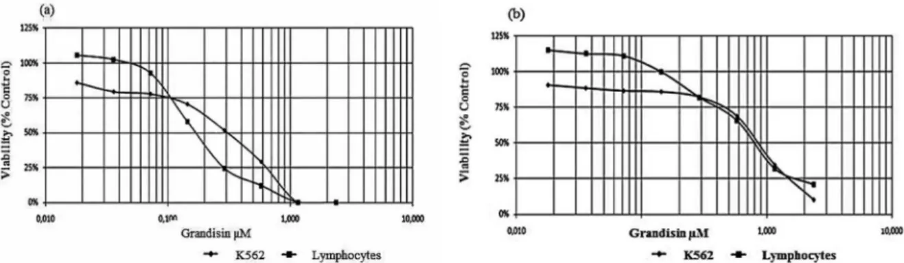
The efect of grandisin on the growth and viability of K562 cells and lymphocytes was examined, using the Trypan blue exclusion method, after 48 hours of exposure in culture. The proliferation of K562 cells and lymphocytes was inhibited in a concentration-dependent manner in response to increased concentrations of grandisin (0.018 to 2.365 µM) (Figure 1a). The 50%
inhibition (IC50) was obtained with a concentration of 0.851 µM for K562 and 0.685 µM for lymphocytes after 48 hours of exposure (Figure 1a). On the other hand, results obtained by the MTT method showed that the 50% inhibition (IC50) values were 0.198 µM for K562 and 0.200 µM for lymphocytes (Figure 1b).
Thus, our data demonstrated that grandisin was more cytotoxic for lymphocytes than for K562 cells in the Trypan blue assay. However, in the MTT assay the cytotoxicity of grandisin was quite similar for both types of cells. Although grandisin proved to be cytotoxic to K562 cells, it was also toxic for lymphocytes in the same range of concentrations tested. These results are important because they demonstrate that even though grandisin exerts anti-leukemic activity, it may act similarly to many other chemotherapeutic agents exhibiting toxicity to normal cells and triggering undesirable side efects, which thus limits its application in the clinical ield (Bhatt, Saleem, 2004). However, the lymphotoxicity of grandisin opens up new treatment ields, such as the immunosuppressive agent similar to PKC412 (CGP41251), a protein kinase inhibitor which has antitumoral and immunosuppressive activities (Ganeshaguru et al., 2002; Miyatake et al., 2007; Kawamoto et al., 2008).
Cell cycle analysis by flow cytometry
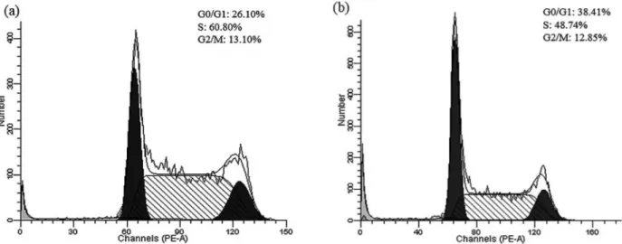
Results from the experiments designed to evaluate whether grandisin interferes with the cell cycle showed that it caused a signiicant G1 phase arrest with an increase of 12.31% in the population of cells and a decrease of 12.06% and 0.25% in the population of cells in the S and G2 phases, respectively, when compared with the control (Figures 2a and 2b).
FIGURE 1 - Cytotoxicity in K562 cells and normal peripheral blood lymphocytes (1 x 10⁶ cells/mL) treated with different
This analysis demonstrated that there was an increase in G1 population and a decline in cell population in the S and G2 phases which indicates that grandisin can interfere with cellular proliferation and with the dynamics of the cell cycle.
Apoptosis induction by grandisin in K562 cells
Apoptosis index
Once the ability of grandisin to induce the death of leukemic cells was detected, we studied the sequence of mechanisms involved in cell death, especially the
apoptotic mechanisms. To understand the mechanism by which grandisin promotes loss of viability in K562 cells, a number of apoptosis related experiments were performed. The efects of grandisin on the morphology of K562 cells were examined after 24 hours of exposure in culture (Figures 3a and b). Results showed a 4-fold increase in the number of cells undergoing apoptosis when compared to the control. As indicated in the photomicrographs, the cells showed morphological characteristics of death by apoptosis such as condensed chromatin, vacuoles, and fragmentation of the DNA and of the cellular membrane. A 40% increase in the number
FIGURE 2 - (a) Analysis of the cell cycle of K562 cells (control) by Flow Cytometry. (b) Analysis of the cell cycle of K562 cells
after treatment with grandisin 0.036 µM for 24 hours.
FIGURE 3 - (a) Apoptosis rate of K562 cells exposed to grandisin 0.036 µM for 24 hours. The rate of apoptosis was determined
of cells undergoing apoptosis was also observed when compared with the control.
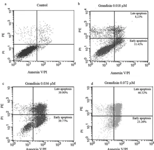
Annexin V-FITC/PI double-staining and analysis by flow cytometry
Once the antitumoral properties of grandisin were determined, we focused on the mechanisms involved in this process. Since morphological changes in K562 after treatment pointed to death by apoptosis, low cytometry analysis with Annexin V/PI stain was conducted. The exposure to grandisin was performed in three concentrations (0.018, 0.036 and 0.072 µM) for 24 hours (Figure 4). In the concentration of 0.018 µM, the increase in cells in early and late apoptosis was 11.42% and 8.22%, respectively. In the intermediate concentration (0.036 µM), the percentage of cells in early apoptosis was 39.77% and 39.90% in late apoptosis. Finally, in 0.072 µM the increase in early and late apoptosis was 23.26% and 46.52%, respectively. The results conirmed that the death mechanism triggered by grandisin was apoptosis.
Caspase activity
A molecular hallmark of apoptosis is the activation of caspases – speciic proteases which bring about cell death through cleavage of multiple protein substrates such as nuclear and cytoskeletal proteins (Bao; Shi, 2007; Elmore, 2007). In this context, caspase-6, -8 and-9 activities were analyzed by colorimetric assays. All caspases analyzed showed increases in activities of 21.4%, 29% and 37% for caspases-6, -8 and -9, respectively (Figure 5).
Our results showed an increase in all caspase activities tested after exposure of K562 to grandisin which suggests that intrinsic and extrinsic cascades were activated. Similar results were observed with apigenin in human breast cancer cells and with emodin in HeLa cells (Choi, Kim, 2009; Yaoxian et al., 2013).
Despite the many advances in cancer research, most antitumor compounds produce undesirable side effects. Thus, significant effort has been invested in inding new phytochemicals with antitumoral properties. Throughout history, natural compounds have been used for the treatment of cancer due to their safety, low toxicity,
FIGURE 4 - Analysis of K562 cells treated with grandisin(1 x 10⁶ cells/mL) for 24 hours. The cells were stained with Annexin
and availability from natural sources (Pratheeshkumar et al., 2012). Grandisin is a lignan extracted from Piper solmsianum which presents antitumoral properties as well as antimalarial and trypanocidal activities (Martins et al., 2003; Bernardes et al., 2006; Valadares et al., 2011). Despite its lymphocytic activities, grandisin did not exert mutagenic efects on the bone marrow cells of exposed mice. Moreover, grandisin proved to be a potent antiangiogenic compound due to the prolonged survival of mice bearing breast carcinoma (Valadares et al., 2011). Other pharmacological properties, such as antinociceptive, anti-inflamatory and antioxidant activities, have also been attributed to grandisin, which thereby shows the therapeutic versatility of this compound (Carvalho et al., 2010).
Thus, this study introduces a new agent capable of inducing apoptosis in a leukemic cell line which presents distinct features of resistance to many drugs. But it is important to highlight that further studies should be conducted to ensure the safety and efficacy of this substance and elucidate more clearly the mechanism which triggers apoptosis.
CONCLUSIONS
The present study suggests that the lignin grandisin has antileukemic activities against the K562 cell line and
that the cell death process occurs via apoptosis. Grandisin also showed immunosuppressant activities indicating new potential uses for immunological pathologies.
ACKNOWLEDGEMENTS
This study was supported by the Brazilian research funding agencies Conselho Nacional de Desenvolvimento Cientíico e Tecnológico (CNPq), Financiadora de Estudos e Pesquisas (FINEP), Coordenação de Aperfeiçoamento de Pessoal de Nível Superior (CAPES), Fundação de Apoio à Pesquisa da Universidade Federal de Goiás (FUNAPE) andFundação de Apoio à Pesquisa do Estado de Goiás (FAPEG).
REFERENCES
BAO, Q.; SHI, Y. Apoptosome: a platform for the activation of initiator caspases. Cell Death Difer., v.14, n.1, p.56-65, 2007.
BARTH, T.; HABENSCHUS, M.D.; MOREIRA, F.L.;
FERREIRA, L.S.; LOPES, N.P.; OLIVEIRA, A.R.M. In
vitro metabolism of the lignan (-)-grandisin, an anticancer drug candidate, by human liver microsomes. Drug Test Anal., v.7, n.9, p.780-786, 2015.
BERNARDES, L.S.; KATO, M.J.; ALBUQUERQUE, S.; CARVALHO, I. Synthesis and trypanocidal activity of 1,4-bis-(3,4,5-trimethoxy-phenyl)-1,4-butanediol and1,4-bis-(3,4-dimethoxyphenyl)-1,4-butanediol. Bioorg. Med. Chem., v.14, n.21, p.7075-7082, 2006.
BHATT, V.; SALEEM, A. Review: Drug-induced neutropenia-pathophysiology, clinical features, and management. Ann. Clin. Lab. Sci., v.34, n.2, p. 131-137, 2004.
BROMBERG, N.; JUSTO, G.Z.; HAUN, M.; DURAN, N.; FERREIRA, C.V. Violacein cytotoxicity on human blood lymphocytes and efect on phosphatases. J. Enzyme Inhib. Med. Chem., v.20, p.449-454, 2005.
CARVALHO, A.A.; GALDINO, P.M.; NASCIMENTO, M.V.; KATO, M.J.; VALADARES, M.C.; CUNHA, L.C.; COSTA, E.A. Antinociceptive and antiinflammatory activities of grandisin extracted from Virola surinamensis.
Phytother Res., v.24, n.1, p.113-118, 2010.
FIGURE 5 - Caspase activities in K562 (2 x 10⁶ cells/mL)
CHOI, E.J.; KIM, G.H. Apigenin induces apoptosis through a mitochondria/caspase-pathway in human breast cancer MDA-MB-453 cells. J. Clin. Biochem. Nutr., v.44, p.260– 265, 2009.
DE MARTINO, L.; D’ARENA, G.; FILOSA, R.; PEDUTO, A.; ZEPPA, R.; DE FEO, V. Natural compounds in anti-leukaemictherapy: a review. Mini Rev. Med. Chem., v.11, n.6, p.492-502, 2011.
E I R I N G , A . M . ; K H O R A S H A D , J . S . ; M O R L E Y, K . ; DEININGER, M.W. Advances in the treatment of chronic myeloid leukemia. BMC Med., v.26, n.9, p.99, 2011.
ELMORE, S. Apoptosis: a review of programmed cell death.
Toxicol. Pathol., v.35, p.495-516, 2007.
FADERL, S.; TALPAZ, M.; ESTROV, Z.; O’BRIEN, S.; KURZROCK, R.; KANTARJIAN, H.M. The biology of chronic myeloid leukemia. N. Engl. J. Med., v.341, n.3, p.164-172, 1999.
FERNANDEZ-LUNA, J.L. Bcr-Abl and inhibition of apoptosis in chronic myelogenous leukemia cells. Apoptosis, v.5, n.4, p.315-318, 2000.
FLEISCHER, A.; GHADIRI, A.; DESSAUGE, F.; DUHAMER, M . ; R E B O L L O , M . P. ; A LV E R E Z - F R A N C O , F. ; REBOLLO, A. Modulating apoptosis as a target for effective therapy. MolImmunol., v.3, n.8, p.1065-1079, 2006.
GANESHAGUR U, K. ; WI C KREMASI NGHE, R . G. ; JONES, D.T.; GORDON, M.; HART, S.M.; VIRCHIS, A.E.; PRENTICE, H.G.; HOFFBRAND, A.V.; MAN, A.; CHAMPAIN, K.; CSERMAK, K.; MEHTA, A.B. Actions of the selective protein kinase C inhibitor PKC412 on B-chronic lymphocytic leukemia cells in vitro.
Haematologica, v.87, n.2, p.167-176, 2002.
JABBOUR, E.; KANTARJIAN, H. Chronic myeloid leukemia: 2014 update on diagnosis, monitoring, and management.
Am. J. Hematol., v.89, n.5, p.547-556, 2014.
KAWAMOTO, T.; AKISUE, T.; KISHIMOTO, K.; HARA, H.; IMABORI, M.; FUJIMOTO, T.; KUROSAKA, M.; HITORA, T.; KAWAGUCHI, Y.; YAMAMOTO, T. Inhibition of PKCalpha activation in human bone and soft tissue sarcoma cells by the selective PKC inhibitor PKC412.
Anticancer Res., v.28, p.825-832, 2008.
KROEMER, G.; GALLUZZI, L.; VANDENABEELE, P.; ABRAMS, J.; ALNEMRI, E.S.; BAEHRECKE, E.H.; BLAGOSKLONNY, M.V.; EL-DEIRY, W.S.; GOLSTEIN, P.; GREEN, D.R.; HENGARTNER, M.; KNIGHT, R.A.; KUMAR, S.; LIPTON, S.A.; MALORNI, W.; NUÑEZ, G.; PETER, M.E.; TSCHOPP, J.; YUAN, J.; PIACENTINI, M.; ZHIVOTOVSKY, B.; MELINO, G. Classiication of cell death: recommendations of the Nomenclature Committee on Cell Death. Cell Death Difer., v.16, n.1, p.3-11, 2009.
MIYATAKE, K.; INOUE, H.; HASHIMOTO, K.; TAKAKU, H.; TAKATA, Y.; NAKANO, S.; YASUI, N.; ITAKURA, M. PKC412 (CGP41251) modulates the proliferation and lipopolysaccharide-induced inflammatory responses of RAW 264.7 macrophages. BiochemBiophys Res Commun., v.7, n.360, n.1, p.115-121, 2007.
MARTINS, R.C.C.; LAGO, J.H.G.; ALBUQUERQUE, S.; KATO, M.J. Trypanocidal tetrahydrofuran lignans from inlorescences of Piper solmsianum. Phytochemistry, v.64, 667-670, 2003.
MOSMANN, T. Rapid colorimetric assay for cellular growth and survival: application to proliferation and cytotoxicity assays. J. Immunol. Methods, v.65, p.55-63, 1983.
O’HARE, T.; ZABRISKIE, M.S.; EIRING, A.M.; DEININGER, M.W. Pushing the limits of targeted therapy in chronic myeloid leukaemia. Nat Rev Cancer, v.12, n.8, p.513-526, 2012.
PICCALUGA, P.P.; PAOLINI, S.; BERTUZZI, C.; ROSTI, G. First-line treatment of chronic myeloid leukemia with nilotinib: critical evaluation. J. Blood Med., v.3, p.151-156, 2012.
PRATHEESHKUMAR, P.; SREEKALA, C.; ZHANG, Z.; BUDHRAJA, A.; DING, S.; SON, Y.O.; WANG, X.; HITRON, A.; HYUN-JUNG, K.; WANG, L.; LEE, J.C.; SHI, X. Cancer prevention with promising natural products: mechanisms of action and molecular targets. Anticancer Agents Med. Chem., v.12, p.1159-1184, 2012.
REN, R. Mechanisms of BCR-ABL in the pathogenesis of chronic myelogenous leukaemia. Nat. Rev. Cancer, v.5, n.3, p.172-183, 2005.
VALADARES, M.C.; CARVALHO, I.C.; OLIVEIRA, J.L.; VIEIRA, M.S.; BENFICA P.L.; CARVALHO, F.S.; ANDRADE, L.V.; LIMA, E.M.; KATO, M.J. Cytotoxicity and antiangiogenic activity of grandisin. J. Pharm. Pharmacol., v.61, p.1709-1714, 2009.
VALADARES, M.C.; OLIVEIRA, L.M.JR.; CARVALHO, F.S.; ANDRADE, L.V.; SANTOS, A.P.; OLIVEIRA, V.; GIL EDE, S.; KATO, M.J. Chemoprotective effect of thetetrahydrofuran lignan grandisin in the in-vivo rodent micronucleus assay. J. Pharm. Pharmacol., v.63, n.3, p.447-451, 2011.
WONG, RS. Apoptosis in cancer: from pathogenesis to treatment. J. Exp. Clin. Cancer Res., v.30, p.87, 2011.
YAOXIAN, W.; HUI, Y.; YUNYAN, Z.; YANQIN, L.; XIN, G.; XIAOKE, W. Emodin induces apoptosis of human cervical cancer Hela cells via intrinsic mitochondrial and extrinsic death receptor pathway. Cancer Cell Int., v.13, n.1, p.71, 2013.
ZHANG, H.J.; TAMEZ, P.A.; HOANG, V.D.; TAN, G.T.; HUNG, N.V.; XUAN, L.T.; HUONG, L.M.; CUONG, N . M . ; T H A O , D . T. ; S O E J A RTO , D . D . ; F O N G , H.H.S.; PEZZUTO, J.M. Antimalarial compounds from
Rhaphidophorade cursiva. J. Nat. Prod., v.64, p.772-777, 2001.
ZIEGLER, U.; GROSCURTH, P. Morphological features of cell death. News Physiol. Sci., v.19, p.124-128, 2004.