http://dx.doi.org/10.1590/s2175-97902017000115218
A
r
*Correspondence: P. C. Ferrari. Departamento de Ciências Farmacêuticas. Universidade Estadual de Ponta Grossa. Avenida General Carlos Cavalcanti, 4748, 84030-900 – Uvaranas - Ponta Grossa, PR, Brazil. Tel. 55-42-3220 3782. E-mail: priscileila@hotmail.com / pcferrari@uepg.br
Floating ability and drug release evaluation of gastroretentive
microparticles system containing metronidazole obtained by spray
drying
Laís Nohemann, Marina Penteado de Almeida, Priscileila Colerato Ferrari*
Department of Pharmaceutical Sciences, State University of Ponta Grossa, Ponta Grossa, Paraná, Brazil
Gastroretentive loating microparticles were developed and evaluated for the controlled metronidazole delivery for treatment of gastric disease. Floating microparticles, varying in proportions of chitosan and hydroxypropyl methylcellulose or ethylcellulose, were obtained by spray drying. Floating microparticles were characterized by physicochemical and in vitro studies, according to their loating ability and drug delivery. Microparticles presented mean diameter from 1.05 to 2.20 µm. The infrared spectroscopy conirmed the drug encapsulation and showed no chemical linkage between microparticles components. X-ray difraction showed changes in the drug`s solid state, from crystalline to amorphous, indicating partial drug encapsulation, due to the presence of some crystalline peaks of metronidazole in microparticles. All microparticles loated immediately in contact of simulated gastric luid and both loating and drug release proiles were dependent of microparticles composition. Microparticles samples constituted by chitosan and hydroxypropyl methylcellulose revealed the best relationship between loating duration and drug release, remaining loating during the occurrence of the drug release, ideal condition for the loating gastroretentive systems.
Uniterms: Floating microparticle.Chitosan. Ethylcellulose. Hydroxypropyl methylcellulose. Controlled drug delivery.
INTRODUCTION
Gastroretentive systems have received signiicant interest in the past few decades, because they are able to
sustain the drug release and to prolong the presence of the
dosage form within the gastrointestinal tract until all the
drug is completely released (Prajapati, Patel, Patel, 2008; Rapolu et al., 2012). Besides being able to continually and sustainably deliver drugs to the small intestinal absorption
window, the improvements provided from gastroretentive
systems include achieving a greater and prolonged therapeutic effect and thus reducing the frequency of
administration periods, and providing a more efective
treatment of local stomach disorders (Chen et al., 2010).
Thereby, various approaches, such as loating, bioadhesive and swelling and expanding systems have been developed
to increase the gastric retention time of dosage forms
(Rajab et al., 2014; Ferrari et al., 2014; Lemieux, Gosselin, Mateescu, 2015; Priyadarshini et al., 2016).
A floating drug delivery system is of particular
interest for drugs, which have a speciic stomach action.
Metronidazole (MT;2-methyl-5-nitroimidazole-1-ethanol; Figure 1) is a drug used for the treatment and prevention of anaerobic microorganism and protozoa infections. MT is an active adjunct in treatment of Helicobacter pylori
(Prasanthi et al., 2011; Emara et al., 2014). It is classiied
as Class I by Biopharmaceutical Classiication System,
has pH independent activity, and it is mainly absorbed in the stomach (Krishnaiah et al., 2003; Abou Youssef et al., 2015).
H. pylori is a gram-negative bacterium that causes infection of the upper gastrointestinal tract (Guimarães,
Corvelo, Barile, 2008). This infection induces a persistent
inlammation of the gastric mucosa with diferent types
of lesions, such as chronic gastritis, peptic ulcer and gastric cancer. The determinants of these outcomes are the
intensity and distribution of inlammation in the gastric
mucosa and the gastritis induced by H. pylori is one of the
most common infections in humans, afecting over 50% of the world population, reaching up to 90 % of infected
individuals in developing countries (Malfertheiner et al., 2012; Garcia et al., 2014; Payão, Rasmussen, 2016).
Microparticles in oral multiparticulate drug delivery system tend to be dispersed in regions of the gastrointestinal tract ensuring a more reliable and reproducible release profile and more uniform drug absorption (Asghar, Chandran 2006). Floating microparticles for the treatment of H. pylori allow the buoyancy of dosage form in the
gastric fluid providing adequate prolongation of drug release near to the ecological niche of the bacterium (Abou Youssef et al., 2015).
Floating dosage forms may be classified into effervescent and non-effervescent systems (Bardonnet
et al., 2006; Barrocas et al., 2007). The effervescent
loating devices are constituted by a matrix of expandable
polymers, polysaccharides or hydrophobic polymers
associated with gas generating compounds such as sodium
bicarbonate. When these systems reach the stomach, carbon dioxide is released, due to the acidity of gastric
contents, and then trapped within the device, causing them to loat. Non-efervescent systems are constituted by hydrocolloids which swell in the acid luid causing the reduction in speciic density, thus allowing the movement towards the top of the liquid, resulting in the luctuation without the gas generator compound device (Barrocas et al., 2007; Ferrari et al., 2014).
The aim of this study was to develop efervescent floating microparticles using polymers with different
properties, such as chitosan (CS; swellable polysaccharide),
hydroxypropyl methylcellulose (HPMC; expandable
polymer) and ethylcellulose (EC; water-insoluble polymer)
for gastric retention of MT for H. pylori treatment; and to evaluate their physicochemical characteristics, the in vitro drug release and loating properties in the simulated
gastric medium.
MATERIAL AND METHODS
Material
Chitosan (low molecular weight; 75-85% deacetylated) was purchase from Sigma Aldrich (São Paulo, Brazil). Metronidazole was purchase from Audaz (São Paulo, Brazil). Ethylcellulose (EthocelTM) and hydroxypropyl methylcellulose (MethocelTM K4M) were a
gift from Colorcon (São Paulo, Brazil). Sodium bicarbonate was purchase from Synth (São Paulo, Brazil). All other reagents and solvents were of analytical grade.
Preparation of floating microparticles
Floating systems were prepared using two diferent
methods according to the polymers solubility. Aqueous
dispersion formulations were prepared to obtain loating microparticles with HPMC, a hydrophilic polymer. Emulsion formulations were prepared to obtain loating microparticles with EC, a water-insoluble polymer. CS was used in both types of formulations.
To both methods, the drug (MT) and CS were dissolved in acetic acid solution (0.1 N) and the sodium bicarbonate (gas generator) was dissolved in puriied water. The composition of samples was described in
Table I. Aiming to evaluate the floating ability and
control of the MT release, the proportions between
TABLE I - Floating systems composition
Formulation
Composition (g)
MT Sodium
bicarbonate HPMC EC CS
Control HPMC:CS - 0.50 1.25 - 1.25
Control EC:CS - 0.50 - 1.25 1.25
HPMC:CS 1:1 1.00 0.50 1.25 - 1.25
HPMC:CS 1:3 1.00 0.50 0.85 - 1.65
HPMC:CS 3:1 1.00 0.50 1.65 - 0.85
EC:CS 1:1 1.00 0.50 - 1.25 1.25
EC:CS 1:3 1.00 0.50 - 0.85 1.65
EC:CS 3:1 1.00 0.50 - 1.65 0.85
HPMC and CS, and between EC and CS, were varied in
1:1, 1:3 and 3:1.
Aqueous dispersion formulations were obtained varying the proportion of HPMC and CS, keeping the
drug and the sodium bicarbonate content equal in all
the samples. Formulations were prepared by mixing all
components under magnetic stirring during 30 minutes.
To prepare the emulsion formulations, two phases were prepared, an aqueous phase composed by CS, MT (both
previously dissolved in acid solution), polyvinyl alcohol
(PVA) at 1.0% (w/v) and sodium bicarbonate, and an
organic phase composed by EC dispersed in acetone. The
two phases were also mixed under magnetic stirring during 30 minutes. The proportions of EC and CS varied. The solid content of all samples was 1.0% (4.0 g), and the total volume prepared was 400 mL, except control formulations (without drug) which were prepared with 3.0 g of solid
content in 300 mL.
Aqueous dispersion formulations and emulsion
formulations were taken to spray dryer (mini, mod MSD
1.0, LABMAQ), under magnetic stirring to obtain the
microparticles. The conditions of the process were inlet
temperature of 100 °C for aqueous dispersion and 80 °C
for emulsion formulations and the sample low was 0.25
L.h-1 for both systems.
Physicochemical characterization of floating microparticles
Particle Size Analysis
The average microparticles diameter was measured by dynamic light scattering method using ZetaSizer®
equipment (Zetasizer Nanoseries, Malvern Instruments, United Kingdom). Microparticles were dispersed into water at a dilution of 1:500 and the homogeneous
suspension could determine the average diameter of the microparticles and the size distribution (Hao et al., 2014).
Scanning electron microscopy (SEM)
SEM was performed using the SSX–550 Superscan (Shimadzu). Samples were brought to the vacuum oven TE 395 (Tecnal) and ixed in a metallic form and coating with gold in the IC-50 equipment Ion Coater (Shimadzu). The micrographs were obtained using accelerating voltages of 15 kV in several magniications.
Fourier Transform Infrared Spectrometry (FTIR)
The FTIR spectra were performed using an IR Prestige-21 (Shimadzu). In order to collect the spectra,
a small amount of microparticles or pure constituents
was mixed with KBr (Merck IR spectroscopy grade)
and compressed to obtain tablets. The FTIR spectra, in
absorbance mode, were obtained in the spectral region of
400-4000 cm-1 using a resolution of 2 cm-1.
X-Ray Diffraction (XRD)
The analyses were performed on an X-ray difractometer (Shimadzu XRD-6000). Monochromatized
CuKα radiation (λ=0.154 nm) was used as the X-ray source
operating at 40 kV. The current low in the tube was at 40 mA and the spectrum was recorded in the range of 3° to 60° 2θ at a scan rate of 2° 2θ.min-1 to check the crystal
pattern of the pure drug and microparticles, as well as the
other constituents of the formulations.
Drug loading
It was performed by pulverization to break microparticles and 100 mg of powder were weighted,
corresponding to 25 mg of MT, and added in 50 mL
lask with HCl 0.1 mol.L-1. The mixture was stirred for 60 minutes on a magnetic stirrer. The obtained samples
were iltered through cellulose acetate membrane (0.45 μm) and analyzed by UV-Vis spectrophotometer at 277
nm (Oh, Heng, Chan, 2015). The assay was realized in sextuplicate. The related concentrations were calculated
using calibration profiles based on absorbance versus
concentration curves previously designed and standardized (range concentration of 0.625 to 25.0 µg.mL-1; y = 0.0369x + 0.0066; r2 = 0.9998).
In vitro characterization of the floating
microparticles
Floating ability
The loating ability of microparticles was evaluated
according an adapted method described by Ferrari et al.
(2014). 50 mg of microparticles were added in 200 mL
of simulated gastric fluid (HCl 0.1 mol.L-1, pH 1.2) at
approximately 40 °C at a bath of water with periodical stirring at 50 rpm. The lag time and the total loating time were visually analyzed.
Drug release
The dissolution studies were performed using a Dissolution Station (Nova Ética® model 299-6A TTS)
based on United States Pharmacopoeia (2007) Apparatus II (paddle method). The acceptor luid was maintained at 37 ± 0.5 °C with the rotation speed set at 50 rpm. The release medium was 400 mL of simulated gastric luid (pH 1.2) for 4 hours. Test was performed in triplicate.
At appropriate time intervals (15, 30, 45, 60, 90,
withdrawn and iltered through cellulose acetate membrane
(0.45 μm). The dissolution medium was replaced with
the same volume maintaining the sink conditions. The filtrate was analyzed by UV spectrophotometer at 277 nm. The concentrations were calculated using calibration proiles based on absorbance versus concentration curves previously designed and standardized (range concentration of 0.625 to 25.0 µg.mL-1; y = 0.0369x + 0.0066; r2=0.9998).
The corresponding drug release proiles were represented
by plots of the cumulative temporal percent amount of drug released (calculated from the total amount of MT contained in each sample).
Difference (f1) and Similarity (f2) Factors
Diference factor (f1) is a measurement of the relative
error between the two dissolution curves and Similarity
factor (f2) measure the closeness between the two
dissolution proiles. The factorswere calculated according to the equations given below:
Eq. 01
Eq. 02
where, n is the number of time points, Rj and Tj are the dissolution values of the reference product and the test product, respectively, at each time point j. In order to
consider the dissolution proiles similar, f1 values should
be between 0 and 15, and f2 values higher than 50 (50-100),
showing the similarity of the dissolution proiles (Costa,
Lobo, 2001).
Statistical analysis
The statistical analyses were realized by Student’s t-test (independent samples) with p<0.05 as the minimal
level of signiicance (Martinac et al., 2005). Data were
analyzed in Microsoft Oice Excel 2007.
RESULTS AND DISCUSSION
Microparticles were obtained by spray drying process. All samples presented a slightly yellow color. The yield of formulations was similar between samples prepared by the same method (or same polymer). Samples prepared with HPMC showed 64.71 (± 10.85)% of yield, while samples with EC showed 47.28 (±4.98)%. These low percentages of yield are due to the equipment, which
has a larger drying chamber and a cyclone chamber in
which microparticles remain adhered. The method used to prepare the formulations also was responsible for the reduction of the percentage of yield, showing lower results
in emulsion formulations.
Physicochemical characterization of floating microparticles
The results of the mean diameter of the microparticles
(Table II) show that all samples presented average particles
size ranging from 1.05 to 2.20 µm. Formulations prepared
with HPMC and CS presented similar size (p>0.05). The particle size of EC and CS formulations was statistically diferent comparing formulations 1:1 and 1:3 (p=0.006), indicating that the larger amount of CS contributed to the
larger size of microparticles.
Polydispersity index is an index ranging from zero,
when all microparticles of the dispersion presenting
TABLE II - Results of microparticles characterization
Formulation Average particle size (µm)
Polidispersity index
Zeta Potential (mV)
Entrapment eiciency (%)
Control HPMC:CS 1.43 (± 0.76) 1.000 (± 0.000) 12.45 (± 2.40)
-Control EC:CS 2.04 (± 0.81) 1.000 (± 0.000) 7.21 (± 3.02)
-HPMC:CS 1:1 1.49 (± 0.54) 1.000 (± 0.000) -2.68 (± 2.87) 31.69 (± 0.73)
HPMC:CS 1:3 1.48 (± 0.70) 1.000 (± 0.000) -6.25 (± 2.94) 42.67 (± 1.84)
HPMC:CS 3:1 1.05 (± 0.31) 0.873 (± 0.219) -4.00 (± 2.42) 26.58 (± 0.72)
EC:CS 1:1 1.09 (± 0.49) 1.000 (± 0.000) -3.71 (± 1.96) 44.64 (± 1.74)
EC:CS 1:3 2.20 (± 0.32) 0.663 (± 0.298) -7.66 (± 2.41) 36.91 (± 2.21)
EC:CS 3:1 2.06 (± 0.63) 0.883 (± 0.101) -5.85 (± 3.53) 66.91 (± 2.01)
relatively the same size, and 1.0, when the average
particle diameter is heterogeneous (Oliveira et al., 2013).
Microparticles composed by HPMC and CS presented index of 1.0 or near of it, indicating low homogeneity of the particle size. EC and CS samples presented index
ranging from 0.663 to 1.0. The particle size could be
related to the method of preparation, in which samples obtained by emulsion were more homogeneous than
microparticles prepared by aqueous dispersion.
Zeta potential is a measure that indicates the
electrical potential that arises when microparticles acquire electric charge on its surface in contact with a liquid.
This electric potential is influenced by changes in the
particle interface with the dispersing medium, due to the
dissociation of functional surface groups or the adsorption of ionic species present in the aqueous dispersion
(Schafazick, Guterres, 2003). The cellulose derivatives
polymers, HPMC and EC, are non ionic materials and the
CS is a positive polysaccharide due to the amino groups, which acquire electrical charge. The drug, MT, contains a nitro group (NO2; Figure 1) presenting negative charge
in aqueous solution. Results of microparticles showed negative charge, which may be related to the presence
of the drug in the microparticles surface, indicating
that the drug was not entirely encapsulated. This partial
encapsulation could be due to a high quantity of drug
added in formulations, in which the proportion of drug and polymers was 1:2.5. These proportions aimed to encapsulate a high amount of drug; however, the potential zeta analysis, as the entrapment eiciency study showed that the drug was not totally encapsulated by polymers
(Table II).
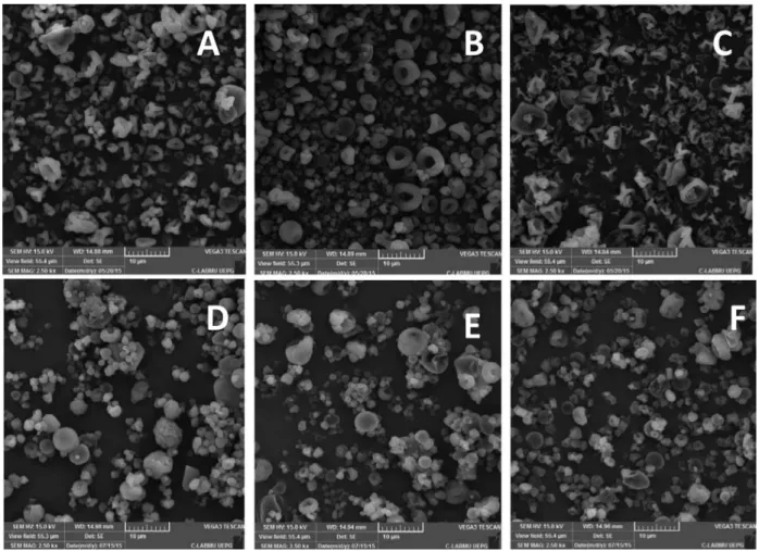
The SEM photomicrographs of microparticles, with magniication of 2500 x, show the morphology and the
surface of samples (Figure 2).
The photomicrographs of the HPMC and CS microparticles’ outer surface (Figure 2 A-C) showed that the size of particles was not uniform, a typical characteristic
of particles produced by spray drying. Photomicrographs
obtained by EC and CS samples (Figure 2 D-F) presented more uniform particles, as compared to HPMC and CS microparticles, but also showed imperfections on their
FIGURE 2 - Photomicrographs of microparticles: (A) HPMC:CS 1:1; (B) HPMC:CS 1:3; (C) HPMC:CS 3:1; (D) EC:CS 1:1;
surfaces, as well as diferent sizes. These characteristics can be due to the conditions of emulsiication process, such as rate of stirring, used solvents and type of emulsiier
(Rinaldi et al., 2009).
According to Stulzer et al., (2007), the morphology of microparticles is probably due to the obtainment
process, which often produces amorphous compounds,
due to the rapid drying process, preventing a uniform organization of particles. The main variables of the drying process are operational, such as inlet and outlet
air temperature, air low pattern, temperature distribution
and humidity and durability time, in addition to structural variables, e.g. the atomizer type. All these variables
can inluence in the shaping of microparticles and also inluence the encapsulation eiciency, as occurred in all
formulations (Table II).
There are many reasons that cause the low entrapment eiciency: (i) duration of spray drying process; (ii) low
viscosity of the sample, which facilitates the internal
circulation of droplets, causing coalescence and resulting in larger droplets. Furthermore, the increase of internal circulation hinders the formation of the membrane during the drying process, decreasing the drug retention into
microparticles, i.e., lower encapsulation eiciency; (iii) emulsion stability, which is related to the encapsulation eiciency, since a larger amount of unencapsulated drug can adhere to the particle’ surface (Barbosa, Borsarelli, Mercadante, 2005; Soottitantawat et al., 2003, 2005). Besides, other operation variables and characteristics of the device itself can contribute to the results found.
Microparticles composed by HPMC and CS showed entrapment eiciency varying from 26.6 to 42.7%, and the sample containing a higher amount of CS yielded a better drug encapsulation. CS is a hydrophilic polymer, soluble
only in acid solutions and HPMC is also hydrophilic,
swellable, soluble in water and pH-independent. The sample composed by HPMC and CS containing a higher amount of CS presented the highest value of encapsulation eiciency, indicating more ability of CS to encapsulate
the MT.
On the other hand, samples prepared with EC and CS containing a higher proportion of CS presented the lowest encapsulation eiciency (36.9%). EC is a hydrophobic polymer, and it`s high amount in the formulation promoted more drug retaining (66.9%), while samples composed by high amounts of CS, in this case, presented low
encapsulation of the MT.
FTIR and XRD studies
FTIR technique allows the identiication of organic
compounds through the functional groups present in the
molecules. Figure 3 shows the spectra of the formulations
and their respective constituents.
The absorption bands at 3223 cm-1 (O-H stretching), 3100 cm-1 (C-H and C=CH stretching), 1535 cm-1 (N-O stretching), 1370 cm-1 (NO
2 symmetric stretching) and 1076 cm-1 (C-O stretching, C-H in plane bending) were assigned as the ingerprints of MT (Wearley, Anthony,
1976; Ramukutty, Ramachandran, 2012), and the MT spectrum shows their characteristics bands: 3200-3600
cm-1 attributed to the O-H stretching, 3221-3101 cm-1
(N-H stretching), 2982-2937 cm-1 (C-H stretching), 1300-1600 cm-1 attributed to the NO
2; 1354-1180 cm
-1 (C-O stretching), 1428-1368 cm-1 (C-H bending, in plane), 1340 cm-1 (C-C stretching), 864 cm-1 (C-H out of plane bending), 825 cm-1 (C-H out of plane bending and C-N stretching), 744 cm-1 ((CH
2)2 rocking).
Spectrums were separated in the range of 1600-600 to better viewing of the peaks. Bands of the MT were
not presented in the spectrum of control microparticles
(without drug), signalized by dotted lines. Figure 3B shows the peak of MT in 1265.35 cm-1, present in the
microparticles, except for the control sample, as well as the peaks in 864.14, 826.53 and 744.55 cm-1, which did not appear in the control sample, confirming the drug
encapsulation into microparticles. Figure 3D shows the MT peaks at 1533.47, 1189.16, 825.56 and 744.55 cm-1, only present in the microparticles containing the drug.
There is a predominance of polymers characteristic bands (Table III) in all microparticles samples, and the
main bands of polymers and the drug were presented in microparticles (with less intensity), indicating no chemical linkage among them.
XRD is an important technique to study the crystal
structure typically found in compounds. Figure 4 shows
the XRD patterns for the different formulations of microparticles.
MT sample shows high crystalline structure, due to the presence of several sharp peaks at 12.02, 13.55,
17.89, 21.27, 24.54, 27.30, 29.07 and 33.57° 2θ. Polymers
showed two peaks: CS at 10.18 and 19.72° 2θ, HPMC at 9.58 and 19.36° 2θ and EC at 7.96 and 20.04° 2θ. These
polymers present crystalline and amorphous phase when pure and in solid state (Suksaeree et al., 2015).
Microparticles showed low peak intensity and baseline shift of the diffractogram was observed, due to the presence of polymers when compared to the drug
pattern, similar to Parida et al. (2016) results. The broad
FIGURE 3 - FTIR spectrums of formulations and pure constituents: (A) HPMC and CS samples: wavelength range: 4000 cm-1
to 500 cm-1; (B) HPMC and CS samples: wavelength range: 1600 cm-1 to 600 cm-1; (C) EC and CS samples: wavelength range:
4000 cm-1 to 500 cm-1; (D) EC and CS samples: wavelength range: 1600 cm-1 to 600 cm-1. MT: metronidazole; HPMC: hydroxypropyl
methylcellulose; EC: ethylcellulose; CS: chitosan.
TABLE III - Characteristics bands of used polymers composing the loating microparticles
Bands (cm-1) CS HPMC EC
3400-2400 - O-H stretching O-H stretching
3180-3350 NH2 stretch -
-2900- 2870 - methoxy group (C-CH3) C-H stretching
1735 - C=O stretching (ester group)
-1650-1665 C=O stretching (amine of the
acetamido)
aromatic ring
-1560-1610 axial deformation
(NH2 group)
-
-1444-1375 - - CH3; CH2
1300-1000 C-O stretching - C-O-C stretching (cyclic ether)
1276 1200-1000 744
- -
-ester group ether group
monosubstituted aromatic ring
some peaks of MT (with lower intensity) were observed in microparticles. HPMC and CS microparticles showed peaks at 9.10, 12.10, 13.74, 19.90, 25.30, 27.43, 33.19°
2θ, and EC and CS microparticles at 7.90, 12.16, 13.66, 19.43, 27.70, 33.24° 2θ, related to the drug and polymers,
showing that microparticles presented semi-amorphous structure, which indicates partial drug encapsulation.
In vitro characterization of the floating
microparticles
The microparticles floating ability is shown in Figure 5. All samples loated immediately when added in contact with the simulated gastric luid. Ten minutes after, 10% of microparticles sinked, except the EC:CS 3:1 formulation (green solid line) which only 50% of
microparticles remained dispersed. After 30 min of assay,
samples of HPMC:CS microparticles were hydrated and remained loating during 2 hours.
HPMC and CS microparticles (dotted lines) showed the same proile, regardless the proportions between CS
and HPMC, indicating that the quantity of polymers did
not inluence the lotation time. In contact with simulated gastric luid, the microparticles immediately loated due to their low density and maintained due to the medium absorption by polymers, which swelled, combined with the gas generator substance (sodium bicarbonate) allowing the lotation for 2 hours.
EC and CS microparticles (solid line) showed diferent loating proiles, however all samples immediately loated in contact with the medium. EC:CS 1:1 and EC:CS 3:1 microparticles remained loating during a reduced time
and after one hour and a half all microparticles stopped
loating. These formulations contain high proportions of EC (equal or more than CS), which is insoluble and not swells. Therefore, the swelling of microparticles was limited and was not suicient to maintain their lotation. EC:CS 1:3 sample, containing a high quantity of CS,
floated during 2 hours; due to the gel layer formation
FIGURE 4 - XRD of microparticles and their constituents: (A) HPMC and CS samples; (B) EC and CS samples. MT: metronidazole;
CS: chitosan; HPMC: hydroxypropyl methylcellulose; EC: rthylcellulose.
FIGURE 5 - Floating ability of microparticles. CS: chitosan;
(by CS), which swelled and sustained the gas generated by sodium bicarbonate allowing the flotation of the
microparticles.
The study of drug release from loating microparticles was presented in Figure 6. MT is a water soluble
drug, belonging to the class I of Biopharmaceutical
Classiication System, i.e. the MT release occurs fast and
its dissolution is not a limiting step for the absorption. The prepared microparticles exhibited prolonged drug release
in the simulated gastric luid.
In the first hour, 75% of the drug were released from HPMC and CS microparticles, and 95% of MT were released in 4 hours. Statistical analysis shows that the drug released from HPMC:CS 1:1 and HPMC:CS
1:3 (p = 0.0122; f1 = 11.82; f2 = 98.07) formulations
were different. HPMC:CS 1:3 microparticles contains more CS and it was responsible for the different drug release proile. It was found that CS decreases the rate of
drug release from microparticles in the beginning of the
dissolution test in simulated gastric luid. The obtained
results in the study conducted by Ritger, Peppas (1987),
showed that the CS hydration and gel formation takes
place more readily at acid pH levels (pH 1.2) than at pH
levels close to neutral, due to its cationic nature (Sahu, Verma, Singh, 2012). Higher amounts of CS resulted in better control of drug release in the irst hour of the test.
HPMC also contributed in prolonging the drug release due
to its swelling, associated with the CS swelling. However,
in formulations containing equal or a higher quantity
of HPMC (than CS) the drug release were similar (p =
0.5950; f1 = 5.96; f2 = 70.06).
EC:CS 1:3 sample presented drug release statistically
diferent from all other samples (p < 0.05; f1 > 15; f2 < 50),
showing 60 % of drug released in 4 hours. This sample also contains a higher quantity of CS. EC is an insoluble polymer that does not swell and reduce the CS swelling (CS absorbs the gastric luid fast, even though it is limited). Probably the association of CS and EC induced a stable and rigid gel layer, able to control the water uptake into the
microparticles and this gel layer controlled the MT release from microparticles. EC, being hydrophobic, has recently
been reported to be an excellent backing material, given its low water permeability and moderate lexibility, due to this insolubility preventing the penetration of water into
microparticles (Remunan-Lopez et al., 1998; Nunthanid et al., 2009).
Formulations containing EC and CS 3:1 were also statistically diferent of all other samples (f1 >15; f2 <50),
showing fast initial drug delivery followed by a plateau. This sample contains a lower quantity of CS and the MT was quickly released. It may be attributed to the CS low amount, because EC has a low water permeability while both CS and the drug are relatively soluble at low pH, therefore this formulation had lower gel formation in acidic pH, allowing the fast drug release by diffusion (Adebisi, Conway, 2014).
MT release from HPMC:CS 1:1 and from EC:CS 1:1 samples showed similar proile until 90 min of dissolution
test (t85%; f1=4.32; f2=64.93) ; however it was signiicantly
different (p=0.0438) after the first second hour, and a
greater quantity of drug was released from HPMC and CS microparticles than from EC and CS microparticles. Samples presented the same proportion between polymers, but in the formulation prepared with EC, the CS swelling was reduced and the medium intake occurred slowly,
prolonging the MT delivery.
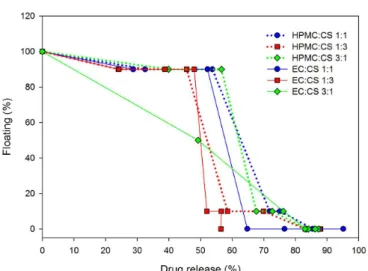
The development of loating gastroretentive systems
requires the in vitro assessment of drug release proiles
as well as the assessment of the loating behavior. Thus, a combined evaluation of formulations, which has no loating lag time and maintains its loating capabilities while releasing the drug was performed (Figure 7).
After completing the drug release, the delivery systems
should lose their loating capability, allowing the gastric
emptying of the microparticles and thus preventing possible accumulation of microparticles in the stomach after multiple administering.
According to Amit et al. (2011), the risk of
microparticles accumulation in the stomach is negligible
following a single administration; however, potential risk
of the aggregation and accumulation of drug-depleted microparticles in the stomach increases after multiple and
sequential administrations. Therefore, the microparticles`
ability to remain loating during the drug release and to sink after drug depletion is the ideal characteristic for loating microparticles.
Results showed that all microparticles remained floating while around 60% of the drug were releasing, except EC:CS 3:1 formulation, in which 50% of microparticles loated during the release of 40 % of the
MT. These results reveal that microparticles remain
loating while most of the drug is being released.
The developed microparticles exhibited diferent
flotation and drug release profiles, according to the
polymer composition. EC:CS 1:3 microparticles remained loating during 100 min, and after 4 hours, only 60 % of drug were released while no microparticles remained loating.
HPMC:CS 1:1 and HPMC:CS 3:1 formulations showed suitable relation between floating ability and MT release, i.e. 90% of microparticles remained loating during 60 % of MT release, and only after 80% of drug release the microparticles stopped to loat. The amount of HPMC did not inluence the relationship between loating and drug release, while the amount of CS caused inluence (red dotted line; Figure 7), because 10% of microparticles remained floating after around 60% of the drug were released from sample containing a greater quantity of CS
than HMPC.
Microparticles of HPMC:CS formulations can be classiied as Type I in the model loating behavior category,
according to Eberle et al. (2016), who refers to the loating
characteristics of hydrophilic eroding systems, in which the loating decrease due to predominant dissolution of the microparticles components. Despite 80 % of the drug were released in 2 hours, the maintenance of the lotation
is essential to promote the microparticles retention in the stomach and after the drug release the microparticles
should stop loating to avoid accumulation.
CONCLUSIONS
Floating microparticles were successfully obtained and characterized to potential gastroretentive efect, and
possible use for gastric disease treatment, such as H. pylori infections. Floating microparticles composed by chitosan and one hydrophilic (HPMC) or one hydrophobic
(EC) polymer were developed. Physicochemical characterization has shown the drug encapsulation and did not indicate chemical linkage occurrence between
components. The microparticles floating ability study
revealed no difference among the HPMC:CS samples, while microparticles containing a higher amount of
EC presented poor flotation. The drug release study
demonstrated better control of the MT release by EC:CS 1:3 sample, and similar profiles among HPMC:CS samples. However, the microparticles composed by HPMC and CS presented the best relationship between
floating ability and controlled drug release and it is
believed to be ideal for loating gastroretentive systems
regarding safe administration.
ACKNOWLEDGMENTS
Authors acknowledge to Complex Multi-User
Laboratories (C-LABMU) of UEPG, to Multi-User Laboratory of the Postgraduate Program in Pharmaceutical
Sciences UEPG/UNICENTRO and to Medicinal
Chemistry and Quality Laboratory of Department of
Pharmaceutical Sciences of UEPG.
REFERENCES
ABOU YOUSSEF, N.A.; KASSEM, A.A.; EL-MASSIK, M.A.; BORAIE, N.A. Development of gastroretentive metronidazole loating raft system for targeting Helicobacter pylori. Int. J. Pharm., v.486, n.1, p.297-305, 2015.
ADEBISI, A.O.; CONWAY, B.R. Lectin-conjugated microspheres for eradication of Helicobacterpylori infection and interaction with mucus. Int. J. Pharm., v.470, n.1, p.28-40, 2014.
AMIT, J.K.; RAMMULRAJSINH, R.; SONALI, D.; KINAL, P.; PRADEEP, A. Hydrodynamically Balanced Systems (HBS): innovative approach of gastroretention: a review.
Int. J. Pharm. Tech. Res., v.3, n.3, p.1495-1508, 2011.
FIGURE 7 - Relation between loating ability and drug release
ASGHAR, L.F.; CHANDRAN, S. Multiparticulate formulation approach to colon specific drug delivery: current perspectives. J. Pharm. Pharm. Sci., v.9, n.3, p.327-338, 2006.
BARBOSA, M.I.M.J.; BORSARELLI, C.D.; MERCADANTE, A.Z. Light stability of spray dried bixin encapsulated with diferent edible polysaccharide preparations. Food Res. Int., v.38, n.8/9, p.989-994, 2005.
BARDONNET, P.L.; FAIVRE, V.; PUGH, W.J.; PIFFARETTI, J.C.; FALSON, F. Gastroretentive dosage forms: overview and special case of Helicobacter pylori. J. Control. Rel.,
v.111, n.1/2, p.1-18, 2006.
BARROCAS, P.M.C.; SANTOS, D.F.G.; FERREIRA, D.C.; COELHO, P.M.B.S.; OLIVEIRA, R.C.S.; VEIGA, F.J.B. Sistemas farmacêuticos gastrorretensivos lutuantes. Rev. Bras. Cienc. Farm., v.43, n.3, p.325-334, 2007.
CHEN, R.-N.; HO, H.-O.; YU, C.-Y.; SHEU, M.-T. Development of swelling/floating gastroretentive drug delivery system based on a combination of hydroxyethyl cellulose and sodium carboxymethyl cellulose for Losartan and its clinical relevance in healthy volunteers with CYP2C9 polymorphism. Eur. J. Pharm. Sci., v.39, p.82-89, 2010.
COSTA, P.; LOBO, J.M.S. Inluence of dissolution medium agitation on release proiles of sustained-release tablets.
Drug Dev. Ind. Pharm., v.27, p.811-817, 2001.
EMARA, L.H.; ABDOU, A.R.; EL-ASHMAWY, A.A.R.; MURSI, N.M. Preparation and evaluation of metronidazole sustained release loating tablets. Int. J. Pharm. Pharm. Sci., v.6, n.9, p.198-204, 2014.
EBERLE, V.A.; HARING, A.; SCHOELKOPF, J.; GANE, P.A.; HUWYLER, J.; PUCHKOV, M. In silico and in vitro
methods to optimize the performance of experimental gastroretentive floating mini-tablets. Drug Dev. Ind. Pharm., v.42, p.808-817, 2016.
FERRARI, P.C.; GROSSKLAUSS, D.B.B.S.; ALVAREZ, M.; PAIXÃO, M.; ANDREIS, U.; CRISPIM, A.G.; CASTRO, A.; EVANGELISTA, R.C.; MIRANDA, J.C.A. A novel automated alternating current biosusceptometry method to characterization of controlled-release magnetic loating tablets of metronidazole. Drug Dev. Ind. Pharm., v.40, n.8, p.1123-1131, 2014.
GARCIA, A.; SALAS-JARA, M.J.; HERRERA, C.; GONZÁLEZ, C. Biofilm and Helicobacter pylori: from environment to human host. World J. Gastroenterol., v.20, n.19, p.5632-5638, 2014.
GUIMARÃES, J.; CORVELO, T.C.; BARILE, K.A. Helicobacter pylori: fatores relacionados à sua patogênese. Rev. Paraense Med., v.22, n.1, p.33-38, 2008.
HAO, S.; WANG, Y.; WANG, B.; ZOU, Q.; ZENG, H.; CHEN, X.; LIU, X.; LIU, J.; YU, S. A novel gastroretentive porous microparticle for anti-Helicobacter pylori therapy: preparation, in vitro and in vivo evaluation. Int. J. Pharm, v.463, p.10–21, 2014.
KRISHNAIAH, Y.S.R.; VEER RAJU, P.; DINESH KUMARI, B.; JAYARAM, B.; RAMA, B.; RAJU V.; BHASKAR P. Pharmacokinetic evaluation of guar gum-based colon-targeted oral drug delivery systems of metronidazole in healthy volunteers. Eur. J. Drug Metab. Pharmacokinet., v.28, n.4, p.287-294, 2003.
LEMIEUX, M.; GOSSELIN, P.; MATEESCU, M.A. Carboxymethyl starch mucoadhesive microspheres as gastroretentive dosage form. Int. J. Pharm., v.496, n.2, p.497-508, 2015.
MALFERTHEINER, P.; MEGRAUD, F.; O’MORAIN, C.A.; ATHERTON, J.; AXON, A.T.R.; BAZZOLI, F.; GENSINI, G.F.; GISBERT, J.P.; GRAHAM, D.Y.; ROKKAS, T.; EL-OMAR, E.M.; KUIPERS, E.J.; EUROPEAN HELICOBACTER STUDY GROUP (EHSG). Management of Helicobacter pylori infection–the Maastricht IV/Florence consensus report. Gut, v.61, p.646-664, 2012.
MARTINAC, A.; FILIPOVIC-GRCIC, J.; VOINOVICH, D.; PERISSUTTI, B.; FRANCESCHINIS, E. Development and bioadhesive properties of chitosan-ethylcellulose microspheres for nasal delivery. Int. J. Pharm., v.291, n.1, p.69-77, 2005.
OH, C.M.; HENG, P.W.S.; CHAN, L.W. Influence of hydroxypropyl methylcellulose on metronidazole crystallinity in spray-congealed polyethylene glycol microparticles and its impact with various additives on Metronidazole release. AAPS PharmSciTech, v.16, n.6, p.1357-1367, 2015.
OLIVEIRA, A.M.; JAGER, E.; JAGER, A.; STEPÁNEK, P.; GIACOMELLI, F.C. Physicochemical aspects behind the size of biodegradable polymeric nanoparticles: a step forward. Colloids Surf., A, v.436, p.1092-1102, 2013.
PARIDA, P.; MISHRA, S.C; SAHOO, S.; BEHERA, A.; NAYAK, B.P. Development and characterization of ethylcellulose based microsphere for sustained release of nifedipine. J. Pharm. Anal., v.6, p.341-344, 2016.
PAYÃO, S.L.M.; RASMUSSEN, L.T. Helicobacter pylori and its reservoirs: a correlation with the gastric infection. World J. Gastrointest. Pharmacol. Ther., v.7, n.1, p.126-132, 2016.
PRAJAPATI, S.T.; PATEL, L.D.; PATEL, D.M. Gastric loating matrix tablets: design and optimization using combination of polymers. Acta Pharmaceut., v.58, p.221-229, 2008.
PRASANTHI, C.H.; PRASANTHI, N.L.; MANIKIRAN, S.S.; RAO, N.R. Focus on current trends in the treatment of Helicobacter pylori infection: an update. Int. J. Pharm. Sci. Rev. Res., v.9, n.1, p.42-51, 2011.
PRIYADARSHINI, R.; NANDI, G.; CHANGDER, A.; CHOWDHURY, S.; CHAKRABORTY, S.; GHOSH, L.K. Gastroretentive extended release of metformin from methacrylamide-g-gellan and tamarind seed gum composite matrix. Carbohydr. Polym., v.137, p.100-110, 2016.
RAJAB, M.; JOUMA, M.; NEUBERT, R.H.H.; DITTGEN, M. Influence of water-soluble polymers on the in vitro
performance of loating mucoadhesive tablets containing metformin. Drug Dev. Ind. Pharm., v.40, n.7, p.879-885, 2014.
RAMUKUTTY, S.; RAMACHANDRAN, E. Crystal growth by solvent evaporation and characterization of metronidazole.
J. Crystal Growth, v.351, n.1, p.47-50, 2012.
RAPOLU, K.; SANKA, K.; VEMULA, P.K.; AATIPAMULA, V.; MOHD, A.B.; DIWAN, P.V. Optimization and characterization of gastroretentive loating drug delivery system using Box-Behnken design. Drug Dev. Ind. Pharm., v.38, p.1-8, 2012.
REMUNAN-LOPEZ, C.; PORTERO, A.; VILA-JATO, J.L.; ALONSO, M.J. Design and evaluation of chitosan/ ethylcellulose mucoadhesive bilayered devices for buccal drug delivery. J. Control. Rel., v.55, n.2, p.143-152, 1998.
RINALDI, A.P.T.; MAZERA, S.K.; PEZZINI, B.R.; ZÉTOLA, M.; BAZZO, G.C. Preparo e caracterização de micropartículas de acetobutirato de celulose e poli(3-hidroxibutirato) contendo piroxicam. Acta Scient. Health Sci., v.31, n.1, p.51-56, 2009.
RITGER, P.L.; PEPPAS, N.A. A simple equation for description of solute release. II: Fickian and anomalous release from swellable devices. J. Control. Rel., v.5, p.37-42, 1987.
SAHU, A.K.; VERMA, A.; SINGH, S.K. Preparation of hydrophilic swelling controlled-release floating matrix tablets containing HPMC and chitosan. Int. J. Pharm. Pharm. Sci. v.4, n.1, p.82-87, 2012.
SCHAFFAZICK, S.R.; GUTERRES, S.S. Caracterização e estabilidade físico-química de sistemas poliméricos nanoparticulados para administração de fármacos. Quim. Nova, v.26, n.5, p.726-737, 2003.
SINGH, V.V.; BEHERA, B.; PRAMANIK, K.; PAL, K. Ultrasonication-assisted preparation and characterization of emulsions and emulsion gels for topical drug delivery.
J. Pharm. Sci., v.104, n.3, p.1023-1044, 2015.
SOOTTITANTAWAT, A.; BIGEARD, F.; YOSHI, H.; FURUTA, T.; OHKAWARA, M.; LINKO, P. Inluence of emulsion and powder size on the stability of encapsulated D-limonene by spray drying. Innov. Food Sci. Emerg. Technol., v.6, n.1, p.107-114, 2005.
SOOTTITANTAWAT, A.; YOSHII, H.; FURUTA, T.; OHKAWARA, M.; LINKO, P. Microencapsulation by spray drying: inluence of emulsion size on the retention of volatile compounds. J. Food Sci., v.68, n.7, p.2256-2262, 2003.
SUKSAEREE, J.; MONTON, C.; MADAKA, F.; CHUSUT, T.; SAINGAM, W.; PICHAYAKORN, W.; BOONME, P. Formulation, physicochemical characterization, and in vitro study of chitosan/ HPMC blends-based herbal blended patches. AAPS PharmSciTech, v.16, p.171-181, 2015.
UNITED STATES Pharmacopoeia: USP 30. Rockville: United States Pharmacopeial Convention, 2007.
WEARLEY, L.L.; ANTHONY, G.D. Metronidazole. Anal.
Profiles Drug Subst., v.5, p.326-344, 1976.
Received for publication on 01st November 2015