70
Braz J Cardiovasc Surg | Rev Bras Cir Cardiovasc
Braz J Cardiovasc Surg 2015;30(1):70-6
Antonio EL, et al. - Are there gender differences in left ventricular remodeling after myocardial infarction in rats?
RBCCV 44205-1616 DOI: 10.5935/1678-741.20140093
Are there gender differences in left ventricular
remodeling after myocardial infarction in rats?
Há diferenças entre os gêneros no remodelamento ventricular esquerdo após infarto do miocárdio em ratos?
Ednei Luiz Antonio
1, MD; Andrey Jorge Serra
2, MsC, PhD; Alexandra Alberta dos Santos
3, MD;
Stella Sousa Vieira
3, MD; Jairo Montemor Augusto Silva
3, MD; Amanda Yoshizaki
4, MD;
Renato Rodrigues Soia
5, MD; Paulo José Ferreira Tucci
3, MD, PhD
1Laboratório de Fisiologia e Fisiopatologia Cardíacas da Disciplina de Cardio-logia da Universidade Federal de São Paulo (Unifesp), São Paulo, SP, Brazil. 2Docente da Universidade Nove de Julho (Uninove), São Paulo, SP, Brazil. 3Laboratório de Fisiologia e Fisiopatologia Cardíacas da Disciplina de Car-diologia da Universidade Federal de São Paulo (Unifesp), São Paulo, SP, Brazil.
4Programa de Pós-graduação em Ciências da Reabilitação da Universidade Nove de Julho (Uninove), São Paulo, SP, Brazil.
5Universidade Nove de Julho (Uninove), São Paulo, SP, Brazil.
This study was carried out at Universidade Federal de São Paulo (Unifesp), São Paulo, SP, Brazil and Universidade Nove de Julho (Uninove), São Pau-lo, SP, Brazil.
Correspondence address: Andrey Jorge Serra
Postgraduate Program in Biophotonics Applied Health Sciences, Universi-dade Nove de Julho (UNINOVE)
Rua Vergueiro, 235 - São Paulo, SP, Brazil Zip code: 01504-001
E-mail: [email protected]
Financial support: Grant number 2009/54225-8, São Paulo Research Foundation (FAPESP).
Article received on September 26th, 2013 Article accepted on June 2nd, 2014 ORIGINAL ARTICLE
Abstract
Objective: An unclear issue is whether gender may inluence at
cardiac remodeling after myocardial infarction (MI). We evaluated left ventricle remodeling in female and male rats post-MI.
Methods: Rats were submitted to anterior descending
coro-nary occlusion. Echocardiographic evaluations were performed on the irst and sixth week post-occlusion to determine myo -cardial infarction size and left ventricle systolic function (FAC, fractional area change). Pulsed Doppler was applied to analyze left ventricle diastolic function using the following parameters: E wave, A wave, E/A ratio. Two-way ANOVA was applied for comparisons, complemented by the Bonferroni test. A P≤0.05
was considered signiicant.
Results: There were no signiicant differences between gen
-ders for morphometric parameters on irst (MI [Female (FE): 44.0±5.0 vs. Male (MA): 42.0±3.0%]; diastolic [FE: 0.04±0.003 vs. MA: 0.037±0.005, mm/g] and systolic [FE: 0.03±0.0004 vs. MA: 0.028±0.005, mm/g] diameters of left ventricle) and sixth (MI [FE: 44.0±5.0 vs. MA: 42.0±3.0, %]; diastolic [FE: 0.043±0.01 vs. MA: 0.034±0.005, mm/g] and systolic [FE: 0.035±0.01 vs. MA: 0.027±0.005, mm/g] of LV) week. Similar indings were reported for left ventricle functional parameters on irst (FAC [FE: 34.0±6.0 vs. MA: 32.0±4.0, %]; wave E [FE: 70.0±18.0 vs. MA: 73.0±14.0, cm/s]; wave A [FE: 20.0±12.0 vs. MA: 28.0±13.0, cm/s]; E/A [FE: 4.9±3.4 vs. MA: 3.3±1.8]) and sixth (FAC [FE: 29.0±7.0 vs. MA: 31.0±7.0, %]; wave E [FE:
85.0±18.0 vs. MA: 87.0±20.0, cm/s]; wave A [FE: 20.0±11.0 vs. MA: 28.0±17.0, cm/s]; E/A [FE: 6.2±4.0 vs. MA: 4.6±3.4]) week.
Conclusion: Gender does not inluence left ventricle remod
-eling post-MI in rats.
Descriptors: Gender and Health. Myocardial Infarction.
Ventricular Remodeling.
Resumo
Objetivo: A inluência do gênero no remodelamento cardíaco
após o infarto do miocárdio é uma questão em intenso debate. Nós avaliamos o remodelamento ventricular esquerdo em ratos infartados de ambos os gêneros.
Métodos: O infarto do miocárdio foi induzido por oclusão da
artéria coronária descendente anterior (fêmeas [FM]; machos [MC]). A ecocardiograia foi realizada na primeira e sexta sema -na pós-oclusão para determi-nar o tamanho do infarto do mio-cárdio e a função sistólica do ventricular esquerdo (mudança na área fracional [FAC]). A função diastólica derivou dos seguintes parâmetros: onda E; onda A; razão E/A. ANOVA duas vias com pós-teste de Bonferroni foi aplicado nas comparações (P≤0,05).
Resultados: Todas variáveis morfométricas foram
simi-lares (P>0,05) entre os gêneros com uma (infarto do miocár -dio [FM: 44,0±5,0 vs. MC: 42,0±3,0, %]; diâmetro diastólico
[FM: 0,04±0,003 vs. MC: 0,037±0,005, mm/g] e sistólico [FM:
71
INTRODUCTION
Myocardial infarction (MI) is an important cause of heart failure and mortality among adults. A number of factors can determine a worsening in prognosis such as infarct expansion, hypertrophy of the non-infarcted myocardium, increased col-lagen deposition in the infarcted and non-infarcted areas, pro-gressive dilatation, geometric changes in chamber shape, and eventual progression to chronic heart failure.
It is well known that premenopausal women are less like-ly to develop coronary heart disease than men. Previous stud-ies have also shown that gender can be a key factor in cardi-ac remodeling post-MI. Thus, studies show gender as a risk factor of unfavorable prognostic[1,2]. Although valuable, these
indings cannot be considered as absolute truth. Several is -sues such as age heterogeneity, drug therapy, associated risk factors, and hemodynamics (e.g., pre- and afterload; blood
volume) may cause dificulties in ensuring that the there is
differential cardiac remodeling between genders post-MI[2,3].
Moreover, some researchers have found higher survival rates in women[4,5] while others show higher mortality in women
due to higher severity of MI[6].
Since possible gender differences in post-MI left ventri-cle (LV) remodeling are not ventri-clear, we conducted the current study using a rat MI model. The MI model by coronary
oc-clusion represented a signiicant advance to provide accurate
control of bias[7-13]. Moreover, coronary occlusion is the most
commonly used experimental model to induce MI in rats and
somewhat reproduces the indings in humans with cardiac
decompensation[14]. We performed paired time evaluations in
the LV with transthoracic echocardiography. This approach has been shown to be readily reproducible in longitudinal as-sessment of morphology and function of LV in rodents[15,16].
METHODS
Animal MI model
The study was approved by the Committee on Ethics from the Federal University of São Paulo and use the “Principles of Laboratory Animal Care formulated by the National
In-[FM: 44,0±5,0 vs. MC: 42,0±3,0, %]; diâmetro diastólico [FM:
0,043±0,01 vs. MC: 0,034±0,005, mm/g] e sistólico [FM: 0,035±0,01 vs. MC: 0,027±0,005, mm/g] do ventricular esquerdo) semanas.
Achado similar ocorreu para os dados funcionais com uma (FAC Abbreviations, acronyms & symbols
FE Female
LV Left ventricle
MA Male
MI Myocardial infarction
stitutes of Health (National Institutes of Health publication number 96-23, revised, 1996; http://bioethics.od.nih.gov/ani-mals.html)”. The MI was induced in three-month-old female Wistar-EPM rats weighing between 180 to 220g. The animals were housed at regular temperature (22°-24°C) on a 12h dark/ light cycle with food and water provided ad libitum. Rats were anesthetized with ketamine (50 mg/kg) plus xylazine (10 mg/ kg) intraperitoneally, intubated, and ventilated with room air (rate: 90 breaths/minute; tidal volume: 2.5 ml on a Harvard rodent respirator [model 683, Harvard Apparatus Co., South Natik, MA, USA]). After thoracotomy, the MI was produced by ligation of the left descending coronary artery as previous-ly described[17,18]. Sham surgery was performed with a similar
process except the suture was tied loosely around the coronary artery. Afterward MI or Sham surgery, the rats returned to their plastic boxes and were kept under observation without any drug therapy. Survivor animals were assigned to the follow-ing groups: (1) Female sham (n=8); (2) Female MI (n=11); (3) Male sham (n=9); (4) Male MI (n=12).
Echocardiographic measurements
Echocardiography has been shown accurate in evaluating cardiac remodeling post-MI[16,19-20]. Echocardiographic
analy-sis was applied on the irst and sixth week post-MI. The rats
were anesthetized as described above and measurements were performed using a 12-MHz transducer connected to an HP Sonos-5500 echocardiograph (Hewlett-Packard, California, USA)[11]. MI size was evaluated by transversal LV
two-dimen-sional view on the basal, mid-transversal, and apical planes. In the diastolic phase, measurements of the endocardial perimeter (EP) and length of the infarcted segment (ISe) for each view were taken. MI size for each segment (ISi), expressed as the proportion of the LV perimeter for each view, was calculated by the following equation: ISi (%) = ISe/EP x 100. MI was
de-ined as a segment with increased echogenicity and/or change
in myocardial thickening or systolic movement.
Only rats with large infarctions (≥ 40% of LV) were includ -ed for evaluation since this is the group that shows the highest severity of the disease[8]. The diastolic (DA) and systolic (SA)
transverse areas of the LV were measured by two-dimensional
[FM: 34,0±6,0 vs. MC: 32,0±4,0, %]; onda E [FM: 70,0±18,0 vs. MC: 73,0±14,0, cm/s]; onda A [FM: 20,0±12,0 vs. MC: 28,0±13,0,
cm/s]; E/A [FM: 4,9±3,4 vs. MC: 3,3±1,8]) e seis (FAC [FM:
29,0±7,0 vs. MC: 31,0±7,0, %]; onda E [FM: 85,0±18,0 vs. MC: 87,0±20,0, cm/s]; onda A [FM: 20,0±11,0 vs. MC: 28,0±17,0 cm/s];
E/A [FM: 6,2±4,0 vs. MC: 4,6±3,4]) semanas.
Conclusão: O gênero não é determinante para o remodela
-mento ventricular esquerdo pós-infarto do miocárdio em ratos.
Descritores: Gênero e Saúde. Infarto do Miocárdio. Remo
72
Braz J Cardiovasc Surg | Rev Bras Cir Cardiovasc
Braz J Cardiovasc Surg 2015;30(1):70-6
Antonio EL, et al. - Are there gender differences in left ventricular remodeling after myocardial infarction in rats?
images on the basal, middle and apical parasternal transverse
planes. The inal value was the arithmetic mean of the mea -surements of the three views. Systolic function was analyzed by the fractional area change (FAC=DA - SA/DA, %) in the three transverse planes (basal, middle, and apical). Pulsed
Doppler at the LV side of the mitral valve provided the low
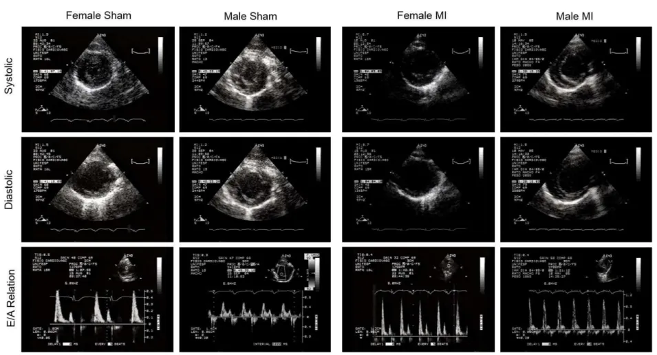
velocity curve to analyze the diastolic function parameters (E and A waves and E/A ratio). Echocardiographic images for cardiac effects of MI are shown in Figure 1.
Statistical analysis
Data were analyzed with GraphPad Prism software 4.0 (San Diego, CA, USA) and values are expressed as mean ± S.D. The Shapiro-Wilk and Levene tests were applied to ver-ify normal statistic distributions and error variances, respec-tively. To determine the effect of time and infarction on the echocardiographic parameters of respective genders, two-way analysis of variance (ANOVA) with repeated measures was performed. To evaluate the difference between genders at each respective time point, regular two-way ANOVA was performed. The Bonferroni post-hoc was carried out for all
analyses and level of signiicance was set at 5%.
RESULTS
To characterize the MI repercussions in both genders, echocardiography analyses were taken into account over six weeks after coronary occlusion. LV morphology and
func-tion were evaluated in the irst and sixth week after ischemic
insult. The sham group was evaluated at the same time. LV morphology data are shown in Table 1. None of the eval-uated parameters changed for female and male sham-operated rats during follow-up. MI size was similar between female and male rats, and we did not see expansion of MI in either gender
during follow-up. Left atrium size was signiicantly higher in
the infarcted rats regardless of gender; moreover, there was not difference in the left atrium size between female and male rats on any assessment time. There was LV dilatation with only a week post-MI; therefore, female and male rats showed a
sig-niicant increase in diastolic and systolic LV diameter when comparing the irst and sixth week. For all follow-ups, LV dil -atation level was similar between genders. When LV diameters were indexed by body weight, LV dilation post-MI was similar
comparing irst to sixth week for both genders.
The LV performance data are shown in Table 2.
Table 2. Echocardiographic functional parameters for female and male rats post-MI.
FAC (%) E (cm/s) A (cm/s) E/A ratio Week 1 Sham 63±4 69±2 24±1 3±1 Week 1 MI 34±6* 70±2 20±1 5±3 Week 6 Sham 67±3 73±6 29±5 3±1 Week 6 MI 29±7* 85±2* 20±1 6±4* Female Week 1 Sham 66±7 59±2 30±5 1±0.5 Week 1 MI 32±4* 73±1 28±1 3±2 Week 6 Sham 63±4 67±4 30±6 2±0.2 Week 6 MI 31±7* 87±2* 28±2 5±3* Male
FAC=fractional area change; E=E wave; A=A wave; Data are shown as means±SD. Two-way ANOVA and Bonferroni tests were applied for comparisons.
*P<0.05 vs. Sham on irst and sixth week
Table 1. Echocardiographic morphology parameters for female and male rats post-MI.
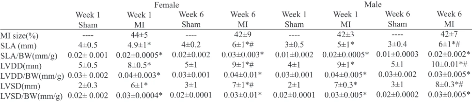
MI size(%) SLA (mm) SLA/BW(mm/g) LVDD(mm) LVDD/BW(mm/g) LVSD(mm) LVSD/BW(mm/g) Week 1 Sham ----4±0.5 0.02± 0.001 5±0.5 0.03± 0.002 2±0.3 0.02± 0.002 Week 1 MI 44±5 4.9±1* 0.02±0.0005* 8±0.5* 0.04±0.003* 6±1* 0.03±0.0004* Week 6 Sham ----4±0.2 0.02±0.002 5±1 0.03±0.001 3±1 0.02±0.0001 Week 6 MI 42±9 6±1*# 0.03±0.003* 9±1*# 0.04±0.01* 7±1*# 0.03±0.01* Female Week 1 Sham ----3±0.5 0.01±0.002 4±1 0.03±0.001 2±1 0.02±0.0001 Week 1 MI 42±3 5±1* 0.02±0.0005* 9±1* 0.04±0.005* 7±0.3* 0.03±0.005* Week 6 Sham ----3±0.4 0.01±0.0003 5±1 0.03±0.002 3±1 0.02±0.0002 Week 6 MI 42±7 6±1*# 0.02±0.002* 10±0.01*# 0.03±0.005* 8±0.3*# 0.03±0.005* Male
MI=myocardial infarction; BW=body weight; SLA=left atrium size; LVDD=left ventricle diastolic diameter; LVSD=left ventricle systolic diameter. Data are shown as means±SD. Two-way ANOVA and Bonferroni tests were applied for comparisons.
73
Bra
z J
Ca
rdi
ova
sc
S
ur
g |
Re
v Bra
s Ci
r Ca
rdi
ova
sc
B
raz
J
Car
di
ov
as
c Sur
g
2015;30(1):70-6
t a
l.
-
A
re
the
re
ge
nde
r di
ffe
re
nc
es
i
n
le
ft
ve
nt
ri
cul
ar
re
m
ode
ling
al
i
nfa
rc
tion i
n ra
ts
?
Fig. 1 - Illustrative example of two-dimensional mode traces of the left ventricle (LV) of the female and male rats. Images for mitral inlow velocity proile determined by pulsed
74
Braz J Cardiovasc Surg | Rev Bras Cir Cardiovasc
Braz J Cardiovasc Surg 2015;30(1):70-6
Antonio EL, et al. - Are there gender differences in left ventricular remodeling after myocardial infarction in rats?
The MI resulted in a signiicant reduction of LV systolic
function within a week of coronary occlusion. The systolic
dysfunction level was not signiicantly different compared to the sixth week post-MI. Our indings indicate that gender did not inluence the deleterious MI effects on LV systolic func
-tion. Both genders showed signiicant increases in E wave
in the sixth week post-MI, whereas the A wave remained unchanged. Thus, female and male rats had a restrictive LV
illing pattern deined as an increased ratio of early (E) to late (A) illing velocities and rapid deceleration of the early ill
-ing wave with six weeks post-MI. There were no signiicant
differences between genders for these parameters (Table 2).
DISCUSSION
We performed this study to evaluate if there are gen-der-related differences in the LV remodeling post-MI. Echo-cardiography serial analyses were performed including sham non-operated rats for paired comparisons. We have included in the study only animals with large infarcts, and this was based on the issue that large infarcts are representative of notable cardiac remodeling[8,21].
The current study showed that several indicators for poor prognosis were seen with only one week of coronary occlu-sion[22-25]. Except for restrictive LV illing pattern (increased in
the sixth week), the left atrium size, LV end-systolic and end-di-astolic dimensions as well as depressed LV systolic function
were increased. These indings are consistent with previous
studies in rats on similar MI sizes and follow-up analysis[3].
As the main interest was the comparison between gen-ders, we directly compared male and female rats with similar infarction sizes. The negative effects of MI on LV morphol-ogy and function were similar for both genders. Therefore, gender was not decisive for LV remodeling post-MI. In respect to LV dilatation and systolic dysfunction, we have shown similar results to other studies[3]. On the other hand,
our results do not corroborate results reported by Litwin et al.[3] in regards to restrictive LV illing pattern. Although Lit
-win et al.[3] showed a higher increase of E wave and E/A ratio
in male rats, we have shown that there was a similar increase in these variables for both genders.
In terms of gender as a determinant of LV remodeling af-ter MI, the reasons for the different pataf-terns in male and fe-male are not clear. Better remodeling of noninfarcted regions in female than in male animals can result in lower operating chamber stiffness; thus female rats may have attenuated the
development of a restrictive LV illing pattern[3]. Lines of
evidence have attributed a key role for sex hormones. This is based, for example, in observations that testosterone is a potent inducer of LV hypertrophy while estradiol has an inhibitory action[26,27]. There is evidence that estrogen reduces collagen
content[28,29] as well as wall stress in late MI[30]. Moreover,
stud-ies showed that estrogen may favor remodeling by preventing
apoptosis[31] and increasing angiogenesis in female[32]. It is also
possible that sexual hormones may indirectly regulate myocar-dial adaptation by vascular or endocrine effects. For example, cardiac preload or afterload may be different between males and females with MI as a result of differences in blood volume regulation, venous or arterial tone[13].
The female rats used in this study were young adults with normal ovaries. This study was not designed to assess the role of sex hormones in post-MI LV remodeling, and we did not monitor the serum hormone changes. Although we can not exclude that there were minor effects of sex hormones on LV functional and echocardiographic parameters, our data do not support the view that the positive effects of the sexual
hormones may spread for a beneicial LV remodeling in fe -male rats with MI.
CONCLUSION
The current study had a preset end point of 6 weeks in which male and female rats showed similar morphological and functional abnormalities. Therefore, we cannot draw conclusions about gender differences in the LV remodeling post-MI. It should be noted that the rats were young adults with no atherosclerotic disorder. It is unlikely that humans would have MI at such a young age. However, this experi-mental model has been widely accepted in studying post-MI LV remodeling.
ACKNOWLEDGMENTS
This study was funded by grant number 2009/54225-8, São Paulo Research Foundation (FAPESP).
Authors’ roles & responsibilities
ELA Data collection and analysis, experimental design and man-uscript writing
AJS Experimental design, statistical analysis and manuscript writing
AAS Collection and analysis of data SSV Collection and analysis of data JMAS Collection and analysis of data AY Collection and analysis of data RRS Study design and manuscript writing
PJFT Experimental design, getting funding for research, critical review of the manuscript
REFERENCES
75
in two different periods (1977-1978 versus 1988-1989). Am J Cardiol. 1993;71(7):518-23.2. Marschner IC, Colquhoun D, Simes RJ, Glasziou P, Harris P, Singh BB, et al.; Long-Term Intervention with Pravastatin in
Ischemic Disease (LIPID) Study. Long-term risk stratiication for
survivors of acute coronary syndromes. Results from the Long-term Intervention with Pravastatin in Ischemic Disease (LIPID) Study. LIPID Study Investigators. J Am Coll Cardiol. 2001;38(1):56-63.
3. Litwin SE, Katz SE, Litwin CM, Morgan JP, Douglas PS. Gender differences postinfarction left ventricular remodeling. Cardiology. 1999;91(3):173-83.
4. Brett KM, Madans JH. Long-term survival after coronary heart disease. Comparisons between men and women in a national sample. Ann Epidemiol. 1995;5(1):25-32.
5. Heer T, Schiele R, Schneider S, Gitt AK, Wienbergen H, Gottwik M, et al. Gender differences in acute myocardial infarction in the era of reperfusion (the MITRA registry). Am J Cardiol. 2002;89(5):511-7.
6. Marrugat J, Sala J, Masiá R, Pavesi M, Sanz G, Valle V, et al. Mortality differences between men and women following
irst myocardial infarction. RESCATE Investigators. Recursos
Empleados en el Síndrome Coronario Agudo y Tiempo de Espera. JAMA. 1998;280(6):1405-9.
7. Fishbein MC, Maclean D, Maroko PR. Experimental myocardial infarction in the rat: qualitative and quantitative changes during pathologic evolution. Am J Pathol. 1978;90(1):57-70.
8. Pfeffer MA, Pfeffer JM, Fishbein MC, Fletcher PJ, Spadaro J, Kloner RA, et al. Myocardial infarct size and ventricular function in rats. Circ Res. 1979;44(4):503-12.
9. Fletcher PJ, Pfeffer JM, Pfeffer MA, Braunwald E. Left ventricular diastolic pressure-volume. Relations in rats with healed myocardial infarction. Effects on systolic function. Circ Res. 1981;49(3):618-26.
10. Baily RG, Lehman JC, Gubin SS, Musch TI. Non-invasive assessment of ventricular damage in rats with myocardial infarction. Cardiovasc Res. 1993;27(5):851-5.
11. Moisés VA, Ferreira R, Nozawa E, Kanashiro RM, Campos O, Andrade JL, et al. Structural and functional characteristics of rat hearts with and without myocardial infarct. Initial experience with Doppler echocardiography. Arq Bras Cardiol. 2000;75(2):125-36.
12. Kanashiro RM, Nozawa E, Murad N, Gerola LR, Moisés VA, Tucci PJ. Myocardial infarction scar plication in the rat. Cardiac mechanics in an animal model for surgical procedures. Ann Thorac Surg. 2002;73(5):1507-13.
13. Cavasin MA, Tao Z, Menon S, Yang XP. Gender differences in cardiac function during early remodeling after acute myocardial infarction in mice. Life Sci. 2004;75(18):2181-92.
14. Tucci PJ. Pathophysiological characteristics of the post-myocardial infarction heart failure model in rats. Arq Bras Cardiol. 2011;96(5):420-4.
15. Litwin SE, Katz SE, Morgan JP, Douglas PS. Serial echocardiographic assessment of left ventricular geometry and function after large myocardial infarction in the rat. Circulation. 1994;89(1):345-54.
16. Santos AA, Helber I, Flumignan RL, Antonio EL, Carvalho AC, Paola AA, et al. Doppler echocardiographic predictors of mortality in female rats after myocardial. J Card Fail. 2009;15(2):163-8.
17. Cosmo S, Francisco JC, Cunha RC, Macedo RM, Faria-Neto JR, Simeoni R, et al. Effect of exercise associated with stem cell transplantation on ventricular function in rats after acute myocardial infarction. Rev Bras Cir Cardiovasc. 2012;27(4):542-51.
18. dos Santos AA, Helber I, Antonio EL, Franco MF, Tucci PJ. Severity of the cardiac impairment determines whether digitalis prolongs or reduces survival of rats with heart failure due to myocardial infarction. Int J Cardiol. 2013;167(2):357-61.
19. Nozawa E, Kanashiro RM, Murad N, Carvalho AC, Cravo SL, Campos O, et al. Performance of two-dimensional Doppler echocardiography for the assessment of infarct size and left ventricular function in rats. Braz J Med Biol Res. 2006;39(5):687-95.
20. dos Santos L, Mello AF, Antonio EL, Tucci PJF. Determination of myocardial infarction size in rats by echocardiography and
tetrazolium staining: correlation, agreements, and simpliications.
Braz J Med Biol Res. 2008;41(3):199-201.
21. Jain M, Liao R, Podesser BK, Ngoy S, Apstein CS, Eberli FR.
Inluence of gender on the response to hemodynamic overload
after myocardial infarction. Am J Physiol Heart Circ Physiol. 2002;283(6):H2544-50.
22. White HD, Norris RM, Brown MA, Brandt PW, Whitlock RM, Wild CJ. Left ventricular end-systolic volume as the major determinant of survival after recovery from myocardial infarction. Circulation, 1987;76(1):44-51.
23. St John Sutton M, Pfeffer MA, Plappert T, Rouleau JL, Moyé LA, Dagenais GR, et al. Quantitative two-dimensional echocardiographic measurements are major predictors of adverse cardiovascular events after acute myocardial infarction. The protective effects of captopril. Circulation. 1994;89(1):68-75.
24. Xie GY, Berk MR, Smith MD, Gurley JC, DeMaria AN. Prognostic value of Doppler transmitral flow patterns in patients with congestive heart failure. J Am Coll Cardiol. 1994;24(1):132-9.
76
Braz J Cardiovasc Surg | Rev Bras Cir Cardiovasc
Braz J Cardiovasc Surg 2015;30(1):70-6
Antonio EL, et al. - Are there gender differences in left ventricular remodeling after myocardial infarction in rats?
complicated by heart failure, left ventricular dysfunction, or both: the VALIANT Echo study. Eur Heart J. 2009; 30(1):56-65.
26. Cabral AM, Vasquez EC, Moysés MR, Antonio A. Sex hormone modulation of ventricular hypertrophy in sinoaortic denervated rats. Hypertension. 1988;11(2 Pt 2):I93-7.
27. Pelzer T, Loza PA, Hu K, Bayer B, Dienesch C, Calvillo L, et al. Increased mortality and aggravation of heart failure in estrogen receptor-beta knockout mice after myocardial infarction. Circulation. 2005;111(12):1492-8.
28. Fischer GM, Swain ML. Effect of sex hormones on blood pressure and vascular connective tissue in castrated and noncastrated male rats. Am J Physiol. 1977;232(6):H617-21.
29. Dubey RK, Gillespie DG, Jackson EK, Keller PJ.
17Beta-estradiol, its metabolites, and progesterone inhibit cardiac
ibroblast growth. Hypertension. 1998;31(1 Pt 2):522-8.
30. Smith PJ, Ornatsky O, Stewart DJ, Picard P, Dawood F, Wen WH, et al. Effects of estrogen replacement on infarct size, cardiac remodeling, and the endothelin system after myocardial infarction in ovariectomized rats. Circulation. 2000;102(24):2983-9.
31. Patten RD, Pourati I, Aronovitz MJ, Baur J, Celestin F, Chen X, et al. 17beta-estradiol reduces cardiomyocyte apoptosis in vivo and in vitro via activation of phospho-inositide-3 kinase/Akt signaling. Circ Res. 2004;95(7):692-9.