Optical density of bone repair
after implantation of homogenous
demineralized dentin matrix in diabetic
rabbits
Abstract: This research evaluated the bone repair process after implan-tation of homogenous demineralized dentin matrix (HDDM) in surgical defects in the parietal bone of rabbits with alloxan-induced diabetes, us-ing a polytetraluorethylene (PTFe) barrier for guided bone regeneration. Thirty-six rabbits were used and divided into four groups: control (C, n = 12), diabetic (D, n = 12, left parietal bone), diabetic with PTFe (D-PTFe, same 12 rabbits, right parietal bone), and diabetic with PTFe asso-ciated to HDDM (D-PTFe+HDDM, n = 12). Bone defects were created in the parietal bone of the rabbits and the experimental treatments were performed, where applicable. The rabbits were sacriiced after 15, 30, 60 and 90 days. The bone defects were examined radiographically and by optical density (ANOVA and Tukey test, p < .05). The radiographic ind-ings showed that the D-PTFe+HDDM group presented greater radiopac-ity and better trabecular bone arrangement when compared to that of the C, D and D-PTFe groups. The statistical analysis showed signiicant differences in the optical density of the newly formed bone among the studied groups. It was possible to conclude that HDDM was biocompat-ible in diabetic rabbits.
Descriptors: Dentin; Bone regeneration; Guided tissue regeneration; Bone density; Diabetes mellitus.
Mônica Fernandes Gomes(a)
Maria Fernanda de Souza Setúbal Destro(b)
Éfani Caroline de Freitas Banzi(c)
Evanice Menezes Marçal Vieira(d)
Aline Rose Cantarelli Morosolli(e)
Maria das Graças Vilela Goulart(f)
(a)PhD, Professor and Chairperson, Special
Health Care Needs Association (ASPE);
(c)DDS, Resident, Special Health Care
Needs Association (ASPE); (f)PhD, Associate
Researcher – Bioscience Center for Special Health Care Needs (CEBAPE), School of Dentistry of São José dos Campos, São Paulo State University (UNESP), São José dos Campos, SP, Brazil.
(b)DDS, Post-doctoral fellow, Department of
Oral Diagnosis, Piracicaba Dental School, State University of Campinas (UNICAMP), Piracicaba, SP, Brazil.
(d)DDS, MS, Professor of Pathology, University
of Cuiabá, Cuiabá, MT, Brazil.
(e)Stomatology Clinic, School of Dentistry,
Pontifical Catholic University of Rio Grande do Sul, Porto Alegre, RS, Brazil.
Corresponding author:
Mônica Fernandes Gomes Av. José Francisco Longo, 777 São José dos Campos - SP - Brazil E-mail: [email protected]
Introduction
Recent studies have shown that homogeneous de-mineralized dentin matrix (HDDM) slices stimulate new bone trabeculae formation, are incorporated into the surrounding bone tissue and are resorbed during the bone remodeling process.1-4
Diabetes mellitus is included in a group of meta-bolic diseases that are characterized by hyperglyce-mia. Diabetes mellitus has been related to long-term increase in blood glucose concentration. Glucose me-tabolites are formed after many cell processes in the tissues. One class of irreversible molecules formed has been termed advanced glycosylation end-prod-ucts (AGEs). AGEs cause qualitative and quantitative changes in extracellular matrix components such as collagen, proteoglycans, laminin, and vitronectin.5
These changes in the extracellular matrix cause spe-ciic alterations in bone formation and bone remodel-ing. Bone formation and bone mineral homeostasis have been shown to be reduced in experimental dia-betic animals. Osseous turnover has also been shown to be depressed as measured by a decreased percent-age of osteoclasts, osteoblasts, and osteoid formation, as well as a decrease in osteocalcin synthesis.6-8
The aim of this work was to evaluate the dynam-ic process of bone repair after HDDM implantation in surgical bone defects created in the parietal bones of diabetic rabbits and occluded with polytetraluor-ethylene (PTFE) barrier.
Material and Methods
Thirty-six New Zealand adult rabbits with an average weight of 3.5 kg were divided into 4 groups: control (C), diabetic (D), diabetic with PTFe barrier (D-PTFe) and diabetic with PTFe barrier associated to HDDM (D-PTFe+HDDM). All animals received human care according to the National Research Council’s criteria and the study protocol had been previously approved by the Committee for Animal Use, School of Dentistry of São José dos Campos.
Diabetes was induced in rabbits by a single intra-venous administration of alloxan monohydrate in the ear vein (dose 90 mg/kg) (Sigma Aldrich Chemical, Saint Louis, MO, USA). In this study, a blood glu-cose level greater than 200 mg/dL indicated hyper-glycemia. NPH Human Insulin (Humulin®N-100,
Eli Lilly do Brasil Ltda., São Paulo, SP, Brazil), and Regular Human Insulin (Biohulin®R-100, BioBRÁS
S.A., Belo Horizonte, MG, Brazil) were administered subcutaneously to the animals daily to maintain a stable serum glucose level between 200 mg/dL and 350 mg/dL. The blood glucose level was monitored three times a day until sacriice of all the animals.
HDDM was obtained by extraction of the central incisor of the rabbits in the control group and then prepared in slices, according to Gomes et al.3,4 (2001,
2002) technique. The pulp tissue was totally removed by the apical foramina and the roots were scaled. After being washed with sterile physiologic serum at 2°C, the teeth were immersed in a 0.6 N hydrochloric acid solution at 2°C, until complete demineralization. The specimens were then washed during 3 hours in distilled water under constant agitation, for total acid removal. The HDDM was cut into slices of approxi-mately 8 µm in thickness with the aid of a freezing microtome (Model CTD, International-Harris Cryo-stat, International Equipment Company, Needham heights, MA, USA). These slices were immersed in a special box illed with ethyl alcohol 70°/gentamicin (5 ml of alcohol per 0.2 ml of gentamicin) and stored at 2°C until the time of implantation.
Treatment protocol
In the control and diabetic groups the bone defects were naturally i lled with blood clot. Subsequently, the periosteum and muscle were sutured, as well as the skin. The animals were treated with an intra-muscular injection of benzylpenicillin (24.000 UI/ kg), streptomycin (10 mg/kg) and dihydrostrep-tomycin (10 mg/kg) (Pentabiótico®; Fort Dodge
Saúde Animal, São Paulo, SP, Brazil) after surgery and with 2.8 mg/kg of Celecoxib, (Celebra®, Pi zer
Pharmacia, São Paulo, SP, Brazil) during 5 days after surgery. Fifteen, 30, 60 and 90 days after surgery, 3 animals of each group were sacrii ced. The bone containing the surgical defect was removed in block, i xed in 10% formalin during 72 hours, and submit-ted to digital radiographic examination.
Optical density and statistical analysis
To obtain digital radiographic images of the spec-imens, a sensor type image plate (Charge-Coupled Device, Trophy Radiologie, Vincennes, France) was utilized and the radiographs were exposed at a 40-cm ffd (i xed focal distance), 7 mAs, and 65 kVp. The sensor plate images were read by the software VixWin 1.9 (Gendex Dental System, Dentsply International, Chicago, IL, USA) and these images were transferred to a computer according to bone density features. The measurements of these digital images were performed by another software (Kodak, Aldus Photo Styler 2.0) which enabled mensuration of 256 grayscale levels. The optical density results were submitted to analysis of variance (ANOVA) and the Tukey test. The level of signii cance used was p < 0.05.
Results
Radiographic features
Defect at 15 days
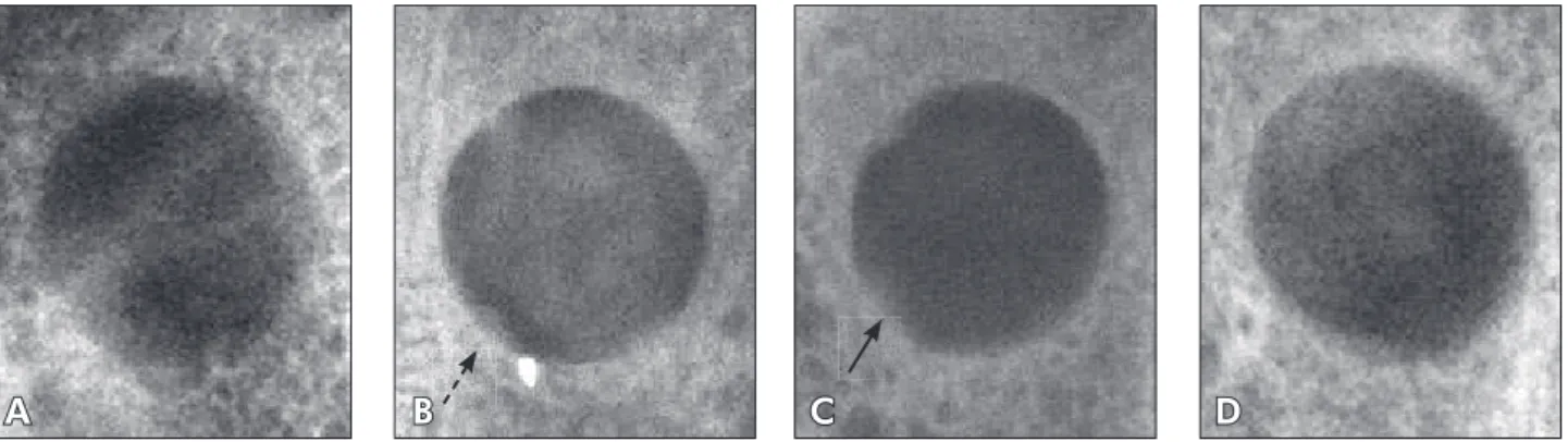
In the control group, the topography of the de-fects was well dei ned, and their limits were irregu-lar (Figure 1A). In the diabetic group, radiolucent images were observed in the bone defect area show-ing well dei ned limits, with higher optical intensity when compared with that of the other groups. In some regions, a discrete radiopaque area was ob-served over the walls of the defect (Figure 1B). In the PTFe-diabetic group, the defects exhibited ra-diolucent images with smooth as well as irregular limits. The radiopaque areas over the defect walls were more widespread and homogeneous when com-pared with that of the other groups (Figure 1C). The D-HDDM+PTFe group showed an accentuated in-crease of radiopacity in the region of the defect. The radiographic intensity was discretely similar to that of the radiopaque images of the surrounding normal bone tissue (Figure 1D).
Defect at 30 days
In the control group, the defect region was not well dei ned and also had irregular limits. The ra-diopaque areas i lling the central portion of the defects were discretely radiolucent, with irregular size and shape next to the bone defect walls. In the diabetic group the topography of the surgical defect still showed irregular limits and signii cant predominance of radiolucent areas. In the PTFe-diabetic group, the bone defect topography was
A B C D
poorly expressed, with imprecise and irregular limits. It was more radiopaque in the peripheral region than in the central region of the surgical de-fect. This radiopacity was similar to that of nor-mal bone. In the diabetic-PTFe group + HDDM, the defect presented a radiopaque image, with less intensity in comparison to that of the surrounding normal bone. These images were homogeneous in the periphery and heterogeneous in the central por-tion of the defect.
Defect at 60 days
In the control group, the bone defects were poor-ly dei ned. In some regions the limits of their walls could be observed, although there was evidence of increasing radiopacity in the limits of the surgical defect. In the diabetic group, the bone defect to-pography was well evidenced, but the pattern of the walls was imprecise and irregular when compared with those of the previous observation times. In the diabetic-PTFe group, the bone defect topography was poorly evidenced and its walls were imprecise and irregular. It was observed that there was a grad-ual increase of radiopacity in the central portion of the defect, but with less intensity when compared with the control group. In the diabetic-PTFe group + HDDM, a shadow was observed at the limits of the previous surgical defects; however, an homogeneous radiopacity was present in the other areas of the de-fect. The radiopacity of the central portion of the defect presented less intensity when compared with the normal surrounding bone tissue.
Defect at 90 days
In the control group, the radiolucent image cor-responding to the defect remained evident, but was more discrete when compared with the previous pe-riods of time (Figure 2A). In the diabetic group, the bone defect topography could still be observed and its walls were more imprecise and irregular when com-pared with the previous observation times of the same groups. The central portion of the defect presented an extensive radiolucent area when compared with the other studied groups (Figure 2B). In the diabetic-PTFe group, the bone defect topography could not be seen anymore, and there was a higher radiopacity area in the peripheral region than in the central region result-ing from centripetal growth durresult-ing the bone repair process. There was a gradual increase of radiopacity in the entire extension of the defect when compared to the control and diabetic groups (Figure 2C). In the diabetic-PTFe+HDDM group, the region of the bone defect was totally replaced by a radiopaque image, with areas presenting higher optical intensity when compared to that of the normal surrounding bone. In some areas, radiopaque lines were observed inter-mixed in the region of the defect, probably character-izing a disorganized arrangement of bone trabeculae formed during the repair process (Figure 2D).
Optical density and statistical analysis
The purpose of the densitometric analysis was to measure the optical density of newly formed bone matrix in the bone defects of the experimental groups, providing the necessary data for statistical
A B C D
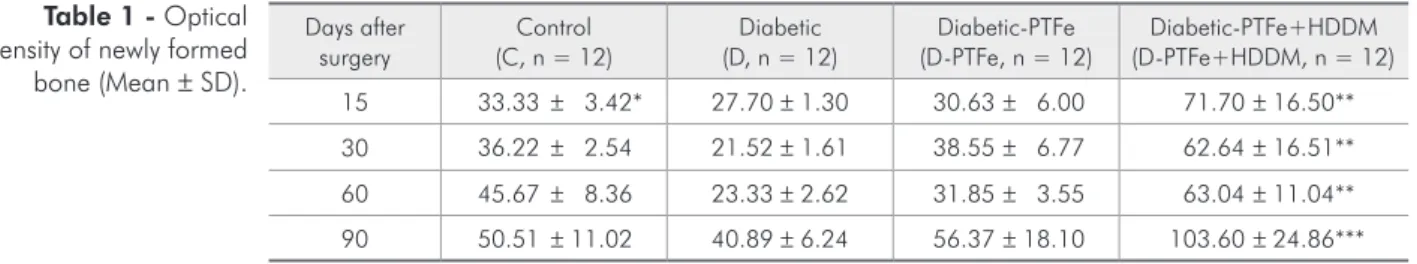
analysis. The optical density results were submitted to analysis of variance (ANOVA). The Tukey test was applied at a 5% signiicant level. A uniformity in the progression of the optical density averages in the Diabetic, D-PTFe and D-PTFe+HDDM groups was not observed (Table 1). We observed that there was a discrete decrease in optical density at 30 and 60 days in the diabetic group, and at 60 days in the D-PTFe group. However, the optical density averages in the D-PTFe+HDDM group showed the same radio-graphic features in all observation times of this study.
At 15 days, signiicant differences in optical densi-ty were observed between the C and D-PTFe+HDDM groups (p < 0.05), the D and D-PTFe+HDDM groups, and the D-PTFe and D-PTFe+HDDM groups (p < 0.01). At 30 and 60 days, the D and D-PTFe+HDDM groups presented statistically sig-niicant optical density differences (p < 0.01). At 90 days, the C and D-PTFe+HDDM groups, the D and PTFe+HDDM groups, and the PTFe and D-PTFe+HDDM groups also presented signiicant dif-ferences in relation to optical density (p < 0.001).
Discussion
During our review of the literature, no work was found using HDDM in diabetic patients. It is known, however, that many chronic complications of Diabetes mellitus patients are related to changes in bone metabolism, since it may impair the bone repair process.5,9
In the last years, researchers have added growth factors to occlusive barriers such as insulin growth factor, growth factor related to platelets, and bone morphogenetic proteins, among others,4,10 to
ac-celerate the bone repair process. Some authors re-ported the presence of these growth factors in bone
matrix and dentin matrix, which can explain their osteoinductive activity.10-12
During all observation periods of the control group, an average optical density that increased gradually and progressively (Table 1) was observed. Moreover, it was noticed that the PTFe barrier pro-moted an osteoconduction property, which inlu-enced bone growth positively. These indings were also observed by Abreu et al.1 (2004), Carvalho et
al.2 (2004) and Gomes et al.3,4 (2001, 2002).
How-ever, a noticeable fact in all the diabetic groups was the discrepant variation in mean optical density values, especially after the 30- and 60-day periods, with an intense decrease in optical density. This fact could be explained by the occurrence of chronic complications in our experiment caused by constant hyperglycemia, which was often dificult to be con-trolled, even using daily human insulin.
In all periods, it was observed that the diabet-ic-PTFe+HDDM group presented a signiicant in-crease in average optical density when compared to the other groups studied (Table 1). After the 15-day period, a not statistically signiicant decrease in optical density in the diabetic groups and in the diabetic-PTFe groups was observed when com-pared with the control group. At 90 days, the group treated with HDDM presented a signiicant increase in optical density when compared with the con-trol, diabetic, and diabetic-PTFe groups (p < .001) (Table 1). Moreover, the radiopaque images in the region of the bone defects implanted with HDDM were radiographically more homogeneous in all of their extension and after every observation period, when compared to the other groups studied. These indings were also observed by Abreu et al.1 (2004).
Therefore, in the diabetic-PTFe+HDDM group,
Days after surgery
Control (C, n = 12)
Diabetic (D, n = 12)
Diabetic-PTFe (D-PTFe, n = 12)
Diabetic-PTFe+HDDM (D-PTFe+HDDM, n = 12) 15 33.33 ± 3.42* 27.70 ± 1.30 30.63 ± 6.00 71.70 ± 16.50** 30 36.22 ± 2.54 21.52 ± 1.61 38.55 ± 6.77 62.64 ± 16.51** 60 45.67 ± 8.36 23.33 ± 2.62 31.85 ± 3.55 63.04 ± 11.04** 90 50.51 ± 11.02 40.89 ± 6.24 56.37 ± 18.10 103.60 ± 24.86***
*p < 0.05 (C/D-PTFe+HDDM). **p < 0.01 (D-PTFe+HDDM/D-PTFe/D). ***p < 0.001 (D-PTFe+HDDM/C/D/D-PTFe).
there was biocompatibility of HDDM, considering its integration with the newly formed bone matrix during all observation periods and the absence of any bone tissue reaction image as suggested images of bone sequestrum, osteomyelitis or others. More-over, it has been demonstrated that HDDM stimu-lates bone tissue osteopromotion precociously and signiicantly, contributing to a more favorable pro-gression of the bone regeneration process.3,4,13
Many advantages of HDDM became evident in the present study, conirming the possibility of its application in clinical dentistry, especially in im-plantodontology and for intra-oral surgeries in Dia-betes mellitus patients which, in general, present high risk of infection and an impaired bone repair process.
Conclusion
It was possible to conclude that homogenous demineralized dentin matrix (HDDM) was
bio-compatible with newly formed bone in diabetic rab-bits. HDDM stimulated an increase in bone defect radiopacity, suggesting new bone formation, and improving an osteopromotion property during the bone regeneration process. There were statistically signiicant differences (p < .001) among the dia-betic-PTFe+HDDM, control, diabetic and diabetic-PTFe groups.
Acknowledgements
This research was supported by the State of São Paulo Research Foundation (FAPESP; grant num-bers 2003/06017-0 and 03/02018-2). The authors are grateful to Johnson & Johnson MD&D Latin America Manufacturing Company, Brazil, for the donation of surgical materials, OneTouch Ultra blood test strips and machine (LifeScan), and to Huber+Suhner Latin America for the inancial sup-port given to the Special Health Care Needs Asso-ciation (ASPE).
References
1. Abreu PP, Morosolli A, Araújo MM, Carvalho VAP, Gomes MF. Effects of autogenous demineralized dentin matrix on dental socket wound healing process in human. Braz Oral Res. 2004;18(Suppl):52.
2. Carvalho VAP, Tosello DO, Salgado MAC, Gomes MF. His-tomorphometric analysis of homogenous demineralized dentin matrix as osteopromotive material in rabbit mandibules. Int J Oral Maxillofac Implants. 2004;19(5):679-86.
3. Gomes MF, Anjos MJS, Nogueira TO, Catanzaro-Guimarães SA. Autogenous demineralized dentin matrix for tissue engi-neering applications: radiographic and histomorphometric studies. Int J Oral Maxillofac Implants. 2002;17(4):488-97. 4. Gomes MF, Silva MJS, Nogueira TO, Catanzaro-Guimarães
SA. Histologic evaluation of the osteoinductive property of autogenous demineralized dentin matrix on surgical bone defects in rabbit skulls using human amniotic membrane for guided bone regeneration. Int J Oral Maxillofac Implants. 2001;16(4):563-71.
5. Guyton AC, Hall JE. Insulin, Glucagon, and Diabetes melli-tus. In: Guyton AC, Hall JE. Textbook of Medical Physiology. 11th ed. Philadelphia: Elsevier; 2006. p. 827-40.
6. Fiorellini JP, Nevins ML, Norkin A, Weber HP, Karimbux NY. The effect of insulin therapy on osseointegration in a diabetic rat model. Clin Oral Implants Res. 1999;10(5):362-8. 7. Lu H, Kraut D, Gerstenfeld LC, Graves DT. Diabetes
inter-feres with the bone formation by affecting the expression of
transcription factors that regulate osteoblast differentiation. Endocrinology. 2003;144(1):346-52.
8. Nevins ML, Karimbux NY, Weber HP, Giannobile WV, Fiorellini JP. Wound healing around endosseous implants in experimental diabetes. Int J Oral Maxillofac Implants. 1998;13(5):620-9.
9. Claro FA, Lima JR, Salgado MAC, Gomes MF. Porous Poly-ethylene for tissue engineering applications in diabetic rats treated with calcitonin: histomorphometric analysis. Int J Oral Maxillofac Implants. 2005;20(2):211-9.
10. Catanzaro-Guimarães SA, Catanzaro Guimarães BP, Garcia RB, Alle N. Osteogenic potential of autogenic demineralized dentin implanted in bony defects in dogs. Int J Oral Maxillofac Surg. 1986;15(2):160-9.
11. Bessho K, Tagawa T, Murata M. Purification of rabbit bone morphogenetic protein derived from bone, dentin, and wound tissue after tooth extraction. J Oral Maxillofac Surg. 1990;48(2):162-9.
12. Nakashima M. Induction of dentine in amputated pulp of dogs by recombinant human bone morphogenetic proteins-2 and -4 with collagen matrix. Arch Oral Biol. 1994;39(12):1085-9. 13. Catanzaro-Guimarães SA. Possibility to reinforce bone