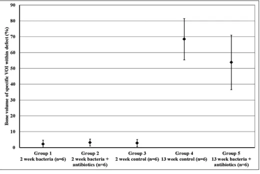
A biodegradable antibiotic-impregnated scaffold to prevent osteomyelitis in a contaminated in vivo bone defect model
Texto
Imagem

Documentos relacionados
First of all we consider a defect consisting of the removal of a single Mo atom from the metal-terminated edge (shown as red line and labeled as Mo1). For this defect, we observe
Figure 2 - Digital radiographic images of bone defect in the parietal bone of rabbit taken at 90 days: ( A ) control group; ( B ) diabetic group; ( C ) diabetic-PTFe group; ( D
According to hard tissue indings, both treatment groups showed signiicant improvements compared to baseline in terms of vertical bone gain, defect ill and defect angle at 6
Pretendeu-se junto destes obter informação privilegiada sobre as dinâmicas relativas à gestão da informação na empresa, como se processa a comunicação interna, a atitude
The remaining dependent vari- ables were as follows: animal model, bone region of the defect, diameter of the defect (in mm), follow-up time after implantation (in weeks),
A presente investigação contribuiu para a reflexão dos fatores que influenciam o bem-estar dos docentes, enfatizou a importância da gestão e regulação emocional, forneceu
Radiographic images of animals in the control group taken immediately after surgery showed grade 0 radiopacity in the defect region, since the defect created remained unfilled
Sob a regência do Professor Doutor Rui Maio, o estágio estava dividido em três partes: uma semana de aulas teóricas e teórico-práticas de conteúdo relacionado com
