A retrospective clinical, radiographic
and microbiological study of periodontal
conditions of teeth with and without
crowns
Um estudo retrospectivo clínico, radiográfico e
microbiológico das condições periodontais de
dentes com e sem coroas
Abstract: The aim of this study was to evaluate retrospectively the periodontal conditions of teeth with ixed crowns that had been in place from 3 to 5 years before the study was conducted. Forty individuals were recalled for a follow-up visit. Full-mouth clinical exam-inations were carried out and Visible Plaque Index (VPI), Gingival Bleeding Index (GBI), Probing Pocket Depth (PPD), and clinical attachment level (CAL) were assessed in 6 sites per tooth. Parallel radiographs were also taken and blindly analyzed by a digital caliper (distance between the apex and the bone crest). BANA tests were performed. A contra-lateral sound tooth was considered the control. Mean values were obtained and Wilcoxon and paired sample t tests were used to compare the test and control sites. Crowns had a mean VPI value of 30.42% as compared to 49.17% for sound teeth. The GBI was 33.33% and 26.25% for test and control teeth respectively. Assessment of PPD revealed values of 2.30 and 2.14 mm, and assessment of CAL revealed averages of 2.02 and 1.89 mm for test and control teeth respectively. The mean values for radiographic distances were 12.73 and 13.67 mm, and for the BANA test, 67.50 and 50.00 for sound and crowned teeth, respectively. Statistically signiicant differences were observed for all parameters except for CAL and for the BANA test. It may be concluded that, with the methods used in the present study, crowns may be associated with more signs of inlammation, however not with periodontal breakdown.
Descriptors: Periodontal diseases; Tooth crown; Periodontics; Prosthodontics.
Resumo: O objetivo deste estudo foi avaliar retrospectivamente as condições periodon-tais de pacientes com coroas ixas colocadas de 3 a 5 anos antes da realização da pesquisa. Quarenta indivíduos foram rechamados. Exames clínicos de toda a boca foram realizados avaliando-se Índice de Placa Visível (IPV), Índice de Sangramento Gengival (ISG), Profun-didade de Sondagem (PS) e Nível Clínico de Inserção (NCI) em 6 sítios por dente. Radio-graias paralelas foram obtidas e analisadas cegamente por paquímetro digital (distância do ápice à crista óssea). Testes BANA foram realizados. Um dente hígido contralateral foi considerado como controle. Valores médios foram obtidos e testes de Wilcoxon e t pareado foram utilizados para comparar os sítios teste e controle. Coroas apresentaram um valor médio de IPV de 30,42%, comparado com 49,17% para dentes hígidos. O ISG foi de 33,33% e 26,25% para dentes com coroas e hígidos, respectivamente. A PS revelou valores de 2,30 e 2,14 mm e a análise do NCI demonstrou médias de 2,02 e 1,89 mm para dentes teste e controle, respectivamente. Os valores médios para as distâncias radiográi-cas foram de 12,73 e 13,67 mm, e para o teste BANA, de 67,50 e 50,00 para dentes hígi-dos e com coroas, respectivamente. Diferenças estatisticamente signiicantes foram obser-vadas para todos os parâmetros, exceto para NCI e para o teste BANA. Pode-se concluir que, com os métodos utilizados no presente estudo, coroas podem ser associadas a mais sinais de inlamação, entretanto não com destruição periodontal.
Descritores: Doenças periodontais; Coroa dentária; Periodontia; Prostodontia. Marcio Dias Giollo(a)
Patrícia Moura Valle(a)
Sabrina Carvalho Gomes(a)
Cassiano Kuchenbecker Rösing(a)
(a) Professors of Prosthodontics, Lutheran University of Brazil, Porto Alegre, RS, Brazil.
Corresponding author:
Cassiano Kuchenbecker Rösing Rua Dr. Valle, 433/801 Porto Alegre - RS - Brazil CEP: 90560-010
E-mail: [email protected]
Introduction
Evidence from different studies points to a very close relationship between the periodontium and crowns and bridges. The vast majority of studies, however, focus on different aspects of the relation-ship between dental materials, surface characteris-tics, adaptation, etc., thus providing guidelines for the dental team.2,4,12
Previous studies looking at this subject have sug-gested that crowns may frequently be a problem to the periodontium.15 At the moment these studies were
performed, however, some confusion between gingi-val inlammation and periodontal breakdown exist-ed, ultimately leading to misleading conclusions.
Even more recent studies evaluating the relation-ship between dental prostheses and the periodon-tium have focused mainly on inlammation and not on periodontal breakdown.5,6,14,15 The success of a
dental prosthesis, however, is only achieved if the restoration remains in place over time and without causing disease.2,4,7,11,12 Evaluating the periodontium
around prosthetic work is thus of utmost impor-tance in clinical dentistry, especially trying to iden-tify risk indicators for adverse events.
Evaluation of periodontal tissues may be per-formed concerning supragingival and subgingival plaque-related parameters. Clinically, plaque indices as well as marginal gingival inlammation relate to supragingival bioilm accumulation. Probeable pocket depth and clinical attachment level relate to the sub-gingival bioilm. Radiographic analysis may reveal signs of alveolar bone destruction. In order to assess microbial composition, the BANA test can be a use-ful tool since it demonstrates the presence of subgingi-val red complex bacteria (Porphyromonas gingivalis,
Treponema denticola and Tannerella forsythia).9,10,16
The aim of the present study was to retrospec-tively evaluate clinical, radiographic and microbio-logical periodontal conditions of teeth with ixed crowns that had been in place from 3 and 5 years before the study was conducted as compared to sound teeth.
Material and Methods
Study population
Forty individuals, 11 male (27.5%) and 29
fe-male (72.5%), aged 30 to 55 years, from a group of 65 patients agreed to participate in this retro-spective study after being invited by the authors. A response rate of 61.5% was achieved. Patients gave their written informed consent and the protocol was approved by the Ethical Committee of the Lutheran University of Brazil.
Inclusion criteria
To be included in the study, subjects should have one tooth with a metaloceramic crown placed at the University Clinic between 3 and 5 years prior to examination (test tooth) and a contra-lateral sound tooth (control tooth).
Exclusion criteria
Subjects with any other prostheses, needing antibiotic therapy in order to be periodontally ex-amined, having any systemic disorder affecting the periodontium, pregnant or breast feeding were not included in the study.
Development of the study
After examining the records of patients that had crowns placed, a telephone call was given in order to invite the patients for examination. From the 40 individuals who agreed to participate, 7 pairs of in-cisors, 7 pairs of canines, 20 pairs or premolars and 6 pairs of molars were examined.
Clinical examination
Periodontal conditions were clinically assessed by a trained and calibrated examiner in terms of Visible Plaque Index (VPI),1 Gingival Bleeding Index
(GBI),1 Probing Pocket Depth (PPD), and Clinical
Attachment Level (CAL). Six sites per tooth were examined (distobuccal, midbuccal, mesiobuccal, distolingual, midlingual and mesiolingual).
VPI: Visible Plaque Index was assessed as a di-chotomic evaluation of the Silness & Löe Plaque In-dex, combining scores 0+1 and 2+3.1
GBI: Gingival Bleeding Index, similarly, com-bined the scores 0+1 and 2+3 of the Löe Gingival Index.1
William’s periodontal probe and were measured from the most apically probeable portion until the gingival margin (PPD) and to the cementum-enamel junction (CAL). Measurements were rounded to the nearest millimeter.10
The clinical location of the crown margin (whether supragingival, at the gingival margin, or subgingival) and its adaptation (whether well-it or maladapted, according to Felton et al.6,1991) were
also assessed by an examiner unaware of the as-sessed clinical periodontal conditions. These assess-ments were performed considering the 6 sites per tooth.
Radiographic examination
Parallel periapical radiographs were taken with the aid of Han-Shin appliances in each test and control tooth and were automatically processed. Measurements of the distance between the apex and the bone crest were performed with the aid of a digital caliper by a calibrated examiner unaware of the clinical conditions at the mesial and distal portions.
Microbiological examination
After removal of supragingival plaque, intra-cre-vicular bioilm was collected with the aid of a sterile Gracey curette and processed in a BANA card dur-ing 15 minutes at a temperature of 55°C, accord-ing to Amalitano et al.3 (1993). Examination of
the results was performed by a calibrated examiner, who did not know whether the tooth was a test or a control tooth, or what the clinical condition of the tooth was.
Reliability
All examinations were performed by an experi-enced periodontist except for the crown adaptation and crown margin location examination, which was performed by a prosthodontist. Concerning the clinical periodontal conditions, the periodontist was trained for VPI and GBI assessment. In relation to PPD and CAL assessment, weighed kappa was test-ed (± 1 mm) and revealed values of 0.85 and 0.71, respectively.
Radiographic measurements were calibrated by
duplicate analyses of 18 radiographs with one week’s interval, without revealing statistically signiicant differences when tested by the paired sample t test (p > 0.05).
The kappa value for the BANA test analysis after double assessment with one week’s interval was of 0.94.
All reliability analyses were performed in indi-viduals not participating in the study, but with char-acteristics similar to those of the ones who did par-ticipate.
Statistical analysis
Relative frequencies of VPI, GBI and BANA test in crowned and sound teeth were compared by the Wilcoxon Test. Mean values of PPD, CAL and Bone Height were calculated and compared be-tween test and control teeth by the paired sample
t test. Spearman correlation coeficients were cal-culated between maladapted and subgingivally lo-cated crowns and the clinical, radiographic and mi-crobiological results. Additionally, total VPI ≥ 50% and GBI ≥ 30% were correlated with the clinical, radiographic and microbiological variables, by Spearman’s correlation coeficient. The alpha level was set at 0.05.
Results
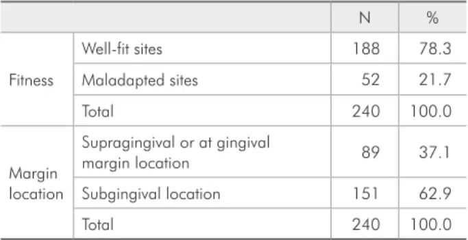
The descriptive data of the characteristics of crowns in terms of margin location and itness are described in Table 1. The majority of the sites of the crowns (78.3%) were considered well-it. Concern-ing margin location, 37.1% of the sites were located supragingivally or at the gingival margin and 62.9% were located subgingivally.
The clinical, radiographic and microbiological parameters of the examined teeth are demonstrated in Table 2. Crowned teeth presented a lower mean value of Visible Plaque (30.42%) as compared with sound teeth (49.17%). Although a large variation was observed in this parameter, statistically signii-cant difference was observed. Conversely, the Gingi-val Bleeding Index was higher (33.33%) in crowned than in sound teeth (26.25%). This difference was also statistically signiicant.
slightly larger in crowned than in sound teeth (2.30
versus 2.14 mm, p = 0.008). Statistically signiicant differences, however, were not observed in Clinical Attachment Level.
The radiographic analysis revealed that the sound teeth had more supporting bone as compared to the crowned teeth (13.67 and 12.73 mm, respec-tively). The microbiological results revealed a mean percentage of BANA positive reactions of 67.50 in crowned and of 50.00% in sound teeth. A statisti-cally signiicant difference could not be demonstrat-ed (p = 0.071).
Table 3 presents the correlations performed be-tween maladapted crowns and the study parameters. All correlation coeficients were relatively low and the only statistically signiicant correlation was observed between maladapted crowns and clinical attachment level (0.15), meaning that maladapted crowns
posi-tively correlate to clinical loss of attachment.
Correlations between subgingival crowns and the study parameters were also calculated (Table 4). Positive correlations were obtained between subgin-gival crowns and all parameters. Statistically signii-cant correlations, however, were observed with the gingival bleeding index (r = 0.14) and a higher cor-relation was observed with the clinical attachment level (r = 0.46), reinforcing the correlation between this parameter and maladaptation of the crowns, as previously demonstrated.
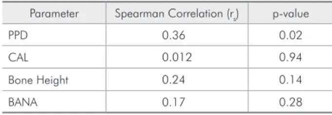
In order to have an idea of the role of supragin-gival plaque control, a dichotomization of the total VPI and total GBI of the individuals was performed. Fifty percent and thirty percent were considered as cut-off points for VPI and for GBI respectively. These were values close to the median. The only statisti-cally signiicant correlation was observed between
Table 2 - Mean (± Standard deviation) of the clinical,
ra-diographic and microbiological parameters in crowned and sound teeth (n = 240 sites, 40 patients).
Parameter Crowned teeth Sound teeth P value
VPI (%) 30.42 (46.10) 49.17 (50.10) 0.000 a
GBI (%) 33.33 (47.24) 26.25 (44.09) 0.035 a
PPD (mm) 2.30 (0.99) 2.14 (0.86) 0.008 b
CAL (mm) 2.02 (1.36) 1.89 (2.00) 0.30 – ns b
Bone Height (mm) 12.73 (2.63) 13.67 (2.38) 0.000 b
BANA (%) 67.50 (47.43) 50.00 (50.64) 0.071 – ns a
a: Wilcoxon rank sign test. b: Paired sample t test. ns: non-significant differ-ence. VPI: Visible Plaque Index, GBI: Gingival Bleeding Index, PPD: Probing Pocket Depth, CAL: Clinical Attachment Level.
Table 3 - Spearman correlation coefficient between
mal-adapted crowns and clinical, radiographic and microbio-logical parameters (n = 240 sites, 40 patients).
Parameter Spearman Correlation (rs) p-value
VPI 0.004 0.95
GBI 0.29 0.65
PPD 0.1 0.12
CAL 0.15 0.019
Bone Height 0.161 0.16
BANA 0.069 0.67
VPI: Visible Plaque Index, GBI: Gingival Bleeding Index, PPD: Probing Pocket Depth, CAL: Clinical Attachment Level.
Table 4 - Spearman correlation coefficient between
sub-gingivally located crowns and clinical, radiographic and mi-crobiological parameters (n = 240 sites, 40 patients).
Parameter Spearman Correlation (rs) p-value
VPI 0.07 0.25
GBI 0.14 0.03
PPD 0.11 0.08
CAL 0.46 0.000
Bone Height 0.14 0.20
BANA 0.22 0.17
VPI: Visible Plaque Index, GBI: Gingival Bleeding Index, PPD: Probing Pocket Depth, CAL: Clinical Attachment Level.
Table 1 - Absolute and relative frequencies of fitness and
margin location of the analyzed crowns (n = 240 sites).
N %
Fitness
Well-fit sites 188 78.3
Maladapted sites 52 21.7
Total 240 100.0
Margin location
Supragingival or at gingival
margin location 89 37.1
Subgingival location 151 62.9
GBI ≥ 30% and probing depth (PPD). VPI did not correlate with any of the clinical, radiographic or mi-crobiological parameters assessed (Tables 5 and 6).
Discussion
The present retrospective study evaluated the periodontal conditions of teeth with ceramic crowns that had been in place for between 3 and 5 years as compared to those of sound teeth. The limita-tions of retrospective analyses, the slightly reduced number of individuals included and the inclusion of different types of teeth must be highlighted when in-terpreting our results. It has to be taken into consid-eration, however, that each crowned tooth was con-trolled by a similar sound tooth. Finding individuals with these conditions is not an easy task. Among the invited individuals, a good response rate was achieved. All the individuals that refused to partici-pate in the study still had their crowns in place and did not want to participate in the study for reasons unrelated to satisfaction with the prostheses. It has to be kept in mind that, as the examined individuals were volunteers, it is more likely that the individuals with a higher standard of healthcare were the ones who showed up for examination.
The comparison employed in the present study (crowned versus contra-lateral sound teeth in the same individual) aimed at minimizing inter-individ-ual differences, so that a more reliable result could be obtained. Smaller numbers of individuals are needed when intra-individual comparisons are pos-sible.2
The clinical examination comprised VPI, GBI, PPD and CAL assessments, which relate to
peri-odontal status. VPI and GBI are considered indi-cators of gingival marginal health status and PPD may indicate both inlammation and destruction, especially if combined with CAL and radiographic examination.10 The microbiological examination in
this study was performed by means of the BANA test. This test is a chair-side evaluation of the pres-ence of putative periodontopathogens (P. gingivalis, T. denticola and T. forsythia).9
An attempt to achieve a better reproducibility was made by having an experienced periodontist or prosthodontist as the examiner. Calibration for PPD, CAL, radiographic measurements and BANA test revealed that the examiners were reproducible, enhancing the reliability of the results.
The presence of supragingival plaque assessed in the present study revealed more plaque in sound than in crowned teeth. This was a surprising ind-ing. The invitation for an examination in patients that had crowns placed, however, could have led to a more careful cleansing of those teeth by the patients before they came in for the consultation. Conversely, marginal inlammation detected by the GBI revealed more inlammation in crowned teeth. This param-eter is more adequate to reveal the patient’s oral hy-giene habit, not just at the time of the examination.12
It should be stressed that, in our study, 62.90% of the crowns were placed subgingivally. This inding conirms those of previous studies.2,7,8,12,13,15 All these
reports afirmed that subgingival restorations are as-sociated with gingival bleeding.
Concerning PPD, the present study observed high-er values for crowned as compared to sound teeth (Table 2). Reitemeier et al.12 (2002) observed
high-Table 5 - Spearman correlation coefficient between total
VPI ≥ 50% and clinical, radiographic and microbiological parameters (n = 240 sites, 40 patients).
Parameter Spearman Correlation (rs) p-value
PPD 0.17 0.27
CAL 0.07 0.66
Bone Height 0.07 0.66
BANA 0.08 0.58
VPI: Visible Plaque Index, PPD: Probing Pocket Depth, CAL: Clinical At-tachment Level.
Table 6 - Spearman correlation coefficient between total
GBI ≥ 30% and clinical, radiographic and microbiological parameters (n = 240 sites, 40 patients).
Parameter Spearman Correlation (rs) p-value
PPD 0.36 0.02
CAL 0.012 0.94
Bone Height 0.24 0.14
BANA 0.17 0.28
er PPD values in subgingivally placed restorations. The study by Valderhaug et al.15 (1993) also showed
slightly greater PPD values in crowned teeth.
In relation to CAL, our results did not show more loss of attachment in crowned than in sound teeth (Table 2). Brunsvold, Lane4 (1990) and
Reite-meier et al.12 (2002) found higher loss of attachment
in places adjacent to maladapted restorations. Other studies8 showed higher values of CAL in
subgingi-vally located restorations, independently of the ob-served adaptation. Although showing higher values of CAL for crowned teeth, Wang et al.17 (1993) did
not ind statistically signiicant differences between crowned and sound teeth. The outstanding study of Schätzle et al.13 (2001), that evaluated 160 patients
during 26 years, did not ind statistically signiicant differences between the study groups, similarly to the results of the present study. These results shed some light into the fact that the higher PPD and GBI values observed for crowned teeth are related to chronic inlammation due to the subgingival place-ment of the majority of the crowns.
Moreover, although with small differences, a con-sistently higher value of bone loss was observed for crowned teeth in the radiographic indings. The exper-imental design (retrospective) does not allow a claim of causality in this respect. The microbiological ind-ings also revealed no differences between teeth with and without crowns. These results are in accordance with the lack of difference in the CAL values.9,10
Correlations between maladapted crowns and the clinical, radiographic and microbiological
pa-rameters evaluated in the present study (Table 3) revealed a statistically signiicant correlation with CAL, a inding that corroborates those of previous studies.2,3,5 Correlations between subgingivally
lo-cated crowns and the parameters evaluated revealed statistically signiicant values for GBI and CAL. This has been demonstrated by Valderhaug et al.15
(1993) and De Backer et al.5 (2006).
The correlations between VPI ≥ 50% and
GBI ≥ 30% were signiicant only between the latter and PPD. This conirms the nature of PPD as a sign of inlammation and was also demonstrated by Re-itemeier et al.12 (2002).
The indings of the present study are challenging both for Periodontology and Prosthodontics. If, on one hand, no differences were observed concerning periodontal destruction, on the other hand, higher degrees of inlammation were observed, especially in subgingivally located crowns. This should be kept in mind, since inlammation is related to a worse quality of oral health, leading do bleeding, redness and halitosis, which today are socially unacceptable. The retrospective design of the present study, the number and diversity of evaluated teeth, as well as the time after installation of the crowns could also account for the observed results.
Conclusion
It may be concluded, taking into consideration the design and limitations of the present study, that crowns may be associated with more signs of inlam-mation, although not with periodontal breakdown.
References
1. Ainamo J, Bay I. Problems and proposals for recording plaque and gingivitis. Int Dent J. 1975;25(4):229-35.
2. Al-Wahadni AM, Mansour Y, Khader Y. Periodontal response to all-ceramic crowns (IPS Empress) in general practice. Int J Dent Hyg. 2006;4(1):41-6.
3. Amalfitano J, De Filippo AB, Bretz WA, Loesche WJ. The effects of incubation length and temperature on the specificity and sensitivity of the BANA (N-benzoyl-DL-arginine-naph-thylamide) test. J Periodontol. 1993;64(9):848-52.
4. Brunsvold MA, Lane JJ. The prevalence of overhanging dental restorations and their relationship to periodontal disease. J Clin Periodontol. 1990;17(2):67-72.
5. De Backer H, Van Maele G, De Moor N, Van den Berghe L, De Boever J. An 18-year retrospective survival study of full crowns with or without posts. Int J Prosthodont. 2006;19(2):136-42. 6. Felton DA, Kanoy BE, Bayne SC, Wirthman, GP. Effect of
in vivo crown margin discrepancies on periodontal health. J Prosthet Dent. 1991;65(3):357-64.
7. Jacobs DJ, Steele JG, Wassel RW. Crowns and extra-coronal restorations: considerations when planning treatment. Br Dent J. 2002;192(5):263-7.
9. Loomer PM. Microbiological diagnostic testing in the treatment of periodontal diseases. Periodontol 2000. 2004;34:49-56. 10. Mombelli A. Clinical parameters: biological validity and
clini-cal utility. Periodontol 2000. 2005;39:30-9.
11. Odman P, Andersson B. Procera AllCeram crowns followed for 5 to 10.5 years: a prospective clinical study. Int J Prostho-dont. 2001;14(6):504-9.
12. Reitemeier B, Hansel K, Walter MH, Kastner C, Toutenburg H. Effect of posterior crown margin placement on gingival health. J Prosthet Dent. 2002;87(2):167-72.
13. Schätzle M, Land NP, Anerud A, Boysen H, Burgin W, Loe H. The influence of margins of restorations of the periodontal tissues over 26 years. J Clin Periodontol. 2001;28(1):57-64.
14. Sjögren G, Lantto R, Tillberg A. Clinical evaluation of all-ceramic crowns (Dicor) in general practice. J Prosthet Dent. 1999;81(3):277-84.
15. Valderhaug J, Ellingsen JE, Jokstad A. Oral hygiene, peri-odontal conditions and carious lesions in patients treated with dental bridges. A 15-year clinical and radiographic follow-up study. J Clin Periodontol. 1993;20(7):482-9.
16. van Winkelhoff AJ, Winkel EG. Microbiological diagnostics in periodontics: biological significance and clinical validity. Periodontol 2000. 2005;39:40-52.