ELECTROPHYSIOLOGICAL EVALUATION
IN MYOTONIC DYSTROPHY
Correlation with CTG length expansion
Beatriz Helena Miranda Pfeilsticker
1, Carmen Sílvia Bertuzzo
2, Anamarli Nucci
1ABSTRACT - In myotonic dystrophy (MD), disease severity has been correlated with expansion of CTG repeats in chromosome 19. The aims of this study were to evaluate efficacy of electromyography in the diagnosis of MD, access the frequency and the characteristics of peripheral involvement in the disease and to verify whether the CTG repeats correlated with the electrophysiological abnormalities. Twenty-five patients and six relatives at risk of carrying the MD gene were examined. Electrical myotonia (EM) was scored. Sensory and motor conduction velocity (CV) were studied in five nerves. Leukocyte DNA analysis was done in 26 subjects. Myopathy and myotonia were found in 27 cases. EM was most frequent in muscles of hand and in tibialis anterior. No significant correlation was found between EM scores and length of CTG expansions. EM scores correlated significantly with the degree of clinical myopathy, expressed by a muscular disability scale. Peripheral neuropathy was found in eight subjects and was not restricted to those who were diabetics.
KEY WORDS: myotonic dystrophy, electromyography, myotonia, myopathy, peripheral neuropathy, CTG repeat.
Avaliação eletrofisiológica na distrofia miotônica: correlação com a expansão de tripletos CTG.
RESUMO - Na distrofia miotônica (DM) a severidade da doença tem sido correlacionada com a expansão de tripletos CTG, no cromossomo 19. Foram objetivos do estudo avaliar a eficácia da eletromiografia no diagnóstico, verificar a freqüência e tipos de neuropatias periféricas e determinar se a expansão CTG correlacionava-se com as anormalidades eletrofisiológicas nesta doença. Examinamos 25 pacientes e seis familiares sob risco de herdar a doença. A miotonia elétrica (ME) foi graduada, cinco velocidades de condução sensitivas e cinco motoras estudadas. O DNA leucocitário foi analisado em 26 indivíduos. Encontrou-se miopatia miotônica em 27 pacientes, sendo a ME mais freqüente nos músculos da mão e no tibial anterior. Não houve correlação significativa entre os graus de ME e o número de repetições CTG. Os graus de ME correlacionaram-se significantemente com a severidade da miopatia clínica, expressa por escala de disfunção muscular. Neuropatia periférica foi encontrada em oito indivíduos, não exclusivamente em diabéticos.
PALAVRAS-CHAVE: distrofia miotônica, eletromiografia, miotonia, miopatia, neuropatia periférica, repetição de tripletos.
1Departments of Neurology and 2Medical Genetics, Faculty of Medicine, University of Campinas (UNICAMP) SP, Brazil. Presented partially
in poster format at the 10th European Congress of Clinical Neurophysiology, Lyon, France, August 2000. Received 20 October 2000, received in final form 23 November 2000. Accepted 27 November 2000.
Dra. Beatriz Helena Miranda Pfeilsticker - Departamento de Neurologia FCM UNICAMP - Caixa Postal 6111 – 13081-970 Campinas SP-Brasil. Email:bhmpfeil@uol.com.br
Myotonic dystrophy (MD) is a systemic disorder, with a variable phenotype including myopathy and myotonia1. Disease severity has been correlated with unstable expansion of CTG repeats in the gene encoding myotonin protein kinase in chromosome 192,3. Electromyography (EMG) is a master tool for the diagnosis, but the incidence of EMG myotonia has varied in different reports because of selection and number of muscles examined and interpretation of electromyographic findings, as well as diagnostic criteria4. Electroneuromyography may also detect peripheral neuropathy (PN) in MD patients5,6.
The aims of this paper were to study prospec-tively the efficacy of EMG in MD diagnosis, access the frequency and the characteristics of peripheral neuropathy in the disease and to correlate the CTGn with abnormalities documented in EMG.
METHOD
and asymptomatic subjects or with minimal MD signs or symptoms belonging to MD families. They were submit-ted to neurological examination, EMG protocol, routine blood analyses to detect common causes of PN and blood sample for DNA analysis. Clinical and EMG results were established before the genetic study. They were classified according to Mathieu et al.7, into a muscular disability
rating scale, as follow: Grade 1 – no clinical muscular im-pairment (diagnosis made by EMG, slit-lamp examination or DNA analysis); Grade 2 - minimal signs (myotonia, jaw and temporal wasting, facial weakness, sternomastoids wasting / weakness, ptosis, nasal speech, no distal weak-ness except isolated digitis flexor weakness); Grade 3 -distal weakness (no proximal weakness, except isolated
triceps brachii weakness); Grade 4 - mild or moderate proxi-mal weakness; Grade 5 - severe proxiproxi-mal weakness (con-fined to wheelchair for short and long distances).
EMG protocol studied the anatomical distribution of electrical myotonia (EM), in 13 muscles and was scored according to Streib and Sun4: Grade 1 - occasional short
run of positive waves following needle movement (inter-preted as nonspecific); Grade 2 - myotonic discharges of more than 0.5 sec duration in two or more muscle areas; Grade 3 - myotonic discharges in most needle locations; Grade 4 - myotonic discharges in each movement in all examined areas. Score for individual muscle: sum of the grades divided by the number of times the muscle was examined. Total EMG myotonia score for a subject: summed grades of EMG-myotonia, divided by the num-ber of muscles examined.
H reflex (tibial nerve), CV in five motor and in five sen-sory nerves were evaluated in each patient. CV techniques and normal values were considered according to Kimura8.
Investigations were performed using a Neuropack equip-ment (Nihon Khoden Co.), superficial electrodes for stimu-lation and recording and monopolar needle for EMG. Tem-perature was controlled by a Cole Palmer Digi Sense Termometer and maintained over 32°C.
Routine blood examination included hemogram, cre-atine kinase, alanine and aspartate aminotransferase, to-tal calcium, potassium, sodium, lactate dehydrogenase, cholesterol total, HDL and LDL-cholesterol, creatinine, glu-cose, triglycerides, urea nitrogen, thyroxine free and thy-rotropin, serum B12 and folic acid.
Genomic DNA was prepared from peripheral blood leukocytes 9,10. The size of the CTG-repeat expansion was
analysed by PCR-Elongase. PCR analyses were performed in a volume of 30 µml, containing 100 ng template DNA,60 mM Tris-SO4 (ph 9.1), 18 mM (NH4)2SO4, 1.1 mM Mg SO4, 10 mM dNTP mix, 10% Dimethylsulfonamide (DMSO), 1 µml Elongaseââ (mixture of Taq and Pyrococcus species GB-D DNA polymerases from Life Technologies, 10480-010), 0.2 µmM of primers 101 and 102 (101: CTTCCCAGGCCTGCAGTTTGCCCATC - 3’; 102: 5’-GAACGGGGCTCGAAGGGTCTTGTAGC 3’). PCR involved
heating to 94°C for 6 min, 65°C for 1 min, 72°C for 1 min and then 30 cycles of 94°C for 1 min, 65°C for 1 min, 72°C for 1 min. Amplified product (2 µml) was mixed with 2 µml of formamide loading buffer, heated for 5 min at 100°C, subjected to electrophoresis in a 2% agarose gel, blotted on to nylon membrane and probed with the 3’-end-la-belled 101 primer.
Statistical analyses were based on linear correlation coefficient of Spearman (non-parametric); Kruskal-Wallis test was used to verify homogeneity between myotonic score and the degree of muscular disability.
RESULTS
Myotonia and myopathy were found in 27 sub-jects, with mean age of 34,4 years. None of them presented congenital MD. The mean grade of mus-cular disability was 2,77. Total EMG-myotonia score, the number of CTG repeats, grade of muscular dis-ability and age of each case is presented in Table 1. Six subjects (Cases 1,2,3,4,13,21,Table 1), belong-ing to three different families with MD, were ini-tially considered relatives at risk to have the abnor-mal gene. They were clinically norabnor-mal (force grade five in all muscles, no clinical myotonia, no muscu-lar atrophy, no other clinical signs of MD). In cases 1,2,3 and 4, EMG and muscle enzimes were also normal. They were daughter (Case 1), son (Case 2) and sisters (Cases 3 and 4) of one patient (Case 26). Cases 13 and 21 were also clinically normal, had mild abnormalities in EMG, and DNA confirmed MD. Case 13 was sister of patients (Cases 22 and 25). The deceased mother of case 21 had a definitive diagnosis of MD.
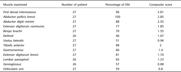
The frequency of EM and composite score for each muscle examined is shown in Table 2.
A significant correlation (0,617), at 0,05 level with Spearman’s test, was found between myotonic score and the grade of muscular disability. There was not a significant correlation (0,232), between CTG ex-pansion and the score of myotonia, neither with the degree of muscular disability (0.026).The p-value in Kruskal-Wallis test was 0,012 for the score of myo-tonia and muscular disability.
Table 1. Casuistic in relation to age, DNA and scores.
Case number Age* DNA Score of muscular disability EMG myotonia score
1 20 R N 0
2 23 R N 0
3 37 R N 0
4 44 R N 0
5 12 5/110 3 0.38
6 14 10/110 2 2
7 19 R 2 1.23
8 20 5/180 2 1.07
9 20 5/200 3 1.38
10 24 5/200 2 1
11 24 5/200 2 2.23
12 27 7/110 3 1.84
13 27 5/200 1 0.15
14 28 7/110 2 0.3
15 35 5/110 4 3.23
16 36 7/200 3 2.15
17 36 5/200 4 2
18 38 15/400 4 2.61
19 39 5/200 4 2.07
20 40 7/100 4 2.15
21 40 5/200 1 0.6
22 42 5/200 3 2.61
23 42 5/100 1 0.15
24 43 7/100 3 1.07
25 45 5/200 3 2.46
26 46 7/150 4 2.15
27 46 5/110 2 2.46
28 46 7/110 2 1.53
29 47 5/80 4 2.15
30 47 5/110 4 2.46
31 56 5/390 3 2.53
*, patient’s ages at the time of EMG; N, diagnosed as normal (force grade five in all muscles, no clinical myotonia, no muscular atrophy, no other clinical
signs of MD); R, DNA analysis was refused by these patients.
Table 2. Percentage of EM and composite score for each muscle examined.
Muscle examined Number of patient Percentage of EM Composite score
First dorsal interosseous 27 96 2.81
Abductor pollicis brevis 27 100 2.85
Abductor digiti minimi 27 88 2.55
Extensor digitorum communis 27 77 1.85
Biceps brachii 27 70 1.55
Deltoid 26 46 1.07
Vastus lateralis 27 51 0.96
Tibialis anterior 27 88 2
Gastrocnemius 27 66 1.4
Extensor digitorum brevis 27 81 1.74
Lumbar paraspinal 26 65 1.23
Genioglossus 26 57 0.88
Table 3. Electroneurographic results in subjects with peripheral neuropathy
Patients
Relative
Diabetic Non diabetic at risk
Case 25 20 19 29 18 22 17 2
Age 45 40 39 47 38 42 36 23
R Fib DL 4,20 4,56 4,02 5.40 5.70 6.36 5.82 3.90
R Fib A 2.20 2.40 1.73 1.93 0.47 1.30 1.40 4.93
R Fib CV 40.2 42.0 37.6 42.0 31.5 30.5 47.0 45.1
R Fib Fw 50.6 51.6 55.0 51.4 ∅ 62.0 46.0 50.8
L PT DL 4.32 4.50 3.30 3.84 4.26 5.64 4.86 4.80
L PT A 5.00 6.00 1.53 3.80 0.93 7.00 4.27 9.83
L PT CV 39.2 40.7 34.5 42.7 35.1 34.8 48.4 48.4
L PT Fw 53.6 51.8 55.8 57.0 57.8 62.2 44.4 51.8
R Med DL 3.06 3.72 3.54 3.30 3.48 3.78 3.06 4.02
R Med A 8.17 5.00 6.50 0.83 7.00 10.0 4.67 12.0
R Med CV 53.3 54.2 51.3 50.2 50.6 51.6 56.4 58.9
R Med Fw 27.4 30.2 31.4 ∅ 30.6 29.4 32.2 27.8
R Uln DL 2.76 2.04 2.58 2.76 2.82 2.76 2.46 2.70
R Uln A 10.0 7.17 11.7 3.33 7.93 10.5 4.93 11.8
R Uln CV 52.4 51.6 50.0 50.6 48.0 45.6 56.8 57.0
R Uln Fw 30.0 29.4 29.6 35.6 31.2 33.2 27.6 28.4
L Med DL 4.80 3.78 3.12 3.60 3.00
L Med A 2.20 5.83 3.87 5.93 4.93
L Med CV 48.5 51.3 47.1 49.8 56.8
L Med Fw 33.8 31.4 31.2 33.4 27.6
Sensitive
R Med DL 2.76 2.40 2.76 2.60 2.80 3.08 2.04 3.16
R Med A 35.3 30.3 17.3 36.0 58.0 44.7 72.7 48.0
R Med CV 53.3 52.1 56.2 57.7 53.6 48.7 61.3 44.3
R Uln DL 2.44 2.56 2.40 2.44 2.60 2.72 1.96 2.60
R Uln A 33.3 23.7 13.3 30.7 44.7 26.0 50.0 34.7
R Uln CV 53.3 60.5 52.5 55.3 51.9 46.0 53.6 51.9
R Rad DL 2.12 1.96 2.28 2.24 3.36 2.36 1.76 2.08
R Rad A 28.0 40.3 15.7 32.7 24.0 30.0 44.0 40.3
R Rad CV 59.0 66.3 57.0 60.3 43.2 53.0 54.0 57.7
R Sur L 3.40 2.84 2.96 2.68 4.64 4.20 2.68 3.32
R Sur A 13.0 14.3 7.67 10.5 18.0 13.0 24.0 17.3
R Sur CV 45.6 52.8 47.3 52.2 37.7 35.7 48.5 43.7
L Sur L 3.60 3.16 3.08 2.48 4.80 4.28 2.72 3.76
L Sur A 12.0 13.3 7.00 14.0 18.1 15.3 27.0 15.0
L Sur CV 40.3 49.1 45.6 50.4 37.5 49.1 46.0 42.6
R H reflex ∅ ∅ ∅ ∅ 33.0 33.2 ∅ 29.5
Small A Small A
L H reflex ∅ ∅ ∅ 33.6 36.8 ∅ ∅ 30.8
Small A Small A
Nerves: FIB, Fibular; PT, Posterior tibial; MED, Median; ULN, Ulnar; RAD, Radial; SUR, Sural. Temperature over 32° C. L, Left; R, Right;
DL, distal latency (ms), measured to the onset of the evoked response; A, amplitude (mV -motor; µmV- sensitive) of the evoked response, measured from baseline to negative peak.
DISCUSSION
Despite the discovery of the gene defect in DM the molecular pathological mechanism in the dis-ease is still not completely known10-13. Currently ex-planations include: deficiency of the DM protein ki-nase14; effect of expanded CTG repeats on neigh-boring genes15-18; interference of the mutant DM protein kinase transcripts with others transcripts (trans RNA interference)19. Studies that are in progress look for new transgenic models that may more closely resemble clinical DM and that provide also a model for anticipation20,21.
The role of DNA analysis to the diagnosis of DM is well establish. In one of the studies that described the genetic mutation in MD13 it was showed that over 98% of MD patients had abnormal CTG repeats in the DMPK gene in chromosome 19. A relation-ship between the size of the CTG repeat in leuko-cyte DNA and the disease severity, in its multisystemic aspects, has been reported in several studies2,3,22. However, the disease is heterogeneous, with clinical variability in all ages considered7 and a given triplet size2. The somatic instability of the CTG repeats has also been reported and raises the question whether expansions in different tissues might account for the lack of precise correlation between the number of CTG repeats in leukocyte DNA and clinical findings23. The CTG expansions are larger in skeletal muscle than in leukocytes24,25, but the pattern of weakness and atrophy in MD do not seem to be related to the dif-ferences in CTG-repeat length between different muscles25.
In MD it may be difficult to access the degree of muscular disability using a functional scale, because it may be influenced by multiple and complex fac-tors7. We used the muscular disability rating scale constructed by Mathieu et al7, based on the clinical muscular involvement and concordant with the usual distal to proximal progression of the muscular in-volvement in MD. In accordance with this author’s observations, we found that the scale allowed reg-istration of significant changes in the degree of muscular deficit. We did not demonstrated a signifi-cant correlation between CTG expansion in leuko-cytes and the EM score, neither with muscular dis-ability. However, patients with CTG equal or over 200 had a tendency to have higher scores of EM. The number of patients examined and clinical variability in all ages are possible explanations for our results. The pathophysiology responsible for the muscle wasting, weakness and for myotonia is not yet
com-pletely defined26,27. Deficiency in muscle anabolism due to multiple causes1, deficiency of MD protein kinase leading to dysregulation of the calcium me-tabolism in muscle fiber26 and reduction of type 2 muscle fiber numbers27 are proposed to explain weakness and wasting. Alteration in the function of slow potassium channels can produce myotonia26. Recently, evidences based on a transgenic mice, sup-port a possible mechanism of RNA gain of function in the pathogenesis of MD15.
In classical cases, myotonia is often widespread and easy to elicit, but in some patients it may be subtle, limited and difficult to be recognized in clini-cal level 1. Electromyography is important specially in these cases and in the evaluation of subjects at risk from MD families4. Detailed EMG was diagnos-tic in two asymptomadiagnos-tic relatives in our data, in whom DNA analysis was confirmatory. Twenty-five cases of MD diagnosed clinically had positive EMG for myopathy and myotonia. DNA study was done in twenty-four of them and CTG expansion was docu-mented in all. Concordance between presence of myotonia on clinical examination and the EMG re-sults was similarly referred by others authors4,28. The selection and number of muscles examined are fun-damental for unequivocal EMG diagnosis, as the sensitivity of diagnosis by EMG exam may vary ac-cording to this parameters4. However, in patients with minimal MD, molecular analysis is the gold stan-dard for diagnosis.
EM was encountered more frequently in hand muscles (88 to 100%) and tibialis anterior (88%) and less in deltoid (46%). The composite score, showing intensity and frequency of EM, was also greater in hand muscles and tibialis anterior. These data are in agreement with the observations of Streib and Sun4. In our series, EM correlated significantly with the degree of myopathy, in accordance with Streib and Sun4, although we used a different muscular disabil-ity scale7. Clinical myotonia may be difficult to elicit in patients with severe wasting and in congenital cases, in the first two years of life29. It should be noticed that no patient in our study had score five for muscular disability and we did not included con-genital cases.
de-scribed6,30. Pathological studies of peripheral nerve showed sensory involvement5,31,32. The abnormalities are probably primary axonal and they do not paral-lel the severity of muscular involvement6. Although the cause of the polyneuropathy remains uncertain, it seems to be one of the manifestations of the dis-ease6, but in some patients diabetes mellitus can be associated. Our findings included seven cases of PN out of 27 patients with MD and one case in a rela-tive at risk, in whom the EMG did not diagnosed MD. Six patients presented a mild to moderate sen-sory-motor axonal polyneuropathy; four were dia-betics, and in two, no other evident etiology was found. Two electrophysiological exams showed a carpal tunnel syndrome, one asymptomatic and as-sociated with axonal sensory-motor neuropathy. The other, in a relative at risk, the carpal tunnel syndrome was symptomatic and associated with a mild axonal sensory neuropathy in lower limbs. Mondelli et al.6 concluded that PN is not rare in MD, involving about 46% of their patient. In our casuistic, 25,9% had PN, with type and intensity similar the encountered in the literature5,6,31,32.
REFERENCES
1. Harper PS, Rüdel R. Myotonic dystrophy. In Engel AG and Franzini-Armstrong C. Myology. 2Ed. New York: McGraw-Hill, 1994;2:1192-1219.
2. Jasper A, Fahsold R, Grehl H, Claus D. Myotonic dystrophy: correla-tion of clinical symptons with the size of the CTG trinucleotide repeat. J Neurol 1995;242:99-104
3. assos-Bueno MR, Cerqueira A, Vainzof M, Marie SK, Zatz M. Myo-tonic dystrophy: genetic, clinical and molecular analysis of patients from 41 Brazilian families. J Med Genet1995;32:14-18
4. Streib EW, Sun SF. Distribution of electrical myotonia in myotonic muscular dystrophy. Ann Neurol 1983;14:80-82
5. Cros D, Harnden P, Pouget J, Pellissier JF, Gastaut JL, Serratrice G. Peripheral neuropathy in myotonic dystrophy: a nerve biopsy study. Ann Neurol 1988;23:470-476
6. Mondelli M, Rossi A, Malandrini A, Della Porta P, Guazzi GC. Axonal motor and sensory neuropathy in myotonic dystrophy. Acta Neurol Scand 1993;88:141-148
7. Mathieu J, De Braekeleer M, Prevost C, Boily C. Myotonic dystrophy: clinical assessment of muscular dystrophy of muscular disability in an isolated population with presumed homogenous mutation. Neurol-ogy 1992;42:203-208
8. Kimura J. Electrodiagnosis in diseases of nerve and muscle: principles and practice. Philadelphia: FA Davis, 1989.
9. Woodhead JL, Fallon R, Figuered H, Longdale J. Malcom AD. Alterna-tive methodology of gene diagnosis. In Davies K.E. Human genetic diseases: a practical approach. Oxford: IRL Press, 1986:51-64. 10. Brook JD, McCurrach ME, Harley HG, et al. Molecular basis of
myo-tonic dystrophy: expansion of trinucleotide (CTG) repeat at the 3’ end
of a transcript encoding a protein kinase family member. Cell 1992;68:799-808
11. Brook JD. Detection of an ustable fragment of DNA specific to indi-viduals with myotonic dystrophy. Nature1992;355:547-548 12. Fu YH, Pizzutti A, Fenwick Jr RG, et al. An unstable triplet repeat in a
gene related to myotonic muscular dystrophy. Science 1992;255:1256-1258
13. Mahadevan M,Tsilfidis C, Sabourin L, et al. Myotonic dystrophy mu-tation: an stable CTG repeat in the 3’ untranslated region of the gene. Science1992;255:1253-1255
14. Timchenko L, Monckton DG, Caskey CT. Myotonic distrophy: an un-stable CTG repeat in a protein kinase gene. Semin Cell Biol 1995;6:13-19. 15. Alwazzan M, Newman E, Hamshere MG, Brook JD. Myotonic dystro-phy is associated with a reduced level of RNA from the DMWD allele adjacent to the expanded repeat. Hum Mol Genet 1999;8:1491-1497 16. Eriksson M, Ansved T, Edstrom L, Anvret M, Carey N. Simultaneous
analysis of expression of the three myotonic locus genes in adult skel-etal muscle samples:the CTG expansion correlates inversely with DMPK and 59 expression leves but not DMAHP levels. Hum Mol Genet 1999;8:1053-1060
17. Korade-Mirnics Z, Tarleton J, Servidei S, et al. Myotonic dystrophy: tissue-specific effect of somatic CTG expansions on allele-specific DMAHP/SIX5 expression. Hum Mol Genet 1999;8:1017-1023 18. Timchenko LT. Myotonic dystrophy:the role of RNA CUG repeats. Am
J Hum Genet 1999;64:360-364
19. Sasagawa N, Takahashi N, Suzuki K, Ishiura S. An expanded CTG tri-nucleotide repeat causes trans RNA interference: a new hypothesis for the pathogenesis of myotonic dystrophy. Biochem Biophys Res Commun 1999;14:264:76-80.
20. Skuk D, Furling D, Bouchard JP, et al. Transplantation of human myo-blasts in SCID mice as a potential muscular model for myotonic dys-trophy. J Neuropathol Exp Neurol 1999;58:921-931
21. Mankodi AK, Logigian EL, Orimo S, Callahan, Thornton C. Transgenic model of myotonic dystrophy. Neurology2000; 54(Suppl 3):A458 22. Genarelli M, Novelli G, Andreasi BF, et al. Prediction of myotonic
dys-trophy clinical severity based on the number of intragenic [[CTG]] n trinucleotide repeats. Am J Med Genet 1996;65:342-347
23. Lavedan C, Hoffmann-Radvanyi H, Shelbourne P, et al. Myotonic dys-trophy: size and sex dependent dynamics of CTG meiotic instability and somatic mosaicism. Am J Hum Genet 1993;52:875-883
24. Thornton CA, Johnson K, Moxley R III. Myotonic dystrophy patients have larger CTG expansions in skeletal muscle than in leukocytes. Ann Neurol 1994;35:104-107
25. Hedberg B, Anvret M, Ansved T. CTG-repeat lenght in distal and proxi-mal leg muscles of symptomatic and non symptomatic patients with myotonic dystrophy: relation to muscle strength and degree of hystopatholigical abnormalities. Eur J Neurol 1999;6:341-346. 26. Behrens MI, Jalil P, Serani A, Vergara F, Alvarez O. Possible role of
apamin-sensitive K+ channels in myotonic dystrophy. Muscle Nerve
1994;17:1264-1270.
27. Tohgi H, Kawamorita A, Utsugisawa K, Yamagata M, Sano M. Muscle histopathology in myotonic dystrophy in relation to age and muscular weakeness. Muscle Nerve 1994;17:1037-1043.
28. Pryse-Phillips W, Johnson GJ, Larsen B. Incomplete manifestation of myotonic dystrophy in a large kinship in Labrador. Arch Neurol 1982;11:582-591
29. Harper PS. Myotonic dystrophy, the clinical picture. In Myotonic dys-trophy. Philadelphia: Saunders, 1979:14-36.
30. Logullo F, Censori B, Danni M, Del Pesce M, Di Bella P, Provinciali L. Peripheral neuropathy in myotonic dystrophy: electrophysiologycal and clinical features.Electromyogr Clin Neurophysiol 1992;32:515-520 31. Pollock M, Dyck PJ. Peripheral nerve morphometry in myotonic
dys-trophy. Arch Neurol1976;33:33-40