MULTIPLE SCLEROSIS
Report on 200 cases from Curitiba, Southern Brazil
and comparison with other Brazilian series
Walter Oleschko Arruda
1, Rosana Herminia Scola
2, Hélio A. G. Teive
1, Lineu C. Werneck
3ABSTRACT – We reviewed the clinical and laboratorial findings of 200 patients in Curitiba, Southern Brazil (25°25’40” S; 49°16’23” W-GR), with multiple sclerosis (MS)according to Poser’s criteria. The patients were classified as: clinically definite (A1 and A2) – 142 patients (71%); laboratory-supported definite – 42 patients (21%); and clinically probable – 16 patients (8%). Relapsing-remitting (RR) form was the most common clinical presentation, with 182 (91%), followed by primary progressive (PP)(16 cases, 8%), and only 2 cases with secondarily progressive form (SP). Nine women and 7 men totalized the 16 PP cases. The mean age of onset was 32.0±9.9 (median 32 years). The gender ratio was female 1.8:1 male. All patients, except 3 African-Brazilian, were white. Seven (3.5%) patients developed a clinical history of Devic´s syndrome. The initial clinical picture included brainstem/cerebellar syndrome in 126 (63%) cases, sensorial findings in 106 (53%)patients, motor (pyramidal) syndrome in 102 (49.5%), and optic neuritis in 79 (39.5%) cases. 122 (61%) patients had a final EDSS score < 3.5; 45 (22.5%) a score between 3.5 and 5.5, and 33 (16.5%) a score ≥ 6.0. There was no significant correlation between the number of relapses or duration of disease with EDSS scores (Spearman’s test). Only 14 (7%) of the total number presented the benign form (EDSS< 3.5 after 10 years of disease). We observed a later age of onset and initial clinical findings with higher frequency of brainstem/ cerebellar syndrome and optic neuritis, when compared to other Brazilian and Western series
KEY WORDS: multiple sclerosis, clinical forms, Brazilian series, EDSS.
Esclerose múltipla: descrição de 200 casos de Curitiba, Paraná e comparação com outras séries brasileiras
RESUMO – Os autores analisaram retrospectivamente 200 pacientes portadores de esclerose múltipla de acordo com os critérios de Poser (1983). Cento e quarenta e dois (71%) dos casos possuíam a forma clinicamente definida, 42 (21%) a forma definida laboratorialmente e 16 (8%) a forma clinicamente provável. A forma recorrente-remitente (RR) foi a mais comumente observada (182 casos, 91%), seguida pela forma progressiva primária (PP)(16 casos, 8%), e somente 2 pacientes com a forma secundariamente progressiva. A idade média de início da doença foi 32,0±9,9 anos (mediana 32 anos).A relação entre os gêneros foi mulheres 1,8:1 homens. Todos os pacientes eram da raça branca, com exceção de 3 pacientes afro-brasileiros. Síndrome de Devic ou neuromielite óptica foi observada em 7 (3,5%) pacientes. Síndrome de tronco cerebral/cerebelar foi a forma mais comum de apresentação inicial da doença, com 126 casos (63%), seguido por achados sensoriais (106 casos, 53%), síndrome motora/piramidal (102 casos, 49,5%), e neurite óptica (79 casos, 39,5%). Nesta série, 122 pacientes (61%) possuíam um EDSS final < 3,5; 45 (22,5%) escore entre 3,5 e 5,5 e 33 (16,5%) um escore ≥ 6.0. Não se observou uma correlação entre o número de surtos ou os anos de doença com o escore EDSS (teste de Spearman). Somente 14 pacientes (7%) possuíam a forma benigna de esclerose múltipla (escore EDSS < 3,5 após 10 anos de doença).Comparada com outras séries brasileiras, a nossa série diferenciou-se em alguns aspectos como idade mais tardia de início da doença e forma clínica de aprediferenciou-sentação inicial com maior frequência de envolvimento de nervo óptico e tronco cerebral/cerebelo. Estas observações devem-se provavelmente a fatores locais de seleção dos pacientes encaminhados ao Hospital Universitário de Curitiba, assim como possivelmente à diferente constituição étnica-racial da população local, quando comparada com populações de São Paulo, Rio de Janeiro e Minas Gerais.
PALAVRAS-CHAVE: esclerose múltipla, formas clínicas, séries brasileiras, EDSS.
Serviço de Neurologia, Departamento de Clínica Médica, Universidade Federal do Paraná, Curitiba: 1Professor Assistente, 2Professor
Adjunto; 3Professor Titular.
Received 21 August 2000, received in final form 20 December 2000. Accepted 23 December 2000.
Multiple sclerosis (MS) is an immune-mediated demyelinating disease of the central nervous system (CNS) that shows a wide range of clinical features and natural history. The variation in prevalence and clinical pattern according to geographical location, probably related to ethnic and environmental fac-tors, have been observed in several studies1,2.
The present report describes 200 patients with diagnosis of MS seen at the University Hospital of the Universidade Federal do Paraná, Curitiba, Brazil. Our findings are discussed in light of other series of MS patients reported in Brazil.
METHOD
The 200 patients were selected from a review of all cases with diagnosis of MS performed in the Hospital de Clínicas, Universidade Federal do Paraná, Curitiba, Brazil. Curitiba is a 1,550,317 people city located in Southern Brazil (approximately Paralel 25 South).
After reviewing all medical records of patients seen in the period between 1975 and 1999, all patients satisfy-ing the Poser criteria3 were included. For primary
progres-sive form (PP), we adopted the criteria recently proposed by Thompson et al.4. All patients with a clinical condition
that could potentially cause the neurological clinical and/ or laboratory findings were excluded (e.g. lupus erythe-matosus, Behçet disease, Sjögren disease, vasculitis, HIV infection, syphilis, CADASIL). Disability was measured us-ing EDSS scores as described by Kurtzke5.
Statistical analysis included t Student test and non-parametric statistics (Spearman’s test).
RESULTS
According to the classification of Poser3, the
pa-tients were distributed as shown as follows:clinically definite (A1 and A2) – 142 patients (71%); labora-tory-supported definite – 42 patients (21%); and clini-cally probable – 16 patients (8%).
Relapsing-remitting (RR) form was the most com-mon clinical presentation, with 182 (91%), followed by primary progressive (PP)(16 cases, 8%), and only 2 cases with secondarily progressive form (SP). Nine women and 7 men totalized the 16 PP cases. The mean age of onset in this group was 32.0±9.9 (me-dian 32.0 years).
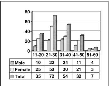
The mean age of onset of the entire group of 200 patients was 32.0±9.9 years (median 32); in women the mean age onset was 30.0±9.9 years (median 28,0) and men had a mean age of onset of 32.5±10.6 years (median 32). Figure 1 shows the distribution of number of cases per decade and ac-cording to gender. There was a larger proportion of women, who represented 64.5% (129 cases), giving a gender ratio of female 1,8:1 male. Only 3 patients
were African-Brazilian; there were no cases of Asi-atic ancestry. All other patients were white. Only 3 patients had a positive family history of MS (each one with one first degree affected relative).
Cerebrospinal fluid (CSF) examination was per-formed in 74 patients. It was normal in 43 cases; mononuclear hypercytosis occurred in 4 cases, in-creased CSF protein is 3 cases, and inin-creased gammaglobulin fraction (cellulose electrophoresis) or increased IgG index in 24 cases. HTLV-1 antibod-ies were tested in 10 patients and were negative in all samples. Blood and CSF VDRL were negative in all cases. Blood VDRL was tested in all 200 cases.
Visual evoked potential were performed in 36 patients, and it was abnormal and in keeping with a demyelinating disorder in 24 cases. Brainstem and somatosensory evoked potentials were obtained in 10 and 6 cases, respectively, and it was abnormal in only 2 cases for each test.
Twenty-one computed tomography (CT) scans were done and the findings were: normal in 9 pa-tients, findings in keeping with a white matter dis-ease in 8, enlarged cerebral sulci in 2, isolated calci-fication 1, and mild ventricular dilatation in 1.
Fourty-seven patients underwent head magnetic resonance imaging (MRI); in 43 (91.5%) the find-ings were compatible with MS6. Fifteen cervical spine
MRI were done and showed demyelinating lesions in 12 (80%). Six patients had both head and cervical MRI examination disclosing demyelinating lesions. In 4 cases, the head MRI was normal, but the cervi-cal spine MRI showed multiple lesions7. In 2 cases,
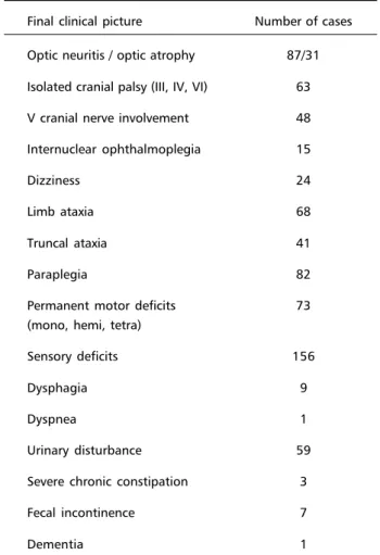
the head MRI was abnormal and the cervical spine examination was normal. Table 1 shows the initial clinical picture of this series. Brown-Séquard syn-drome at level T8 was the clinical initial picture in
only one patient. Seven (3.5%) patients developed a clinical history of Devic´s syndrome. Two patients developed their first relapse during pregnancy and puerperal period (each one). In six (3%) patients, bilateral optic neuritis occurred at onset.
Table 2 depicts the clinical findings at the last evaluation. More than one finding might have been present at that time.
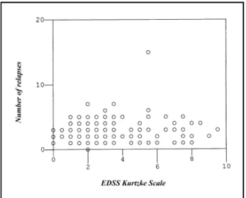
We observed that 122 (61%) patients had a final EDSS score < 3.5; 45 (22.5%) a score between 3.5 and 5.5, and 33 (16.5%) a score ≥ 6.0. There was
no correlation between the number of relapses or duration of disease with EDSS scores (Spearman’s test) (Figs 3 and 4). This observation did not change after the exclusion of benign cases. Only 14 (7%) of the total number presented the benign form accord-ing to the definition proposed by Weinshenker8
(EDSS< 3.5 after 10 years of disease).
Table 1. Initial syndromic presentation in 200 cases of MS.
Symptoms/signs Number of cases (%)
Brainstem/cerebellar 126 (63)
Sensory 106 (53)
Motor 99 (49.5)
Optic neuritis 79 (39.5)
Table 2. Clinical findings: last evaluation.
Final clinical picture Number of cases
Optic neuritis / optic atrophy 87/31
Isolated cranial palsy (III, IV, VI) 63
V cranial nerve involvement 48
Internuclear ophthalmoplegia 15
Dizziness 24
Limb ataxia 68
Truncal ataxia 41
Paraplegia 82
Permanent motor deficits 73 (mono, hemi, tetra)
Sensory deficits 156
Dysphagia 9
Dyspnea 1
Urinary disturbance 59
Severe chronic constipation 3
Fecal incontinence 7
Dementia 1
Table 3. Clinical features of other Brazilian series and present series.
Nr. Gender ratio Mean age Race Clinical form Poser criteria F:M of onset
Callegaro9 120 1.6:1 27.9±8.9* 79.2% W 85% RR Definite MS
10% M 10.2% PP
10% B 4.2% SP
0.8% Y
Leite10 51 2.1:1 34.5±13.9 62.7% W 47% RR 45 Definite MS
37.4% non-white 19.7% PP 6 Probable 33.3% “mixed”
Lana-Peixoto11 67 2.3:1 28.9±10.4** 76% W - Definite MS
19.4% M 4.5% B
Papaiz-Alvarenga12,13 88 3:1 27.9±11.3* 68.2% W 88.6% RR A - 74%
31.8% B 4.5% PP B - 5.3%
6.8 SP C - 17.8%
Tilbery14 214 2.9-1 28 (RR) 96% W 82 % RR Definite MS
36 (PP) 4.2%B 18% PP
0.5% Y
Oliveira15 50 2:1 32.5±9.6 64% W 60% RR 76% definite
34% M+B 30% PP 24% probable
2% Y 10% SP
Moreira16 302 3.1:1 37.7 94% W 72% RR Definite MS
5% B 14% PP
1% Y 14% SP
Present series 200 1.8:1 32.0±9.9 98.5% W 91% RR A - 61%
1.5% B 8% PP B – 21%
1% SP C – 8%
W, White; B, Black; M, Mulato; Y, Asian; RR, Relapsing-remitting form; PP, primary progressive form; SP, secondarily-progressive form. t Student test: *p<0,0005; **p<0.05, when compred with the present series.
Fig 3. Number of relapses and EDSS score. Spearman´s test showed no statistically significant correlation.
DISCUSSION
Table 3 and Figure 2 depicts some features from other Brazilian series and ours.
Comparison of clinical data from several retro-spective series of MS patients may be a daunting task9,10 for several reasons: first, ascertainment bias
probably play a major role in some observed ences; the reasons for that is, to some extent, differ-ent criteria of inclusion (though all series mdiffer-entioned the Poser criteria), different local characteristics of the Services, e.g. the study of Belo Horizonte11 is from
a strong referal Service of Neuro-ophthalmology; and different racial composition of these centers. For in-stance, the series from Rio de Janeiro12,13 has a much
larger proportion of black patients (Afro-Brazilian), whilst our own series showed a clear preponderance of white patients. In all series there is a higher pro-portion of female to male patients. Due to the marked racial miscigenation, it is even difficult to categorize some patients. In all series, a distinction of white Latinos and white Caucasian is not made. Nevertheless, there is a clear predominance of white race, as well as a similar female to male ratio. The relapsing-remitting form (RR) is the most common form, followed by primary progressive form. There is a marked difference of frequency in the PP forms – again, it is conceivable that this different is more related to bias of selection than other environmen-tal or racial factors. It is also conceivable that the use of different clinical criteria may explain to some extent this difference. In all series, including ours, there is a tendency for a later age of onset in the PP forms, and the female:male proportion tends to be equal.
Our group of patients presented a higher mean age of onset when compared to three other series11,13,14.This might be related to a longer interval
of time between onset of disease and clinical diag-nosis. The increasing availability of MR imaging tends to decrease this time gap.
In terms of clinical presentation, pyramidal find-ings and optic neuritis occur in approximately equal proportions in all series, except for a much higher frequency of pyramidal syndrome and lower fre-quency of optic neurits at disease’s onset in Oliveira’s series15. Perhaps their larger number of patients with
PP form may partially account for this difference (30% versus 8 to 19% in other series).
Brainstem/cerebellar syndromes and sensorial findings occured in much variable proportions in the
other series. Our high frequency of brainstem/cere-bellar symptomatology (63%) at disease’s onset in our patients might be owing to different registering methods of clinical findings rather than implying a true clinical heterogeneity between the clinical se-ries.
Neuromyelitis optica form has been reported to occur more frequently in African American and Asian type MS patients16,17. Papaiz-Alvarenga12,13, who had
the series with the largest proportion of African Bra-zilian (31.8%) patients reported 5.6% with this clini-cal form, Leite et al.10 had 15.6%, whereas
Lana-Peixoto11, with 24 % of African Brazilian patients,
reported a prevalence of 12% with Devic´s syndrome and suggested that his patients thus presented a clinical form more alike the Asian MS patients. We observed only 3.5% of our patients with this clinical form, and this observation may be explained by the very low proportion of either African-Brazilian and Asiatic elements. On the other hand, we had a high number of patients with optic neuritis as their initial symptom (39.5%). This frequency is somewhat higher than in other Western series and similar to some Brazilian series10,11 and Asian series. There is
no explanation for this observation based on the current available genetic racial data in our series. It seems pointless, at this time, to try defining our se-ries as having a Western or Asian pattern. Apart the methodological difficulties already mentioned, other racial/genetic and environmental factors may play a role in leading to these observations. In order to delineate the clinical profile of MS in Brazil, a pro-spective study about the natural history of MS is being conducted by the Brazilian Committee for Treatment and Research in Multiple Sclerosis (BCTRIMS).
The variation in the clinical presentation of MS and the course of the disease is well recognised. In most patients the condition has a RR course at first, but after a variable period progressive disability oc-curs, often with superimposed relapses (secondary progressive disease). Our extremely low number (1%) of patients with SP form conceivably reflects a short time of follow-up and bias of selection.
In about 30% of patients the disease follows a fairly benign course and appreciable disability has not developed 10 to 15 years after its onset. How-ever, using different criteria18, Moreira et al.16 found
19.8% of their patients with the benign form, com-pared to only 7% in our series8. The diagnosis of
time of individual follow-up in different Services may account for such differences.
The poor correlation of EDSS scale and time of disease and number of relapses observed in our se-ries reflects the difficult issue of prediction of up-coming disability and the therapeutic decisions de-rived from it19. EDSS Kurtzke scale remains by far
the most widely used scoring system in MS despite some perceived problems20,21. The development of
combined analysis of clinical rating systems, envolving some aspects of neurological function in more detail than the EDSS, with more reliable and consistent MRI techniques may improve the evalua-tion of progression of the disease20-22.
Kira et al.23 observed that DR2-associated
DRB1*1501 allele and DRB5*0101 allele were asso-ciated with Western-type MS, but not with Asian-type MS, in their Japanese MS patients. Alves-Leon24
studied 44 African-Brazilian MS patients living in Rio de Janeiro, and observed a significantly higher pro-portion of HLA-DQB1*0602 allele (MS 45% versus 17% controls). Papais-Alvarenga et al.25 recently
sho-wed that DR2 haplotype (DRB1*1501-DQB1*0602) also confered genetic suceptibility to white MS Bra-zilian patients in Rio de Janeiro. Finally, the mode of transmission of genetic susceptibility to MS is com-plex and might include other genes than the HLA system (a polygenic disease)26-28.
Acknowledgments - Cláudia S. Kamoy Kay, Hipólito Carraro Jr, Patrícia Coral, and Fábio Iwamoto contribute in collecting and organizing the clinical data
REFERENCES
1. Matthews WB, Compston A, Allen IV, Martyn CN (eds). Mc Alpine´s multiple sclerosis. 2 Ed. Edinburgh: Chuchill Livingstone, 1991. 2. Paty DW, Ebers GC. Multiple Sclerosis. Philadelphia: F.A Davis
Com-pany, 1998
3. Poser CM, Paty DW, Scheinberg L, et al.. New diagnostic criteria for multiple sclerosis: guidelines for research protocol. Ann Neurol 1983;13:227-231.
4. Thompson AJ, Montalban X, Barkhof F, et al. Diagnostic criteria for primary progressive multiple sclerosis: a position paper. Ann Neurol 2000;47:831-835.
5. Kurtzke JF. Rating neurologic impairment in multile sclerosis: na ex-panded disability status scale. Neurology 1983;33:1444-1452.
6. Paty DW, Noseworthy JH, Ebers GC. Diagnosis of multiple sclerosis. In: Paty DW, Ebers GC. Multiple sclerosis, Philadelphia: F.A. Davis Compay, 1998:48-123
7. Thorpe JW, Kidd D, Moseley IF, et al. Spinal MRI in patients with suspected multiple sclerosis and negative brain MRI. Brain 1996;119:709-714. 8. Weinshenker BG. The natural history of multiple sclerosis. Neurol Clin
1995;13:119-146.
9. Callegaro D. Contribuição ao estudo clínico evolutivo da esclerose múltipla: análise de 120 pacientes. Thesis, Universidade de São Paulo. São Paulo, 1989.
10. Leite ACCB, Andrade C, Novis S. Esclerose múltipla no Rio de Janeiro: apresentação clínica em 51 casos. Arq Neuropsiquiatr 1990;48(Supl):66A 11. Lana-Peixoto M, Lana-Peixoto MIV. Is multiple sclerosis in Brazil and
Asia alike? Arq Neuropsiquiatr 1992;50: 119-125.
12. Papais-Alvarenga RM, Santos CMM, Colin DD, et al. Esclerose múltipla: influência do sexo e etnia no perfil clínico de 88 pacientes no município do Rio de Janeiro. Rev Bras Neurol 1995;31:89-98.
13. .Papais-Alvarenga RM, Santos CMM, Abreu JS, et al. Esclerose múltipla: perfil clínico e evolutivo no município do Rio de Janeiro. Rev Bras Neurol 1995;31:75-87.
14. Tilbery CP, Felipe E, Baldauf CM, Peres MFP. Esclerose múltipla. Análise clínica e evolutiva de 214 casos. Arq Neuropsiquiatr 1995;53:203-207.
15. Oliveira EML, Annes M, Oliveira ASB, Gabbai AA. Esclerose múltipla.: estudo clínico de 50 pacientes acompanhados no Ambulatório de Neurologia UNIFESP-EPM.Arq. Neuropsiquiatr 1999;57:51-55. 16. Moreira MA, Felipe E, Mendes MF, Tilbery CP. Esclerose múltipla:
estudo descritivo de suas formas clínicas em 302 casos. Arq Neuropsiquiatr 2000;58:460-466.
17. Kuroiwa Y, Igata A, Itahara K, et al. Natiowide survey of multiple sclerosis in Japan: clinical analysis of 1,084 cases. Neurology 1975;25:845-848 18. Phillips PH, Newman NJ, Lynn MJ. Optic neuritis in African
Ameri-cans. Arch Neurol 1998;55:186-192
19. Thompson AJ, Hutchinson M, Brazil J, et al. A clinical and laboratory study of benign multiple sclerosis. Q J Med 1986;58:69-80.
20. Confraveux C, Vukusic S, Moreau T, Adeleine P. Relapses and progres-sion of disability in multiple sclerosis. N Engl J Med 2000;343:1430-1438. 21. Confraveux C, Vukusic S, Grimaud J, Moreau T. Clinical progression and decision making process in multiple sclerosis. Multiple Sclerosis 1999,5:212-215.
22. Amato MP, Ponziani G. Quantification of impairment in MS: discus-sion of the scales in use. Multiple sclerosis 1999,5:216-219.
23. Kira J, Knai T, Nichimura Y, et al. Western versus Asian types of mul-tiple sclerosis: immunogenetically and clinically distinct disorders. Ann Neurol 1996;40:569-574.
24. Alves-Leon SV. HLA DQB1*0602 confere susceptibilidade genética para esclerose múltipla numa população de pacientes Afro-Brasileiros da cidade do Rio de Janeiro, Thesis, Universidade Federal do Rio de Janeiro. Rio de Janeiro, 1999.
25. Papais-Alvarenga R, Alves-Leon SV, Caballero A, Alonso A, Fernadez O. DR2 haplotype confers genetic susceptibility to multiple sclerosis in white Brazilian patients. Communication. XIX Brazilian Congress of Neurology, Salvador, 2000.
26. Chataway J, Feakes R, Coraddu F, et al. The genetics of multiple scle-rosis: principles, background and updated results of the United King-dom systematic genome screen. Brain 1998;121:1869-1887.
27. Oturai A, Larsen F, Ryder LP, et al. Linkage and association analysis of susceptibility regions on chromosomes 5 and 6 in 106 Scandinavian sib-ling pair families with multiple sclerosis. Ann Neurol 1999;46:612-616. 28. Noseworthy JH, Lucchinetti C, Rodriguez M, Weinshenker BG.