Effe ct o f all-trans re tino ic acid
o n ne wly diagno se d acute
pro m ye lo cytic le uke m ia patie nts:
re sults o f a Brazilian ce nte r
Serviço de Transplante de Medula Ó ssea, Hospital de Clínicas, Universidade Federal do Paraná, Curitiba, PR, Brasil
B.C. de-Medeiros, E. Strapasson, R. Pasquini and C.R. de-Medeiros
Abstract
Thirty-seven patients with acute promyelocytic leukemia (APL) were treated with all-trans retinoic acid (ATRA). Patients received 45 mg m-2 day-1po of ATRA until complete remission (CR) was achieved, defined as: a) presence of less than 5% blasts in the bone marrow, with b) white blood cells >103/mm3, c) platelets >105/mm3 and d) hemoglobin concen-tration >8 g/dl, with no blood or platelet transfusions. Thirty-one (83.7%) patients achieved CR by day 50, and 75% of these before day 30. Correction of the coagulopathy, achieved between days 2 and 10 (mean, 3 days), was the first evidence of response to treatment. Only one patient had been previously treated with chemotherapy and three had the microgranular variant M3 form. Dryness of skin and mucosae was the most common side effect observed in 82% of the patients. Thrombosis, hepatotoxicity and retinoid acid syndrome (RAS) were observed in 7 (19%), 6 (16%) and 4 (11%) patients, respectively. Thirteen (35%) patients had to be submitted to chemotherapy due to hyperleukocytosis (above 40 x 103/mm3)and six of these presented with new signs of coagulopathy after chemotherapy. Four (11%) patients died secondarily to intracerebral hemorrhage (IH) and two (5.4%) dropped out of the protocol due to severe ATRA side effects (one RAS and one hepatotox-icity). RAS and IH were related strictly to hyperleukocytosis. The reduced use of platelets and fresh frozen plasma probably lowered the total cost of treatment. We conclude that ATRA is an effective agent for inducing complete remission in APL patients.
Co rre spo nde nce C.R. de-Medeiros
Serviço de Transplante de Medula Ó ssea, Hospital de Clínicas, UFPR R. General Carneiro, 181, 15o- andar 80060-900 Curitiba, PR
Brasil
Fax: + 55-41-264-5472
Received January 15, 1998 Accepted August 5, 1998
Ke y wo rds
•All-trans retinoic acid •Acute promyelocytic
leukemia
•Retinoid acid syndrome
Intro ductio n
Acute promyelocytic leukemia (APL), or M3 in the French-American-British classifi-cation, represents 5-15% of cases of acute myeloid leukemia (AML) (1). This distinct subset of AML is associated with the pres-ence of abnormal promyelocytes in the blood
promy-elocytic leukemia gene with the gene for retinoic acid receptor alpha (3). As demon-strated initially by Huang et al. (4), patients with APL have a high response rate to differ-entiation therapy with all-trans retinoic acid (ATRA), even though complete remission (CR) has not been sustained by ATRA therapy alone (5). One major advantage of the ATRA schedule is that it results in rapid disappear-ance of DIC (4), mainly through cytodiffer-entiation rather than immediate lysis of leu-kemic cells. However, some new life-threat-ening retinoid-induced complications, such as leukocytosis, retinoid acid syndrome (RAS) and thromboembolic events, have been demonstrated in a significant number of cases (6). In spite of several studies re-porting the efficacy of the differentiating therapy with ATRA, no reports have been published thus far in the Brazilian literature. The authors present 37 patients with APL from two major cancer centers from Curitiba, Brazil, where induction therapy, toxicity and treatment costs of ATRA were assessed.
Patie nts and Me tho ds
Patie nts and e ligibility crite ria
Between March 1992 and November 1995, 37 patients (Table 1) diagnosed with APL from Hospital de Clínicas and Hospital Nossa Senhora das Graças, Curitiba, were included in a clinical trial to evaluate the efficacy of ATRA in inducing CR and its toxicity. Patients or their legal representa-tives gave informed consent to participate in the experimental study, which was approved by the Ethics Commissions of both hospi-tals. Inclusion criterion was a diagnosis of APL based on FAB group morphology crite-ria (7). Although cytogenetic evaluation for t(15;17) was attempted for most patients, demonstration of the PML-RARα protein was not required as a criterion for eligibility. Prior to the beginning of ATRA therapy, complete coagulation studies and normal
hepatic and renal function tests were required.
Pro to co l de sign
Patients received 45 mg m-2 day-1po of
ATRA (Tretinoína, Roche Pharmaceuticals, São Paulo, Brazil) until day 50 of ATRA or refractoriness was demonstrated. When the coagulopathy was still present beyond the 10th day of treatment, ATRA was increased to 90 mg m-2 day-1po for 5 additional days. If
no correction of DIC occurred, aggressive chemotherapy was started. CR was defined on days +30 or +50 of ATRA therapy on the basis of peripheral blood counts and bone marrow aspiration and biopsy studies as: 1) presence of less than 5% blasts in the bone marrow, with 2) white blood cells >103/
mm3, 3) platelets >105/mm3 and 4)
hemoglo-bin concentration >8 g/dl, with no blood or platelet transfusions. Red blood cell transfu-sion was used to maintain hemoglobin levels above 8 g/dl and platelet transfusion was performed to maintain platelet count above 20 x 103/mm3. Fresh frozen plasma and
cryo-precipitate transfusions were performed in patients with any evidence of coagulopathy (6). Patients with severe thrombotic phe-nomena such as pulmonary embolism and large vein thrombosis received heparin 5000 U iv q4h for 10 days, followed by heparin 5000U sc bid until the end of the ATRA therapy. Those who reached white blood cell count (WBC) above 40 x 103/mm3 were
managed with cytosine arabinoside (500 mg/ m2, iv) and daunomycin (60 mg/m2, iv) in
Labo rato ry fo llo w-up
Daily complete blood cell counts (CBC) and coagulation studies were performed af-ter initiating the ATRA therapy until stabili-zation of leucocytes and correction of coagulopathy, respectively. The studies were repeated once a week until day +30 or +50 of ATRA therapy. A bone marrow aspirate was obtained on days +30 and +50 to check for CR. Hepatic and renal function tests were performed weekly during hospitalization and twice a month thereafter.
Endpo ints
The major endpoints of this study were: 1) intolerance to ATRA, 2) maintenance of the coagulopathy beyond day 15 of ATRA therapy, 3) no CR after day 50, and 4) resis-tance to ATRA.
Re sults
Re spo nse to the rapy
CR was achieved in 31 (83.7%) patients, by day 30 of ATRA treatment in 23 of them and by day 50 in 8, and no patient had the drug discontinued because of resistant dis-ease. Correction of the coagulopathy was the first evidence of response to ATRA and was detected in 34 patients between days 2 and 10 (mean, 3 days). Thirteen patients had to be submitted to chemotherapy due to el-evated WBC (above 40 x 103/mm3)and six
of these presented with new signs of coagu-lopathy after chemotherapy. Four (10.8%) patients died secondarily to central nervous system bleeding on days +4, +5, +7 and +18. Two had early death while on ATRA therapy only and two died after chemotherapy due to elevated WBC, and all four had leucocytes above 40 x 103/mm3 at the time of death.
Two patients dropped out of the protocol due to severe ATRA side effects (RAS in one and hepatotoxicity in the other). Median
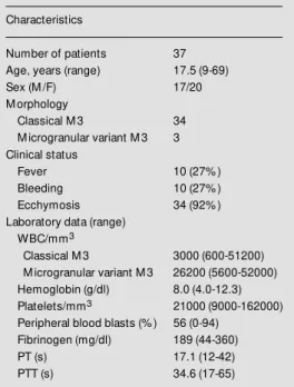
Table 1 - Characterization of the patient population.
Characteristics
Number of patients 37
Age, years (range) 17.5 (9-69)
Sex (M /F) 17/20
M orphology
Classical M 3 34
M icrogranular variant M 3 3 Clinical status
Fever 10 (27% )
Bleeding 10 (27% )
Ecchymosis 34 (92% )
Laboratory data (range) WBC/mm3
Classical M 3 3000 (600-51200) M icrogranular variant M 3 26200 (5600-52000) Hemoglobin (g/dl) 8.0 (4.0-12.3) Platelets/mm3 21000 (9000-162000)
Peripheral blood blasts (% ) 56 (0-94) Fibrinogen (mg/dl) 189 (44-360)
PT (s) 17.1 (12-42)
PTT (s) 34.6 (17-65)
WBC was 2.4 x 10³/mm3 (range 0.6 to 52 x
103/mm3) for all patients at diagnosis,
reach-ing a maximum of 24.3 x 10³/mm3 (range 4.2
to 100.6 x 10³/mm3) in all patients alive
between days +6 and +19 (mean, +15 days) after initiating ATRA treatment.
To xicity
The most common side effect of ATRA therapy was dryness of skin and mucosae in 82% (28) of the patients, which developed from the first to the third week. Severe throm-bosis was observed in 7 (19%) patients, but only one had a WBC above 20 x 103/mm3.
RAS was diagnosed in 4 (11%) patients on days +3, +5, +10 and +15 of ATRA treat-ment. One of these patients received chemo-therapy ordered by the attending physician instead of the proposed RAS treatment, and was excluded from the protocol. These pa-tients presented a maximum median WBC of 48 x 103/mm3, compared to 22.5 x 10³/mm3
severe intrahepatic cholestasis with liver function tests reaching values 8 times higher than the normal upper limit and was with-drawn from the protocol.
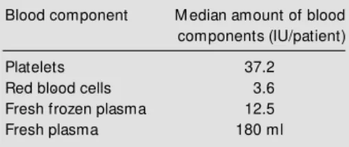
Suppo rtive care and ho spitalizatio n
Hospitalization varied from 0 to 50 days (mean, 19 days). The amount of blood com-ponents utilized during this time is summa-rized in Table 2. The amount of platelets and fresh frozen plasma transfused in the ATRA group was significantly less than for the chemotherapy group, 31.7 IU versus 135 IU (P = 0.05) and 100 ml versus 1,330 ml (P<0.01), respectively. Thirteen patients had to be managed with antibiotics (one due to periodontitis, one due to tonsillitis, another due to scrotal sac cellulitis and the rest due to fever of unknown origin during the leuko-penic period).
D iscussio n
APL is a distinct subgroup of AML with specific clinical and molecular features. Over the last few years, the introduction of ATRA has opened new perspectives for the treat-ment and understanding of several of its leukemic mechanisms. This study shows the differentiating capacity of ATRA as an in-ductor of CR, with 83.7% of the newly diag-nosed patients achieving CR during the course of treatment. Such remission rates are similar to those obtained in several other randomized trails with ATRA (4,10) and also to the historical results obtained with
standard chemotherapy (11).
DIC is the most common complication of APL and its severity and frequency are often aggravated by chemotherapy, as was the case for 6 of our patients. Although patients sub-mitted to chemotherapy frequently reach CR, they usually need intensive blood support to prevent bleeding, attributed to the release of both procoagulatory and fibrinolytic sub-stances from damaged malignant cells, low fibrinogen levels and worsening of thrombo-cytopenia. In our own service, we compared historical data of 28 patients treated with chemotherapy alone from 1985 to 1992 with those for the group treated with ATRA. The first evidence of a response to ATRA in our patients was correction of the coagulopathy (mean, 3 days), with a rapid resolution of fibrinolysis and fibrinogenopenia, conse-quently reducing the amount of blood com-ponents needed and the possible implica-tions of hemorrhagic diathesis, as reported by several other authors (9,12,13).
Leukocytosis, an adverse effect of ATRA therapy in APL (14), which occurred in 37% of our patients, is due to an increase in circulating maturing blast cells secondary to cytodifferentiation rather than leukemic cell lysis, and is not observed during con-ventional intensive chemotherapy. The high number of circulating blasts increases blood viscosity and is associated with leukoblastic emboli, resulting in leukostasis of the small vessels. Furthermore, the leukemic blasts can infiltrate the arteriolar endothelial walls and cause a secondary hemorrhage (15). Therefore, a major concern about APL pa-tients treated with ATRA is early death secondary to hyperleukocytosis during in-duction treatment (11,16), especially due to RAS and intracerebral hemorrhage. In our study, 4 of 37 patients (11%) had an early death due to intracerebral hemorrhage, with a higher median WBC than in the group that did not develop this lethal complication (48 x 103/mm3 versus 22.5 x 10³/mm3). RAS
also occurred only in patients whose
leuko-Table 2 - Utilization of blood components in ATRA patients.
Blood component M edian amount of blood components (IU/patient)
Platelets 37.2
Red blood cells 3.6
Fresh frozen plasma 12.5
cytes were above 40 x 103/mm3 (4 of 13
patients).
In order to avoid hyperleukocytosis and its complications, the French group designed a protocol whose major goal was to prevent the increase in leukocyte counts by the early use of chemotherapy in combination with ATRA (17,18), but this strategy barely im-proved their CR rates. Vahdat et al. (19), analyzing the concept of full-dose chemo-therapy based on the peripheral blood leuko-cyte count proposed by the French group, demonstrated that chemotherapy actually contributes to excess early mortality due to the reappearance of the bleeding diathesis, a fact that was also observed in the present study. Indicating how hard it is to establish an ideal leukocyte parameter for the combi-nation of early chemotherapy with ATRA, several Chinese reports have found a very low incidence of RAS despite very high leukocyte counts (4,5,20). In agreement with Fenaux et al. (21), we may speculate about the presence of genetic predisposition to RAS among different populations, explain-ing these intriguexplain-ing differences.
In our group, 3 of the 4 patients with RAS were managed with a short course of high-dose dexamethasone and had their abnor-malities corrected, just as demonstrated by others (8,22). Based on these results, the serious complications of RAS have been almost entirely eliminated with a liberal policy of steroid administration, substan-tially reducing mortality. We note, however, that this approach mandates careful patient monitoring, particularly with respect to fluid balance and renal function. As a step ahead in the management of these patients, how would one avoid the occurrence of RAS? Strategies such as prophylactic corticoste-roids (22,23), concomitant use of ATRA and hydroxyurea (22) and lower doses of ATRA (20) (15 to 25 mg/m2) have been applied
with acceptable results, but not yet evaluated in randomized trials.
As in other subtypes of acute leukemias,
APL patients show a strict relationship be-tween leukocytosis and severe hemorrhage. In our group, 4 patients died of intracerebral hemorrhage and all had leukocyte counts above 40 x 103/mm3 at the time of bleeding,
versus a 22 x 103/mm3 leukocyte count in
patients who did not present this complica-tion. Since all our patients were submitted to the same approach in terms of platelet trans-fusion and the coagulation abnormalities had been corrected at time of death, we conclude that hyperleukocytosis is probably involved in the etiology of retinoid-induced intracere-bral hemorrhage.
ATRA therapy rapidly corrects the bleed-ing diathesis and normalizes the fibrinogen levels of APL patients. However, a proco-agulant tendency persists due to the exist-ence of low grade DIC which accounts for the thrombotic events seen as late as 1 month after ATRA therapy (24) and for the 10% hemostatic death rate reported among these patients (25). Furthermore, patients treated with conventional chemotherapy alone rarely develop thromboembolic events (2). Seven patients in whom we clinically detected these phenomena during ATRA therapy were suc-cessfully managed with heparin. In spite of some reports implicating thromboembolism in leukocytosis (26,27), no relationship was noticed between these complications in our patients, and only one had leukocytes above 20 x 103/mm3 at the time of thrombosis.
Some authors even recommend the prophy-lactic administration of low molecular weight heparin for one month in order to avoid thrombotic events in these patients (24).
be considered predictable, so that avoiding leukocytosis above 20 x 103/mm3 would
sig-nificantly reduce the possibility of RAS and probably of intracerebral hemorrhage. Care-ful clinical monitoring of these patients would also allow an early diagnosis of thrombotic
Re fe re nce s
1. Stone RM & M ayer RJ (1990). The unique aspects of acute promyelocytic leukemia. Journal of Clinical Oncology, 8: 1913-1921.
2. Tallman M S & Kw aan HC (1992). Reas-sessing the hemostatic disorder associ-ated w ith acute promyelocytic leukemia. Blood, 79: 543-548.
3. Takatsuki H, Umemure T, Sadamura S, Yamashita S, Goto T, Abe Y, Yufu Y, Inaba S, Nishimura J & Naw ata H (1995). Detec-tion of minimal residual disease by verse transcriptase polymerase chain re-action for the PM L/RARα fusion mRNA: a study in patients w ith acute promyelo-cytic leukemia follow ing peripheral stem cell transplantation. Leukemia, 9: 889-892.
4. Huang M E, Ye YC, Chen SR, Chai JR, Lu JX, Zhao L, Gu LJ & Wang ZY (1988). Use of all-trans retinoic acid in the treatment of acute promyelocytic leukemia. Blood, 72: 567-572.
5. Chen ZX, Xue YQ, Zhang R, Tao RF, Xia XM , Li C, Wang W, Zu WY, Yao XZ & Ling BJ (1991). A clinical and experimental study on all-trans-retinoic acid-treated acute promyelocytic leukemia patients. Blood, 78: 1413-1419.
6. Fenaux P, Le Deley M C, Castaigne S, Archimbaud E, Chomienne C, Link H, Guerci A, Duarte M , Daniel M T, Bow en D, Huebner G, Bauters F, Fegueux N, Fey M , Sanz M , Low enberg B, M aloisel F, Auzanneau G, Sadoun A, Gardin C, Bas-tion Y, Ganser A, Jacky E, Dombret H, Chastang C & Degos L (1993). Effect of all transretinoic acid in new ly diagnosed acute promyelocytic leukemia. Results of a multicenter randomized trial. Blood, 82: 3241-3249.
7. Bennet JM , Cat avsky D, Daniel M T, Flandrin G, Galton D, Gralnick M & Sultan C (1976). Proposals for the classification of the acute leukemias. British Journal of Haematology, 33: 451-459.
8. Frankel SR, Eardley A, Lauw ers G, Weiss M & Warrel RP (1992). The ‘retinoic acid syndrome’ in acute promyelocytic leuke-mia. Annals of Internal M edicine, 117:
292-296.
9. Fleiss JL (1981). Statistical M ethods for Rates and Proportions. 2nd edn. John Wiley and Sons, New York.
10. Warrel RP, Frankel SR, M iller W, Itri L, Andreeff M , Jabulow ski A, Gabrilove J, Gordon M S & Dmitrovsky E (1991). Dif-ferentiation therapy of acute promyelo-cytic leukemia w ith tretinoin (all trans retinoic acid). New England Journal of M edicine, 324: 1385-1394.
11. Cordonnier C, Vernant JP, Brun B, Heilm ann M G, Kuentz M , Bierling P, Farcet JP, Rodet M , Duedari N, Imbert M , Jouault H, M annoni P, Reyes F, Dreyfus B & Rochant H (1985). Acute promyelocytic leukemia in 57 previously untreated pa-tients. Cancer, 55: 18-28.
12. Leong KW, Bosco JJ & Teh A (1994). Advantage of induction therapy w ith all-trans retinoic acid in acute promyelocytic leukemia in a country w ith limited transfu-sion resources: a M alaysian experience. European Journal of Haematology, 53: 237-241.
13. Eardley AM , Heller G & Warrell Jr RP (1994). M orbidity and costs of remission induction therapy w ith all-trans retinoic acid compared to standard chemotherapy in acute promyelocytic leukemia. Leuke-mia, 8: 934-939.
14. Castaigne S, Chomienne C, Daniel M T, Berger R, Fenaux P & Degos L (1990). All trans-retinoic acid as a differentiation therapy for acute promyelocytic leukemia. I. Clinical results. Blood, 80: 2176-2181. 15. M ckee Jr LC & Collins RD (1974).
Intra-vascular leukocyte thrombi and aggre-gates as a course of mortality and morbid-ity in leukemia. M edicine, 52: 463-468. 16. Drapkin RL, Timothy SG, Dow ling M D,
Arlin Z, M ackenzie S, Kem pin S & Clarkson B (1978). Prophylatic heparin therapy in acute promyelocytic leukemia. Cancer, 41: 2484-2491.
17. Fenaux P, Castaigne S, Chomienne C, Dombret H & Degos L (1992). All-trans retinoic acid treatment for patients w ith acute promyelocytic leukemia. Leukemia, 6: 64-67.
18. Fenaux P, Cast aigne S, Dom bret H, Archimbaud E, Duarte M , Chomienne C & Degos L (1992). All-trans retinoic acid fol-low ed by intensive chemotherapy gives a high complete remission rate and may prolong remissions in new ly diagnosed acute promyelocytic leukemia: a pilot study on 26 cases. Blood, 80: 2176-2181. 19. Vahdat L, M aslak P, M iller Jr WH, Eardley A, Heller G, Scheinberg DA & Warrel Jr RP (1994). Early mortality and the retinoic acid syndrome in acute promyelocytic leu-kemia: Impact of leukocytosis, low -dose chemotherapy, PM N/RAR-α isoform, and CD13 expression in patients treated w ith all-trans retinoic acid. Blood, 84: 3843-3849.
20. Chen GQ, Shen ZX, Wu F, Han JY, M iao JM , Zhong HJ, Li XS, Zhao JQ, Zhu J, Fang ZW, Chen SJ, Chen Z & Wang ZY (1996). Pharmacokinetics and efficacy of low -dose all-trans retinoic acid in the treat-ment of acute promyelocytic leukemia. Leukemia, 10: 825-828.
21. Fenaux P, Chastang C & Degos L for the French APL group (1994). Treatment of new ly diagnosed acute promyelocytic leu-kemia (APL) by a combination of all-trans retinoic acid (ATRA) and chemotherapy. Leukemia, 8: S42-S47.
22. Tallman M S, Andersen JW, Schiffer CA, Appelbaum FR, Feusner JH, Ogden A, Sheperd L, Willman C, Bloomfield CD, Row e JM & Wiernik PH (1997). All-trans retinoic acid in acute promyelocytic leuke-mia. New England Journal of M edicine, 337: 1021-1028.
23. Wiley JS & Firkin FC (1995). Reduction of pulmonary toxicity by prednisolone pro-phylaxis during all-trans retinoic acid treat-ment of acute promyelocytic leukemia. Leukemia, 9: 774-778.
24. Degos L (1994). Is acute promyelocytic leukemia a curable disease? Treatment strategy for a long-term survival. Leuke-mia, 8: S6-S8.
25. Rodeghiero F & Castaman G (1994). The pathophysiology and treatment of hemor-rhagic syndrome of acute promyelocytic leukemia. Leukemia, 8: S20-S26.
26. Escudier SM , Kantarjian HM & Estey EH (1996). Thrombosis in patients w ith acute promyelocytic leukemia treated w ith and w ithout all-trans retinoic acid. Leukemia and Lymphoma, 20: 435-439.
27. Wang ZY, Chen Z, Huang W, Li XS, Lu JX,
Huang LA, Zhang FQ, Gu LJ, Ouyang RR & Chen SJ (1993). Problems existing in differentiation therapy of acute promyelo-cytic leukemia (APL) w ith all-trans retinoic acid (ATRA). Blood Cells, 19: 633-641. 28. Tallman M S, Andersen JW, Schiffer CA,
Brasil
Carlos Eduardo Rocha-Miranda
Vice President, Academia Brasileira de Ciências Rua Anfilófio de Carvalho, 29, 3º andar 20030-060 Rio de Janeiro, RJ, Brasil Tel.: 220-4794/Fax: 240-4695 E-mail: cerm@abc.org.br
Argentina
Israel Algranati
Instituto de Investigaciones Bioquímicas Fundación Campomar Av. Patricias Argentinas, 435 1405 Buenos Aires Tel: 863-4018/Fax: 865-2246 e-mail: algra@iris.iib.uba.ar
Mexico
Hugo Aréchiga
Facultad de Medicina UNAM Ciudad Universitaria 04510 Mexico, D.F.
Tel: 622-0725/Fax: 550-8859 e-mail: arechiga@servidor.unam.mx
Chile
Manuel A. Kukuljan, Ph.D.
Universidad de Chile
Departamento de Fisiologia y Biofisica Casilla 70005 Santiago 7, Chile Tel: 2-678-6310/Fax: 2-777-6916 e-mail: kukuljan@bitmed.med.uchile.cl
All other countries
Silvia Montano de Jiménez
The Pew Latin American Fellows Program 3333 California Street, Suite 410 San Francisco, CA 94118
Deadline for applications: O ctober 1, 1999
The Pew Latin Am erican Fellows Program in the Biom edical Sciences is providing support for young scientists from Latin Am erica for post-doctoral training in the United States.
Ten Fellows will be selected in
1999. An award of $50,000 will be provided as a salary stipend for the fellow during the period of training (2 years) and will be administered by the sponsoring U.S. institution. The sponsoring institution is expected to supplement the stipend with at least $5,000 a year and provide medical benefits for the fellow. Following the two year fellowship, the Program will issue an additional $35,000 award to the sponsoring institution to purchase equipment and supplies for the fellow to establish a laboratory in his or her home country.
Applicants must have held a Ph.D. and/or M.D. degrees, or equivalent, for no more than five years as of July 1, 1999. Strong preference will be given to those applicants with no previous postdoctoral training outside of their home country. Applicants are not required to have a commitment of a position and laboratory space after the fellowship. However,
applicants must submit a written statement of intent to return to Latin America. Fellows must have a confirmed position and laboratory space in their home country by the end of the fellowship period in order to obtain the $35,000 portion of the award.
Fellows will be selected on the basis of their promise as outstanding investigators, as well as the scientific merit of their research proposal, their record of training and how well their interests coincide with the laboratory of their sponsor in the United States. If potential applicants need assistance with the identification of an appropriate sponsoring laboratory in the United States, they may contact the Program O ffice before August 1, 1999. The Program will accept applicants from Mexico, Central and South America. Applications may be obtained from the Regional Committee contact listed here for your country or from our website at
http://futurehealth.ucsf.edu/pewlatin.htm l
The application deadline is O ctober 1, 1999. Winners will be notified in April 2000 and the fellowship should begin no later than August 2000.
FELLOWS
PROGRAM
in the
BIOMEDICAL
S . C . I . E . N . C . E . S
PEW
Latin American