online | memorias.ioc.fiocruz.br
Bioactive endophytic fungi isolated from
Caesalpinia echinata
Lam.
(Brazilwood) and identification of beauvericin as a
trypanocidal metabolite from
Fusarium
sp.
Fernanda Fraga Campos1,2, Policarpo A Sales Junior2, Alvaro José Romanha2,3, Márcio SS Araújo2, Ezequias P Siqueira2, Jarbas M Resende4, Tânia MA Alves2, Olindo A Martins-Filho2,
Vera Lúcia dos Santos5, Carlos A Rosa5, Carlos L Zani2, Betania Barros Cota2/+
1Departamento de Ciências Biológicas e da Saúde, Universidade Federal dos Vales do Jequitinhonha e Mucuri, Diamantina, MG, Brasil 2Centro de Pesquisas René Rachou-Fiocruz, Belo Horizonte, MG, Brasil 3Departamento de Microbiologia, Imunologia e Parasitologia,
Universidade Federal de Santa Catarina, Florianópolis, SC, Brasil 4Departamento de Química, Instituto de Ciências Exatas 5Departamento de Microbiologia, Instituto de Ciências Biológicas, Universidade Federal de Minas Gerais, Belo Horizonte, MG, Brasil
Aiming to identify new sources of bioactive secondary metabolites, we isolated 82 endophytic fungi from stems and barks of the native Brazilian tree Caesalpinia echinata Lam. (Fabaceae). We tested their ethyl acetate extracts in several in vitro assays. The organic extracts from three isolates showed antibacterial activity against Staphylococcus aureus and Escherichia coli [minimal inhibitory concentration (MIC) 32-64 μg/mL]. One isolate inhibited the growth
of Salmonella typhimurium (MIC 64 μg/mL) and two isolates inhibited the growth of Klebsiella oxytoca (MIC 64 μg/
mL), Candida albicans and Candida tropicalis (MIC 64-128 μg/mL). Fourteen extracts at a concentration of 20 μg/mL
showed antitumour activities against human breast cancer and human renal cancer cells, while two isolates showed anti-tumour activities against human melanoma cancer cells. Six extracts were able to reduce the proliferation of hu-man peripheral blood mononuclear cells, indicating some degree of selective toxicity. Four isolates were able to inhibit
Leishmania (Leishmania) amazonensis and one isolate inhibited Trypanosoma cruzi by at least 40% at 20 μg/mL. The
trypanocidal extract obtained from Fusarium sp. [KF611679] culture was subjected to bioguided fractionation, which
revealed beauvericin as the compound responsible for the observed toxicity of Fusarium sp. to T. cruzi. This depsipep-tide showed a half maximalinhibitory concentration of 1.9 μg/mL (2.43 μM) in a T. cruzi cellular culture assay.
Key words: endophytic fungi - bioactive - Caesalpinia echinata Lam. - Fabaceae - Trypanosoma cruzi - beauvericin
Neglected tropical diseases (NTDs) and cancer are disorders that generate a high global burden and novel therapies for these disorders are needed (Ehrenberg & Ault 2005, Farmer et al. 2010). Although a large number of antibiotics have saved hundreds of millions of lives over the last few years, the increase of opportunistic in-fections and antimicrobial resistance to drugs used in the clinic have contributed to the challenges faced by medi-cine in curing infectious diseases (Fauci & Morens 2012). The treatment of some cancers is palliative and this is not only a problem for the developed world (Farmer et al. 2010). The influence of small-molecules approved as drugs between 1981-2010, as natural products (N), and small-molecules directly derived from N (ND), is quite marked in the treatment of cancers (N = 11.1%, ND = 32.3%) and infectious diseases (N = 6.2%, ND = 40.9%) (Newman & Cragg 2012). NTDs, especially Chagas dis-ease and leishmaniasis, affect poor and vulnerable groups
doi: 10.1590/0074-02760140243
Financial support: CNPq, FAPEMIG, FIOCRUZ + Corresponding author: [email protected] Received 9 September 2014
Accepted 28 November 2014
and working to discover new medicines is not an attrac-tive endeavour for pharmaceutical companies (Ehrenberg & Ault 2005). Several challenges in the treatment of these diseases, such as the ability of the drugs used clinically to cause toxic effects and their effectiveness at chronic stages, have not been overcome (Feasey et al. 2010).
The Caesalpinia genus (Leguminosae, Caesalpin-ioideae) includes approximately 130 species occurring in the tropics (Larsen et al. 1980, Lewis 1998). Caesal-pinia echinata Lam. (Fabaceae) is an endangered species occurring in a highly threatened ecosystem. C. echinata
is a native tree from Brazil that was the main source of red pigment in the XVI century during colonisation by Portugal and its popular name was given to the new land discovered when Portuguese navigators arrived in South America (Oliveira et al. 2002).
Endophytic fungi colonise all plant tissues (Petrini et al. 1992, Rodriguez et al. 2009, Vaz et al. 2009, Campos et al. 2011) and based on estimates, many fungal spe-cies and their secondary metabolites have not yet been described (Hyde & Soytong 2007, Hyde et al. 2007). The analysis of data recorded by PubMed and SciFinder in the last five years has revealed promising drug candi-dates from endophytic fungi that could be useful for dif-ferent therapeutic applications.
fo-cused on the investigation of the endophytic fungi living in the tissues of C. echinata as sources of bioactive natu-ral products that could be used against some neglected diseases. In this work, we describe the taxonomic identi-fication of the fungal isolates that produced biologically active extracts and the identification of beauvericin as the trypanocidal component of Fusarium sp. extract.
SuBJECTS, MATERiALS And METhOdS
Plant material - Healthy stems and barksof C. echi-nata were collected in Zoo-Botanical Foundation, Belo Horizonte (FZB-BH), state of Minas Gerais, Brazil, in March 2008. A voucher specimen was deposited at the FZB-BH Herbarium under the code BHZB-6458.
Endophytic fungi isolation and storage - Plant samples were collected in plastic bags and taken to the laboratory for processing. Plant material was washed in tap water, allowed to dry at room temperature (RT) and cut into pieces of approximately 1 × 1 cm. The surface of the fragments were sterilised by immersion in 70% ethanol (1 min) and 2% sodium hypochlorite (3 min), followed by one wash with sterile distilled water (2 min) (Collado et al. 1996). The fragments were plated onto potato dextrose agar (PDA) (Difco, USA) plates (Merck) containing 0.1 g/L chloramphenicol (Sigma, USA). The plates were incubated for up to 60 days at 25ºC and indi-vidual colonies were transferred to PDA. After complete growth, these colonies were photographed. Stock fungal cultures were deposited in the Culture Collection of croorganisms and Cells of the Federal University of Mi-nas Gerais. Fungal mycelial pieces were preserved at RT in sterile distilled water containing 30% v/v of glycerol.
Cultivation and extraction of the fungal cultures - Pieces of fungi mycelia (5 mm diameter) were transferred to five Petri dishes containing 20 mL of malt extract agar (PDA, Difco) medium (malt extract 1%, glucose 1%, pep-tone 0.1% and agar 1% in 1 L of purified water) and were cultured for 14 days at 28ºC. The biomass of the fungi mycelia were extracted by maceration with ethyl acetate for 48 h at RT. After passing through filter paper, the sol-vents were evaporated under reduced pressure using a ro-tary evaporator at 45ºC. Residual solvent in the extracts was eliminated in a vacuum centrifuge at 40ºC.
Antimicrobial activity assays - Antimicrobial activity was evaluated using the following microorganisms from the American Type Culture Collection (ATCC) (USA):
Staphylococcus aureus ATCC 25295, Escherichia coli
ATCC 18804, Bacillus cereus ATCC 11778, Klebsiella oxytoca ATCC 49131, Salmonella typhimurium ATCC 14028, Pseudomonas aeruginosa ATCC 27853, Candida albicans ATCC 18804 and Candida tropicalis ATCC 750.
Bacterial strains were maintained on brain heart in-fusion agar (Difco). Yeast strains were maintained on Sabouraud dextrose agar (Oxoid, UK).
Culture media and inocula - Mueller Hinton Broth (Himedia, India) was prepared in accordance with the Clinical and Laboratory Standards Institute (CLSI) document M7-A6 for minimal inhibitory concentra-tion (MIC) bacterial assays (NCCLS 2003). Inocula of
all bacteria were obtained using the spectrophotomet-ric method prescribed by CLSI M7-A6, with final con-centrations of 5 x 10⁵ colony-forming unit (CFU)/mL. Candida cultures were grown at 35ºC and their inocula were prepared from fresh cultures according to the CLSI document M27-A2 (CLSI 2008). For the susceptibility tests, the final concentration was 1.5 x 10³ CFU/mL.
Af-ter homogenisation by vortexing, the transmittance was measured at 520 nm and was adjusted to 69-70%.
Susceptibility test - The broth microdilution tests for bacteria and yeast were performed following the CLSI guidelines of M7-A6 and M27-A2, respectively. Susceptibility to antimicrobial agents was determined by the microbroth dilution method performed in sterile flat-bottom 96-well microplates (Difco). Fungal extracts were dissolved in dimethyl sulphoxide (DMSO) followed by the addition of Mueller Hinton Broth for bacterial as-says and RPMI for yeast asas-says. Eight serial dilutions (2-256 µg/mL) were prepared using the corresponding media as the diluents and maintaining a constant volume of 1 mL in each tube. For each dilution, aliquots of 0.1 mL were distributed in the microplates. For growth and sterility control, media alone was used without the addi-tion of extract and solvent. As a control for toxicity of the solvent, culture with DMSO was used. Chlorampheni-col (Sigma-Aldrich) (0.78-100 µg/mL) was used as the positive antibacterial control and amphotericin B (AMB) (Sigma-Aldrich) (0.03-15 µg/mL) was used as the positive antifungal control. After the assembly of the plates, each bacterial and fungal strain was inoculated and the plates were incubated at 37ºC for 24 h for bacteria and 48 h for
Candida species. Endpoints were determined visually by comparing the growth in the sample wells to the growth in drug-free control wells. MIC measurements were de-fined as the lowest sample concentration for which the well was optically clear and were expressed in µg/mL.
cytocidal from 100-200%. Etoposide (ETO) at 1.6 µg/ mL, culture medium without samples and culture me-dium with DMSO 1% (v/v) were used as controls.
In vitro assay with human peripheral blood mononu-clear cells (PBMC) - PBMC isolation from venous blood
- Venous blood from healthy adult volunteers was col-lected in heparinised tubes and centrifuged over a Ficoll-Hypaque cushion (Histopaque, Sigma). PBMC were col-lected from the Ficoll-Hypaque interphase and washed three times with RPMI-1640 medium (Gibco, USA). An aliquot of the cells was incubated with trypan blue (0.4% in NaCl 0.9%) and the viability of the cells was evaluated by visual inspection under a microscope. The cell sus-pensions were adjusted to 1.5 x 10⁶ cell/mL and cultured
in RPMI-1640 medium supplemented with 5% (v/v) heat-inactivated, pooled human sera type AB (Flow Labora-tories, Royaune-UNI) and L-glutamine (2 mM) (Gibco). An antibiotic/antimycotic solution containing 1 mg/mL penicillin, 1 mg/mL streptomycin and 25 µg/mL fungi-sone (Sigma) was added to control for fungal and bacte-rial contamination. In vitro cellular proliferation (blasto-genesis) was assessed as previously described (Gazzinelli et al. 1983). Briefly, 1.5 x 10⁵ cells were cultured in
com-plete RPMI-1640 in flat-bottomed microtitre plates (Co-star, tissue culture treated polystyrene # 3512, Sigma). The cultures were stimulated with 2.5 µg/mL of lectin from Phaseolus vulgaris phytohaemagglutinin (PHA) (Sigma) and incubated for 72 h at 37°C in a humidified atmosphere containing 5% CO2. Cell proliferation was determined using Alamar Blue according to the manu-facturer’s recommendations (Invitrogen, cat. DAL1100). The experiments were repeated three times using differ-ent samples of blood. Allopurinol and dexamethasone at 20 µg/mL were used as controls on PHA stimulated PBMC cultures. The results were expressed as percent inhibition of the PHA stimulated lymphocyte prolifera-tion in relaprolifera-tion to the control (no extracts added).
In vitro assay with human PBMC - The assay was performed as above, except that the culture was not stimulated with PHA. Cell toxicity was determined us-ing Alamar Blue followus-ing the manufacturer’s protocol (Invitrogen, cat. DAL1100). ETO at 20 µg/mL was used as the positive (toxic) control. The cytotoxic activity was evaluated by comparing the PBMC cultures with and without fungi extracts.
Assays with Leishmania (Leishmania) amazonensis amastigotes-like forms - Leishmanicidal activity was de-termined against amastigote-like forms of the parasite, which were obtained as previously described (Callahan et al. 1997). Briefly, promastigotes of L. (L.) amazonensis
(strain IFLA/BR/196/PH-8) were obtained from lesions of infected hamsters. The parasites were grown at 26ºC in Schneider’s medium (pH 7.2) and then stimulated to differentiate into amastigote forms by raising the temper-ature (32ºC) and lowering the pH (6.0) of the Schneider’s medium. After seven days under these conditions, 90% of the promastigotes were transformed into amastigote-like forms, verified by microscopy and they were then used in the bioassays. The amastigote density was adjusted to 1 x
108 parasites per mL and 90 μL was added to each well of the 96-well plates. Solutions of the test samples at 200 μg/
mL containing DMSO 1% (v/v) in water were performed
for each sample and then, 10 μL of the solution were
added to each well of the 96-well plates. The plates were incubated at 32ºC for 72 h and then, cell viability was determined using the methyl thiazolyl tetrazolium assay (Teixeira et al. 2002). The results were calculated from the measured absorbencies using the formula [1-(Abs exp/Abs contr) x 100], which expresses the percentage of parasite death in relation to the controls without drug.
AMB at 0.02 μg/mL (Fungison®, Bristol-Myers Squibb,
Brazil) was used as a positive drug control.
Assays with Trypanosoma cruzi amastigote and try-pomastigote forms - This assay was performed using T. cruzi (Tulahuen strain) expressing E. coli β-galactosidase as a reporter gene (Buckner et al. 1996, Romanha et al. 2010). Infective trypomastigote forms were obtained by monolayer culture of mouse L929 fibroblasts in RPMI-1640 medium (pH 7.2-7.4) without phenol red (Gibco) and with 10% foetal bovine serum and 2 mM glutamine. For
the bioassay, 4,000 L929 cells in 80 μL of supplemented
medium were added to each well of a 96-well microtitre plate. After an overnight incubation, 40,000
trypomas-tigotes in 20 μL were added to the cells and incubated for
2 h. The medium containing extracellular parasites was
replaced with 200 μL of fresh medium and the plate was
incubated for an additional 48 h to establish the infection. The medium was then replaced with solutions of the test
samples at a concentration of 20 μg/mL in DMSO (< 1%
in aqueous RPMI-1640 medium) and the plate was in-cubated for 96 h. To determine the half maximal inhibi-tory concentration (IC50) values, the cells were exposed to active samples at serial decreasing dilutions starting at
20 μg/mL and the IC50 values were calculated by linear interpolation. After this period, 50 μL of 500 μm chloro -phenol red β-D-galactopyranoside in 0.5% Nonidet P-40 was added to each well and the plate was incubated for 16-20 h, after which the absorbance at 570 nm was mea-sured. Controls with uninfected cells, untreated infected cells, infected cells treated with benznidazole (BNZ) at
1 μg/mL (positive control) and cells treated with DMSO
1% were used. Tetraplicates were run in the same plate and the experiments were repeated at least once.
The cytotoxicities of beauvericin and BNZ on unin-fected mouse L929 fibroblasts were obtained. The IC50 values were calculated by linear interpolation and the se-lectivity index (SI) values were determined based on the ratio of the IC50 value in the host cell divided by the IC50 value of the parasite (Romanha et al. 2010).
Statistical analysis - The samples were tested in qua-druplicate in the T. cruzi assays and in triplicate in the other assays. At least two independent experiments were per-formed. Values represent the mean ± variation coefficient.
was based on the internal transcribed spacer-ribosomal DNA (ITS-rDNA) sequences. The pair of primers ITS1
(sequence: 5′-TCCGTAGGTGAACCTGCGG-3′) and ITS4 (5′-TCCTCCGCTTATTGATATGC-3′) was used
for ITS-rDNA amplification (White et al. 1990). The se-quences were generated using MEGABACE (Amersham Biosciences, USA), which were used to feed PHRED-PHRAP software to find the consensus sequence. The sequence was then compared with those deposited in GenBank using BLASTN software to identify the isolate down to the genus level. All fungal ITS-rDNA sequenc-es obtained in this work were deposited in the GenBank with accessions KF611676-KF611689 (Table I).
Mass cultivation and extraction of Fusarium sp. [working code (WC) 9] - Pieces of mycelia (5 mm di-ameter) were transferred to 30 Petri dishes containing 20 mL of PDA and were cultured for 14 days at 28ºC. The fungal biomass was placed in Erlenmeyer flasks and extracted by maceration with ethyl acetate for 48 h. The suspensions were filtered through filter paper and the filtrate evaporated to dryness under reduced pres-sure using a rotary evaporator at 45ºC. The residue was transferred to 20 mL flasks and the residual solvent was removed in a vacuum centrifuge at 40ºC for 18 h.
Bioassay-guided fractionation of the Fusarium sp. extract - The extract obtained (110 mg) was dissolved in 1 mL of a mixture of methanol (MeOH) and water (MeOH: H2O, 75:25 v/v). After centrifugation, the solu-tion was injected into a semi-preparative reverse phase high-performance liquid chromatographic (RP-HPLC) column [250 mm × 4.6 mm internal dimension (i.d.)], 5
μm particle diameter) using a Shimadzu chromatograph
(Shimadzu Corp, Japan) equipped with a LC6AD pump
and manual injection valve (Rheodyne™ 7125, Rheo-dyne Co, USA) using a fixed 1.000 µL sample loop and a dual-wavelength detector (SPD M10A) controlled by LCsolution software v.1.25 (Shimadzu Corp). The sam-ple was purified with a linear gradient of water (A) and MeOH (B) using 10%B-100%B for 50 min and 100%B for 10 min. The eluent was pumped at 7 mL/min and the
effluent absorption measured at λ 220 nm and 254 nm.
Ten fractions corresponding to different peaks were col-lected and tested in the T. cruzi assay. Fraction (Fr)-5 (17 mg, 95% pure by HPLC) was the most active.
Spectral data of the trypanocidal Fr-5 - Proton (1H)
and carbon nuclear magnetic resonance (NMR) spectra, distortionless enhancement by polarisation transfer, het-eronuclear single quantum coherence and hethet-eronuclear multiple bond coherence (HMBC) experiments were performed on a Brucker DRX 400 spectrometer using the pulse programs provided by the manufacturer. The substance was dissolved in perdeuterated solvents doped with 0.1% tetramethyl silane as the internal standard.
Liquid chromatography-diode-array detection-mass spectrometry (LC-DAD-MS) analysis of Fr-5 was per-formed in a Thermo Surveyor Plus (Thermo Fisher Sci-entific, USA) chromatograph equipped with a Finnigan Surveyer PDA Plus diode-array detector and C18 column
(Atlantis C18, Waters, USA) (3 μm particle diameter, 150 mm × 2.1 mm i.d.). A flow rate of 200 μL/min was used
and the effluent entirely directed the Bruker ETD-maX-is quadrupole TOF (Bruker Daltonics, Germany) for electrospray ionisation (ESI) in the positive ion mode. The LC-DAD-MS was conducted in a gradient system using a mixture of water (A) and MeOH (B) with 0.1% formic acid, 1%B-100%B for 13 min, 100%B for 4 min, 100%B-1%B for 0.5 min, 1%B for 11.5 min.
TABLE I
Identification of endophytic fungi isolated from Caesalpinia echinata Lam. (Fabaceae) using primers internal transcribed spacer (ITS)1 and ITS4
WC Closest related species
Similarity (%)
Base pairs analysed (n)
Identification and GenBank accessions
25 Aspergillus sp. [KF367538.1] 100 523 Aspergillus sp. [KF611682]
45 Epicoccum sorghi [KC106717.1] 100 461 E. sorghi [KF611685]
46 E. sorghi [KC106698.1] 100 509 E. sorghi [KF611686]
9 Fusarium sp. [JQ905668.1] 100 357 Fusarium sp. [KF611679]
58 Fusarium sp. [HM631978.1] 99 481 Fusarium sp. [KF611688]
2 Nectria pseudotrichia [JN995626.1] 100 495 N. pseudotrichia [KF611677]
6 N. pseudotrichia [JF832647.1] 100 360 N. pseudotrichia [KF611678]
33 N. pseudotrichia [JN995626.1] 100 509 N. pseudotrichia [KF611683]
24 Talaromyces sp. [JX898040.1] 100 566 Taralomyces sp. [KF611681]
1 Xylaria arbuscula [JN601145.1] 99 497 X. arbuscula [KF611676]
11 Xylaria sp. [DQ322134.1] 96 515 Xylaria sp. [KF611680]
41 Xylaria berteri [JQ936300.1] 100 411 X. berteri [KF611684]
55 Xylaria sp. [JQ862693.1] 100 473 Xylaria sp. [KF611687]
84 Xylaria sp. [KC507252.1] 99 516 Xylaria sp. [KF611689]
The mass detector was set to the mass-to-charge ra-tio(m/z) range of 50-1500 atomic mass units. The instru-ment was operated under the following conditions: end plate offset, -500 voltage (V); capillary V, 4.500 V; nebu-liser pressure, 0.4 bar; dry gas (nitrogen) flow rate, 4.0 L/min; dry temperature, 180ºC; collision-induced disso-ciation energy, 25 eV; collision energy, 7 eV; ion cooler
radio-frequency, 25 excitation V; transfer time, 45 μs.
Fr-5 (beauvericin): white powder; specific optical ro-tation = + 47 [concentration at g/100 mL (c) 0.8, MeOH]; ultraviolet (UV) (DAD, MeOH) maximum wavelength
(λmax) 202 nm.
1H NMR [deuterated MeOH (CD 3OD),
400 megahertz (MHz)] chemical dislocation in ppm (δ)H 7.26-7.24 [12H, multiplet (m), H-10/H-11/H-13/H-14], 7.17 (3H, m, H-12), 5.45 [3H, doublet of doublets (dd), coupling constant(J) = 10.9 and 3.9, Hz, H-7], 4.92 [3H, dublet (d), J = 8.6 Hz, H-1], 3.36 (3H, dd, J = 14.5 and 5.0 Hz, H-8b), 2.99 [9H, singlet (s), H-6], 2.97 (3H, dd, J
= 11.8 Hz, H-8a), 2.01 (1H, td, J = 21.2, 6.8 and 6.7 Hz, H-2), 0.78 (9H, d, J = 6.6 Hz, H-3), 0.43 (9H, d, J = 6.7 Hz, H-4). Carbon-13 (13C) NMR (CD
3OD, 100 MHz): δC
170.2 (C, C-5), 169.7 (C, C-15), 136.9 (C, C-9), 129.1 and 128.8 (CH, C-10/C-11/C-13/C-14), 127.0 (CH, C-12), 75.7 (CH, C-1), 57.6 (CH, C-7), 35.0 (CH2, C-8), 32.6 (CH3, C-6), 29.9 (CH, C-2), 18.5 (CH3, C-3), 17.7 (CH3, C-4); high resolution-ESI-MS m/z 784.4180 [M + H]+ (calcd.
for C45H57N3O9, 783.4095).
High performance LC coupled to an UV detec-tor (HPLC-DAD) analysis - The ethyl acetate extract from Fusarium sp. (WC 9)and Fr-5 (beauvericin) were analysed by HPLC-DAD in a Shimadzu chromatograph (Shimadzu Corp) equipped with a LC10AD pump and manual injection valve (Rheodyne™ 7725i, Rheodyne Co) using a fixed 20 µL sample loop, a CTO-20A ther-mostat-controlled oven compartment and a SPD-M20A diode array detector (190-800 nm) controlled by LCsolu-tion software. A Shim-pack® C18 column (5 μm, 250 mm
× 4.6 mm i.d.) maintained at 40ºC was used in the chro-matographic analysis. The separations were conducted in a gradient system, using a mixture of water (A) and acetonitrile (ACN) (B) with 0.1% trifluoroacetic acid (TFA), 10%B-100%B for 30 min and 100%B for 10 min as the mobile phase, at a flow rate of 1.0 mL/min. The ethyl acetate extract (5 mg/mL) and Fr-5 (beauvericin) (200 µg/mL) were dissolved in MeOH. The particulates were removed by centrifugation and the sample injection
volume was 20 μL for each sample.
RESuLTS
Isolation and molecular identification of endophytic fungi from C. echinata Lam. - Eighty-two endophytic fungal strains were isolated from plant bark (10 samples) and stems (13 samples) (Table II). Fourteen fungal isolates
yielded extracts that were active in vitro at 20 μg/mL in
at least one biological assay. Based on the results of their ITS1-5.8S-ITS4 partial sequences, these isolates were sub-mitted to the GenBank to obtain their accession numbers and the closest related species were achieved by BLAST analysis. The results (Table I) show that all sequences had more than 96% similarity with the species in GenBank. Most sequences presented 99% (n = 3) or 100% (n = 10) similarity to the closest related species in GenBank.
All 14 isolates belong to the Ascomycota phylum, with 10 from the Sordariomycetes class, two from the Eurotiomycetes class and two from the Dothideomycet-es class. Among the fungi that produced active extracts, five were from the Xylaria genus (35%) and three were from the Nectria genus (21%). Fusarium and Epicoccum
afforded two active extracts each (14%) and Taralomy-ces and Aspergillus afforded one active extract each (7%) (Tables III, IV).
Biological activities of extracts from endophytic fungi isolated of C. echinata Lam. - The ethyl acetate extracts of 82 fungal isolates from C. echinata Lam.
were tested in in vitro biological assays to predict their leishmanicidal, trypanocidal, cytotoxic and antimicrobi-al activities. Forty-four fungantimicrobi-al isolates were considered
active (≥ 40% of the inhibition of growth) in at least one
biological assay (Tables III, IV).
The extracts from Talaromyces sp. (WC 24), Aspergil-lus sp. (WC 25) and Epicoccum sorghi (WC 45) showed antibacterial activity against positive and Gram-negative bacterial species (S. aureus and E. coli) with MIC
values ranging from 32-64 μg/mL. Aspergillus sp.extract
(WC 25) showed an MIC value of 64 μg/mL against S. ty-phimurium and K. oxytoca. Talaromyces sp. extract (WC
24) showed an MIC of 64 μg/mL against K. oxytoca. An-tifungal activity was only observed for the extracts from
Fusarium sp. (WC 9)and Nectria pseudotrichia (WC 33), which inhibited C. albicans and C. tropicalis at
concen-trations ranging from 64-128 μg/mL (Table III).
Four isolates, Fusarium sp. (WC 9), Xylaria sp. (WC 11), N. pseudotrichia (WC 33) and Fusarium sp. (WC 58), were able to inhibit the growth (45-77%) of the amastigote forms of L. (L.) amazonensis. However, only Fusarium sp. (WC 9) inhibited (92%) the amastigote and trypomastig-ote forms of T. cruzi when tested at 20 μg/mL (Table IV).
Fourteen fungal isolates exhibited cytotoxicity to-ward MCF-7 and TK-10 cell lineages, inhibiting their growth by at least 40%. In addition, the extracts of N. pseudotrichia (WC 33) and E. sorghi (WC 45) inhibited the growth of UACC-62. The extracts of tree fungi ( Xy-laria sp., WC 11; Aspergillus sp. WC 25 and N. pseudo-trichia,WC 33) displayed cytocidal activity at 20 μg/ mL; in other words, the number of viable cells was less than in the initial inoculum (Table IV). Although all 14 isolates showed some degree of cytotoxicity against
three tumour cell lineages at 20 μg/mL,only one was
TABLE II
Number of isolates from different plant parts
Plant part
Samples (n)
Fungi isolated (n)
Morphotypes (n)
Barks 10 34 14
Stems 13 48 10
-cytotoxic to human PBMCs and six were able to reduce the PHA stimulated proliferation of PBMCs (Table IV).
Chemical characterisation of the trypanocidal com-pound of the Fusarium sp. extract and HPLC-DAD anal-ysis - To identify the trypanocidal component of Fusa- rium sp. (WC 9), the ethyl acetate extract from this strain was produced at a larger amount and was submitted to semi-preparative RP-HPLC fractionation. The fractions were tested in the intracellular T. cruzi assay and only Fr-5 was able to kill 100% of the T. cruzi amastigote and
trypomastigote forms at a concentration of 5 μg/mL. The
HPLC chromatogram of Fr-5 showed a single peak (Fig. 1A) with UV purity index of approximately 95%. The spectral data of Fr-5 were in full agreement with those reported for beauvericin (Fig. 1B) (Newman 2008, Hu & Rychlik 2012). The UV spectra exhibited absorptions
between 196-230 nm (λmax 202 nm), which is consistent
with previous reports (Monti et al. 2000, Mahnine et al. 2011). The HRMS of Fr-5showed a quasi-molecular [M + H]+ ion peak at m/z 784.4180, which is consistent with
the molecular formula of: C45H57N3O9 (calcd. 783.4095) (Fig. 1C). The 13C NMR spectrum showed 15 carbon
sig-nals, which together with the mass spectra analysis sug-gested a symmetrical structure for Fr-5. The 1H and 13C
NMR spectra of Fr-5 showed signals of aromatic carbons and 1H (δ
H 7.26-7.17, δC 127.0-129.1) from a benzyl moi-ety (δH 3.36, dd, J = 14.5 and 5.0 Hz/δC 35.0) due to the
phenylalanine residues. The ESI-(+)-LC-MS/MS spectra of Fr-5 (Fig. 1C) showed fragment ions at m/z 262.1439 and 244.1324 produced by the cleavage of the phenyla-lanine amide bond followed by loss of H2O, as observed in beauvericin (Sewram et al. 1999, Hu & Rychlik 2012). The hydroxy-isovaleryl moiety showed 1H signals at δ
H
4.92 (1H, d, J = 8.6 Hz/ δC 75.7), δH 2.01 (1H, td, J = 21.2,
6.8 and 6.7 Hz/ δC 29.9), δH 0.78 (3H, d, J = 6.6 Hz/δC 18.5) and δH 0.43 (3H, d, J = 6.7 Hz/δC 17.7). In addition,
the N-methylamino acid moiety of beauvericin matches
the signals of Fr-5 at δ 2.99 (3H, s, δC 32.6, N-CH3).
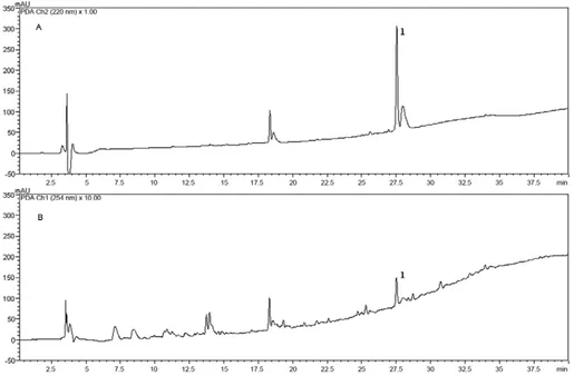
The ethyl acetate extract from Fusarium sp. (WC 9) was analysed using RP-HPLC-DAD and beauvericin (1) eluted after 27.5 min (Fig. 2) using a mixture of ACN and water with 0.1% TFA.
Trypanocidal and cytotoxicity activities of the beau-vericin - While the crude extract of Fusarium sp. WC 9 showed an IC50 of 30 μg/mL in the assay with T. cruzi
forms expressing the β-galactosidase gene, Fr-5 (beau-vericin) showed an IC50 value 15 times smaller (1.9 μg/
mL, 2.43 μM). This compound showed cytotoxic activ
-TABLE III
In vitro antimicrobial activities of extracts from endophytic fungi of Caesalpinia echinata Lam. (Fabaceae)
Fungal isolate (WC)
Microorganisms
Minimal inhibitory concentration (MIC)
(μg/mL)
SA EC BC ST PA KO CA CT
Aspergillus sp. (25) 32 64 > 256 64 > 256 64 > 256 > 256
Epicoccum sorghi (45) 64 32 > 256 > 256 > 256 > 256 > 256 > 256 E. sorghi (46) > 256 > 256 > 256 > 256 > 256 > 256 > 256 > 256
Fusarium sp. (9) > 256 > 256 > 256 > 256 > 256 > 256 64 128
Fusarium sp. (58) > 256 > 256 > 256 > 256 > 256 > 256 > 256 > 256 Nectria pseudotrichia (2) > 256 > 256 > 256 > 256 > 256 > 256 > 256 > 256 N. pseudotrichia (6) > 256 > 256 > 256 > 256 > 256 > 256 > 256 > 256
N. pseudotrichia (33) 64 > 256 > 256 > 256 > 256 > 256 128 128
Talaromyces sp. (24) 32 64 > 256 > 256 > 256 64 > 256 > 256
Xylaria arbuscula (1) > 256 > 256 > 256 > 256 > 256 > 256 > 256 > 256 Xylaria sp. (11) > 256 > 256 > 256 > 256 > 256 > 256 > 256 > 256 Xylaria berteri (41) > 256 > 256 > 256 > 256 > 256 > 256 > 256 > 256 Xylaria sp. (55) > 256 > 256 > 256 > 256 > 256 > 256 > 256 > 256 Xylaria sp. (84) 64 > 256 > 256 > 256 > 256 > 256 > 256 > 256
Controls
Amphotericin B NT NT NT NT NT NT 0.12 1.2
Chloramphenicol 16 8 16 16 8 8 NT NT
BC: Bacillus cereus; CA: Candida albicans; CT: Candida tropicalis; EC: Escherichia coli; KO: Klebsiella oxytoca; NT: not tested; PA: Pseudomonas aeruginosa; SA: Staphylococcus aureus; ST: Salmonella typhimurium; WC: working code. Values in
ity against the host cell (mouse L929 fibroblasts) used in the T. cruzi assay, showing an IC50 of 5 μg/mL (6.38 μM). Thus, under our assay conditions, beauvericin showed an SI of only 2.7. BNZ was used as the standard and showed an IC50 value of 3.8 μM and an SI value of 625 against mouse L929 fibroblasts.
diSCuSSiOn
In our previous work (Cota et al. 2011), we found that the crude ethanol extract of C. echinata Lam. kills 90% of the amastigote-like forms of L. amazonensis at a con-centration of 20 µg/mL. This promising result prompted us to continue to study C. echinata as a host plant of potential bioactive endophytic fungi. Previous work by other groups identified 43 taxa belonging to Hyphomy-cetes and three belonging to CoelomyHyphomy-cetes in leaf lit-ter of C. echinata Lam. (Grandi & Silva 2003, 2006). They reported the presence of Epicoccum nigrum as an
anamorphic fungi (Grandi & Silva 2006). In the present study, we identified two isolates (E. sorghi; WC 45 and 46) of the same genus as bioactive endophytic fungi.
Few reports are available on the biological activi-ties of fungi growing in C. echinata. Machado (2009) isolated Botryosphaeria rhodina, Xylaria multiplex and
Pestalotiopsis sp. as endophytic fungi from the leaves and stems of C. echinata. Although none of these iso-lates were active against Enterococcus faecalis, P. aeruginosa or S. aureus by agar diffusion assay (100 μg and 1,000 μg), they were able to inhibit the growth of the
phytopathogens Pythium debaryanum and Phytoththora palmivora (Machado 2009).
Most of the fungi identified in the present work have been previously reported as endophytic in other plants.
Xylaria, Nectria and Aspergillus genera are found in
Piper aduncum (Piperaceae) and many other plants in Brazilian savannas (Martínez-Luis et al. 2011, Vaz et
TABLE IV
In vitro antiprotozoan, cytotoxic and antiproliferative activities of extracts from endophytic fungi of Caesalpinia echinata Lam. (Fabaceae)
Fungal isolate (WC)
Tumour cell lineages (%)
PBMC (%)
Protozoan (%)
UACC-62 TK-10 MCF-7 Mortality
Proliferation
decreased LA TC
Aspergillus sp. (25) - 103 ± 9 - - - -
-Epicoccum sorghi (45) 75 ± 7 47 ± 10 67 ± 9 - - -
-E. sorghi (46) - 97 ± 5 60 ± 17 - 15 ± 7 -
-Fusarium sp. (9) - 98 ± 5 88 ± 4 - 15 ± 8 45 ± 0 92 ± 4
Fusarium sp. (58) - - 48 ± 10 49 ± 26 - 45± 4
-Nectria pseudotrichia (2) - 54 ± 14 68 ± 4 NT NT -
-N. pseudotrichia (6) - 95 ± 7 93 ± 0 NT NT -
-N. pseudotrichia (33) 102 ± 7 96 ± 15 60 ± 11 - 37 ± 15 77 ± 3
-Talaromyces sp. (24) - 95 ± 9 60 ± 7 - 20 ± 13 -
-Xylaria arbuscula (1) - 60 ± 1 57 ± 9 - - -
-Xylaria sp. (11) - 113 ± 3 47 ± 8 NT NT 51 ± 1
-Xylaria berteri (41) - 43± 6 - - 20 ± 15 -
-Xylaria sp. (55) - 92 ± 4 58 ± 0 - - -
-Xylaria sp. (84) - 51±9 48 ± 8 NT NT -
-Controls
AMB NT NT NT NT NT 82 ± 3 NT
BNZ NT NT NT NT NT NT 86 ± 8
ETO 176 ± 9 185 ± 9 100 ± 5 33 ± 14 NT NT NT
DEX NT NT NT 18 ± 13 - NT NT
ALL NT NT NT - 21 ± 14 NT NT
all extracts were tested at 20 μg/mL. Results were expressed in terms of percentage of the inhibition. ALL: allopurinol tested at 20 μg/mL; AMB: amphotericin B tested at 0.02 μg/mL; BNZ: benznidazole tested at 1.0 μg/mL = 3.8 μM; DEX: dexamethasone tested at 20 μg/mL; ETO: etoposide tested at 1.6 μg/mL in tumour cell lineages and at 20 μg/mL in human peripheral blood
Fig. 2: high performance liquid chromatographic coupled to an ultraviolet (UV) detector profile of ethyl acetate Fusarium sp. (working code 9). UV detection at 220 nm (A) and 254 nm (B). Column RP-18, 250 mm × 4.6 mm i.d.; mobile phase (A: H2O; B: acetonitrile) with 0.1% trifluoroacetic
acid; 10%B-100%B in 30 min, 100%B in 10 min, flow rate of 1.0 mL/min. The ethyl acetate extract (5 mg/mL) and beauvericin (1,200 µg/mL). Fig. 1: total-ion chromatogram (A) of beauvericin (B). Column RP-18, 150 mm × 2.1 mm i.d.; mobile phase [A: H2O; B: methanol) with 0.1%
formic acid; 1%B-100%B in 13 min, 100%B in 4 min, 100%B-1%B in 0.5 min, 1%B in 11.5 min flow rate 200 μL/min-1. Electrospray
ionisation-(+)-MS/MS of (B) (precursor m/z 784.4179 [M + H]+) and main fragments (C).
al. 2012). In addition, Fusarium species are the most frequent endophytes (Liang et al. 2012). The other two genera described in this paper, Taralomyces and Epicoc-cum, were recently found to be endophytic (Fávaro et al. 2012, Bara et al. 2013).
Our results show that, overall, approximately 17% of our fungal isolates were active in at least one of the four bioassays performed. Recently, Higginbotham et al.
(2013) showed that fungi isolated from the plant family Fabaceae (Fabales) had a high percentage of highly tive genotypes and were associated with moderate ac-tivity against Plasmodium falciparum and MCF-7 cells (breast cancer cell line) when tested at a concentration of 10 µg/mL. Moreover, extracts from fungi of Aspergillus
against Leishmania donovani, T. cruzi and MCF-7 cells (Higginbotham et al. 2013). The results of our biological assays lead us believe that all isolates tested, except for the isolate Fusarium sp. (WC 58), which showed high toxicity to the PBMC in vitro, are potential sources of compounds useful in the development of drugs against infectious agents and immunomodulatory metabolites.
In the present study, the fungi Fusarium sp. [KF611679] was the only one that showed activity against T. cruzi amastigote and trypomastigote forms and exhibited the best MIC values against C. albicans
and C. tropicalis. Several Fusarium species isolated from plants are known to produce secondary metabo-lites, such as terpenoids, alkaloids and mycotoxins, with promising biological activities (Hyde & Soytong 2007, Hyde et al. 2007, Campos et al. 2012).
The trypanocidal activity of the fungi extract [KF611679] was attributed to beauvericin. Beauveri-cin is a mycotoxin produced by many fungi, including
Fusarium spp (Wang & Xu 2012). Beauvericin displays insecticidal (Hamill et al. 1969), antitumour (Cheng et al. 2009), antibacterial, antifungal and antiviral activities (Zhan et al. 2007, Shin et al. 2009, Meca et al. 2010, Xu et al. 2010). Beauvericin was also reported to have leish-manicidal activity (EC50 1.86 μM) against promastigotes of Leishmania braziliensis (Nascimento et al. 2012). To the best of our knowledge, this is the first report on the trypanocidal activity of this cyclic hexadepsipeptide. Our
results support those of previous studies (Klarić et al.
2008, Nascimento et al. 2012) that also showed that this compound was cytotoxic; we obtained an IC50 of 5 μg/mL
(6.38 μM) against the host cell (mouse L929 fibroblasts)
used in a T. cruzi assay. Under our assay conditions, beau-vericin showed an SI of only 2.7, a value that, according to current guidelines (Romanha et al. 2010), is too low for beauvericin to be considered for pre-clinical studies.
Notwithstanding, according to a recent review (Feud-jio et al. 2010), beauvericin-mediated cytotoxicity to-wards various mammalian and cancer cell lines is only partially understood and involves several cellular targets and molecular mechanisms. Furthermore, only a few studies have addressed the effects of beauvericin in ani-mals and those studies have found only minor acute toxic effects. The authors emphasised that the consequences of chronic exposure and of pharmacologically active doses of beauvericin in humans/animals have not been explored in detail. Therefore, the biological activities of beauveri-cin on mammalian cancer cells and protozoan parasites suggest that beauvericin is a potential drug candidate for the treatment of cancers and infectious diseases. There is a need for further studies to determine the efficacy and safety of beauvericin in animals infected with T. cruzi.
In conclusion, this work demonstrated the in vitro leishmanicidal, trypanocidal, antimicrobial and cytotox-ic activities of crude extracts prepared from endophytcytotox-ic fungi isolated from stems and barks of C. echinata. In ad-dition, the bioassay-guided fractionation of Fusarium sp. (WC 9) extract using the T. cruzi assay allowed us to iden-tify the cyclic hexadepsipeptide mycotoxin beauvericin as the trypanocidal component produced by the fungus.
ACknOwLEdgEMEnTS
To the PDTIS-Fiocruz, for use of its facilities, to FZB-BH, to provide the plant material, mainly Juliana O Rego, Inês R de Andrade, Albina CO Nogueira and Maria Guadalupe C Fernandes, and to Daniela NB Maia, Priscila AM Ferreira and Markus Kohlhoff, for the technical support.
REFEREnCES
Bara R, Aly AH, Pretsch A, Wray V, Wang B, Proksch P, Debbab A 2013. Antibiotically active metabolites from Talaromyces wort-mannii, an endophyte of Aloe vera. J Antibiot (Tokyo) 66: 491-493.
Buckner FS, Verlinde CL, La Flamme AC, Van Voorhis WC 1996. Efficient technique for screening drugs for activity against Try-panosoma cruzi using parasites expressing β-galactosidase.
Anti-microb Agents Chemother 40: 2592-2597.
Callahan HL, Portal AC, Devereaux R, Grogl M 1997. An axenic amastigote system for drug screening HL. Antimicrob Agents Chemother 41: 818-822.
Campos FF, Johann S, Cota BB, Alves TMA, Rosa LH, Caligiorne RB, Cisalpino PS, Rosa CA, Zani CL 2011. Antifungal activity of trichothecenes from Fusarium sp. against clinical isolates of
Paracoccidioides brasiliensis. Mycoses 54: 122-129.
Campos FF, Siqueira EP, Cota BB 2012. Natural products from
Fusarium. In FR Rios, ER Ortega, Fusarium: epidemiology, en-vironmental sources and prevention, Nova Science Publishes, New York, p. 1-74.
Cheng CK, Chang KC, Lee YJ 2009. Antiproliferative effect of beau-vericin on retinoblastoma. Fu-Jen J Med 7: 161-169.
CLSI - Clinical and Laboratory Standards Institute 2008. Reference method for broth dilution antifungal susceptibility testing of yeast. Approved Standard M27-A3, CLSI, Wayne, 25 pp.
Collado J, Platas G, Peláez F 1996. Fungal endophytes in leaves, twigs and bark of Quercus ilex from Central Spain. Nova Hedwigia 63: 347-360.
Cota BB, Oliveira DM, Siqueira EP, Souza-Fagundes EM, Pimenta AM, Santos DM, Rabello AL, Zani CL 2011. New cassane diter-penes from Caesalpinia echinata. Fitoterapia 82: 969-975.
Ehrenberg J, Ault S 2005. Neglected diseases of neglected popula-tions: thinking to reshape the determinants of health in Latin America and the Caribbean. BMC Public Health 5: 119.
Farmer P, Frenk J, Knaul FM, Shulman LN, Alleyne G, Armstrong L, Atun R, Blayney D, Chen L, Feachem R, Gospodarowicz M, Gralow J, Gupta S, Langer A, Lob-Levyt J, Neal C, Mbewu A, Mired D, Piot P, Reddy KS, Sachs JD, Sarhan M, Seffrin JR 2010. Expansion of cancer care and control in countries of low and mid-dle income: a call to action. Lancet 376: 1186-1193.
Fauci AS, Morens DM 2012. The perpetual challenge of infectious diseases. N Engl J Med 366: 454-461.
Fávaro LCL, Sebastianes FLS, Araújo WL 2012. Epicoccum nigrum
P16, a sugarcane endophyte, produces antifungal compounds and induces root growth. PLoS ONE 7: e36826.
Feasey N, Wansbrough-Jones M, Mabey DCW, Solomon AW 2010. Neglected tropical diseases. Br Med Bull 93: 179-200.
Feudjio FT, Dornetshuber R, Lemmens M, Hoffmann O, Lemmens-Gruber R, Berger W 2010. Beauvericin and enniatin: emerging toxins and/or remedies? World Mycotoxin J 3: 415-430.
periph-eral blood mononuclear cells from treated, but not active cases of schistosomiasis. J Immunol 130: 2891-2895.
Grandi RAP, Silva TV 2003. Hyphomycetes sobre folhas em decom-posição de Caesalpinia echinata Lam.: ocorrências novas para o Brasil. Rev Bras Bot 26: 489-493.
Grandi RAP, Silva TV 2006. Fungos anamorfos decompositores do folhedo de Caesalpinia echinata Lam. Rev Bras Bot 29: 275-287.
Hamill RL, Higgens GE, Boaz HE, Gorman M 1969. The structure of beauvericin, a new desipeptide antibiotic toxic to Artemia salina.
Tetrahedron Lett 49: 4255-4258.
Higginbotham SJ, Arnold AE, Ibañez A, Spadafora C, Coley PD, Kursar TA 2013. Bioactivity of fungal endophytes as a function of endophyte taxonomy and the taxonomy and distribution of their host plants. PLoS ONE 8: e73192.
Hu L, Rychlik M 2012. Biosynthesis of 15N3-labeled enniatins and beauvericin and their application to stable isotope dilution assays.
J Agric Food Chem 60: 7129-7136.
Hyde KD, Bussaban B, Paulus PW, Crous S, Mckenzie EHCL, Photita W, Lumyong S 2007. Diversity of saprobic microfungi. Biodivers Conserv 16: 7-35.
Hyde KD, Soytong K 2007. Understanding microfungal diversity - a critique. CryptogamMycol28: 281-289.
Klarić MS, Rumora L, Ljubanović D, Pepeljnjak S 2008. Cytotoxic
-ity and apoptosis induced by fumonisin B (1), beauvericin and ochratoxin A in porcine kidney PK15 cells: effects of individual and combined treatment. Arch Toxicol 82: 247-255.
Larsen K, Larsen S, Vidal JE 1980. Legumineuses-Caesalpinioides. In A Aubréville, JF Leroy, Flore du Cambodge du Laos et du Vietnam, Vol. 18, Muséum National d’Histoire Naturelle, Paris, p. 1-227.
Lewis GP 1998. Caesalpinia: a revision of the Poincianella - Erythors- temon group, Royal Botanic Gardens, Kew, 233 pp.
Liang H, Xing Y, Chen J, Zhang D, Guo S, Wang C 2012. Antimi-crobial activities of endophytic fungi isolated from Ophiopogon
japonicus (Liliaceae). BMC Complement Altern Med 12: 238-243. Machado MABL 2009. Isolamento, caracterização e avaliação da
atividade antimicrobiana de fungos endofíticos de Caesalpinia echinata Lam. (Leguminosae-Caesalpinioideae), PhD Thesis, Universidade Federal de Alagoas, Maceió, 126 pp.
Mahnine N, Meca G, Elabidi A, Fekhaoui M, Saoiabi A, Font G, Mañes J, Zinedine A 2011. Further data on the levels of emerg-ing Fusarium mycotoxins enniatins (A, A1, B, B1), beauvericin and fusaproliferin in breakfast and infant cereals from Morocco.
Food Chem 124: 481-485.
Martínez-Luis S, Cherigo L, Higginbotham S, Arnold E, Spadafora C, Ibañez A, Gerwick WH, Cubilla-Rios L 2011. Screening and evaluation of antiparasitic and in vitro anticancer activities of Panamanian endophytic fungi. Int Microbiol 14: 95-102.
Meca G, Sospedra I, Soriano JM, Ritieni A, Moretti A, Manes J 2010. Antibacterial effect of the bioactive compound beauvericin pro-duced by Fusarium proliferatum on solid medium of wheat. Toxi-con 56: 349-354.
Monks A, Scudiero D, Skehan P, Shoemaker R, Paull K, Vistica D, Hose C, Langley J, Cronise P, Vaigro-Wolff A 1991. Feasibility of a high-flux anticancer drug screen using a diverse panel of cul-tured human tumor-cell lines. J Nat Cancer Inst 83: 757-766.
Monti SM, Fogliano V, Logrieco A, Ferracane R, Ritieni A 2000. Si-multaneous determination of beauvericin, enniatins and fusapro-liferin by high performance liquid chromatography. J Agric Food Chem 48: 3317-3320.
Nascimento AM, Conti R, Turatti ICC, Cavalcanti BC, Costa-Lotufo LV, Pessoa C, Moraes MO, Manfrim V, Toledo JS, Cruz AK,
Pupo MT 2012. Bioactive extracts and chemical constituents of two endophytic strains of Fusarium oxysporum. Rev Bras Far-macogn 22: 1276-1281.
NCCLS - National Committee for Clinical Laboratory Standards 2003. Methods for dilution antimicrobial susceptibility tests for bacteria that grow aerobically. Approved Standard M7-A6, 6th ed., NCCLS, Wayne, 53 pp.
Newman DJ, Cragg GM 2012. Natural products as sources of new drugs over the 30 years from 1981 to 2010. J Nat Prod 75: 311-335.
Newman K 2008. Marine-derived fungi: a source for structurally new and bioactive secondary metabolites, MsD Thesis, Univer-sität Bonn, Bonn, 153 pp.
Oliveira LFC, Edwards HGM, Velozo ES, Nesbitt M 2002. Vibration-al spectroscopic study of brazilin and brazilein, the main con-stituents of brazilwood from Brazil. Vib Spectrosc 28: 243-249.
Petrini O, Sieber TN, Toti L, Viret O 1992. Ecology, metabolite pro-duction and substrate utilization in endophytic fungi. Nat Toxins 1: 185-196.
Rodriguez RJ, White Jr JF, Arnold AE, Redman RS 2009. Fungal endo-phytes: diversity and functional roles. New Phytol 182: 314-330.
Romanha AJ, de Castro SL, Soeiro MNC, Lannes-Vieira J, Ribeiro I, Talvani A, Bourdin B, Blum B, Olivieri B, Zani C, Spadafora C, Chiari E, Chatelain E, Chaves G, Calzada JE, Bustamante JM, Freitas-Junior LH, Romero LI, Bahia MT, Lotrowska M, Soares M, Andrade SG, Armstrong T, Degrave W, Andrade ZA 2010. In vitro and in vivoexperimental models for drug screening and develop-ment for Chagas disease. Mem Inst Oswaldo Cruz 105: 233-238.
Rosa LH, Vaz ABM, Caligiorne RB, Campolina S, Rosa CA 2009. Endophytic fungi associated with the Antarctic grass Deschamp-sia antarctica (Poaceae). Polar Biol 32: 161-167.
Sewram V, Nieuwoudt TW, Marasas WFO, Shephard GS, Ritieni A 1999. Determination of the Fusarium mycotoxins, fusaproliferin and beauvericin by high-performance liquid chromatography - electro-spray ionization mass spectrometry. J Chromatogr A 85: 175-185.
Shin CG, An DG, Song HH, Lee C 2009. Beauvericin and enniatins H, I and MK1688 are new potent inhibitors of human immunode-ficiency virus type-1 integrase. J Antibiot 62: 687-690.
Teixeira MC, Santos RJ, Sampaio RB, Pontes-de-Carvalho L, dos Santos WL 2002. A simple and reproducible method to obtain large numbers of axenic amastigotes of different Leishmania spe-cies. Parasitol Res 88: 963-968.
Vaz ABM, Brandão LR, Vieira MLA, Pimenta RS, Morais PB, Sobral MEG, Rosa LH, Rosa CA 2012. Diversity and antimicrobial ac-tivity of fungal endophyte communities associated with plants of Brazilian savanna ecosystems. Afr J Microbiol Res 6: 3173-3185.
Vaz ABM, Mota RC, Bomfim MRQ, Zani CL, Rosa CA, Rosa LH 2009. Antimicrobial activity of endophytic fungi associated with Orchidaceae in Brazil. Can J Microbiol 55: 1381-1391.
Wang Q, Xu L 2012. Beauvericin, a bioactive compound produced by fungi: a short review. Molecules 17: 2367-2377.
White TJ, Bruns T, Lee S, Taylor J 1990. Amplification and direct se-quencing of fungal ribosomal RNA genes for phylogenetics. In MA Innis, DH Gelfand, JJ Sninsky, TJ White, PCR protocols: a guide to methods and applications, Academic Press, San Diego, p. 315-322.
Xu Y, Zhan J, Wijeratne EMK, Burns AM, Gunatilaka AAL, Molnár I 2010. Cytotoxic and antihaptotactic beauvericin analogues from precursor-directed biosynthesis with the insect pathogen Beau- weria bassiana ATCC 7159. J Nat Prod 70: 1467-1471.