Article
ISSN 0102-695X doi: 10.1590/S0102-695X2011005000111 Received 13 Dec 2010 Accepted 15 Feb 2011 Available online 17 Jun 2011
Blepharocalyx salicifolius
Ezequias P. Siqueira,
*,1Djalma M. Oliveira,
2Susana Johann,
3Patrícia S. Cisalpino,
3Betania B. Cota,
1Ana Rabello,
1Tânia
M. A. Alves,
1Carlos L. Zani
11Centro de Pesquisa René Rachou, Brazil,
2Universidade Estadual do Sudoeste de Bahia, Campus de Jequié, Brazil,
3Universidade Federal de Minas Gerais, Instituto de Ciências Biológicas,
Brazil.
Abstract: Blepharocalyx salicifolius (Kunth) O. Berg, Myrtaceae, is an endemic species that occurs at Southern America. This species was studied to intend to isolation of the active compounds that could be used in vitro model against leishmaniosis, tumoral cell and paracoccidioidomycosis. After Gel Permeation Chromatography, the ethanolic extract from leaves yielded sixteen fractions. Five compounds were isolated and assayed, showing activity against tumoral cells, from
3.33 to 12.83 μg.mL-1; Leishmania (Leishmania) amazonensis from 2.19 to 20.80
μg.mL-1 and Paracoccidioides brasiliensis from 3.10 to 12.5 μg.mL-1.
Keywords:
biological assays
Blepharocalyx salicifolius
chalcone flavonoid Myrtaceae triterpene
Introduction
Blepharocalyx salicifolius (Kunth) O. Berg, Myrtaceae, is an endemic species that occurs at Southern America, especially in countries like Brazil, Paraguay, Argentina and Uruguay. It is a bush that grows until 15 m length. The popular culture has presented several therapeutic indications for this species (Mors et al., 2000). Biological activities, by means of infusion from leaves, have been described as antibacterial (with Staphylococcus aureus and
Echerichia coli), anti-inflammatory, antinociceptive,
antispasmodic and intestinal transit models (Limberger
et al., 2001). Five compounds were isolated and assayed against the pathogenic agents of the leishmaniasis, paracoccidioidomycosis and additionally against human cancer cell lines. It is necessary to improve the therapeutic arsenal against these diseases because the actual drugs are insufficient, very toxic or the parasites have presented resistance (Pecoul et al., 1999). This work is part of the ongoing research project for this species, in order to discover compounds, from natural source, which can be used for this purpose. (Siqueira et al., 2010).
Material and Methods
General
Gel Permeation Chromatography (GPC) was carried out using SephadexTM LH-20 gel (GE Healthcare,
USA). Ethanol was used as mobile phase. Thin layer chromatography (TLC) was developed using silica HF254 plates (Merck). The spots were visualized after
spraying the plates with vanillin-sulfuric acid or NP/PEG
(diphenylborinic acid ethanolamine ester - polyethylene glycol). Adsorptive column chromatography (ACC) was carried out using silica-gel 60-230 mesh (Merck) or
Silica-gel HF 60 (Merck). Reverse Phase HPLC (RP-HPLC) was carried out by Shimadzu HPLC System, using a
Shim-pack® C18 column (5 μm, 250 mm x 20 mm i.d.). Gas
Chromatography/Mass Spectrometry (GC/MS) analyses were performed on a Shimadzu QP-5050A instrument,
equipped with a with a PTE™-5 column (30 m, 0.25 mm,
0.25 µm, Supelco, USA), using helium as the carrier gas. Nuclear Magnetic Resonance (NMR) experiments (1H and 13C, DEPT, HMQC, HMBC and COSY) were recorded
on a Brucker DRX 400 spectrometer using standard
Bruker pulse sequences and appropriate solvents. Electron impact (70 eV) low-resolution mass spectra (EI-MS) were
Crude extract
Leaves of Blepharocalyx salicifolius (Kunth) O. Berg, Myrtaceae, species were collected at Parque
Estadual do Rio Preto, Minas Gerais State, Brazil, in November 2006. Exsiccates of the species were committed at UFMG Herbarium (BHCB 87484).
Crude extract was obtained by means of maceration of the B. salicifolius leaves. The material was dried using an oven at 35 ºC for two weeks. The leaves were grounded and conditioned in ethanol (analytical grade).
Extraction process was performed three times. The raw
extract was obtained after filtration and evaporation to dryness, using a rotary evaporator.
Isolation of the compounds
Raw extract (about 3.74 g) was dissolved
in ethanol (20 mL) and injected in the GPC system.
It was obtained 190 fractions that were grouped by
means of TLC, according to chemical profile, using
dichloromethane/methanol (95/5 v/v) as eluent and revealed with vanillin/sulfuric acid to give sixteen new fractions named F1 to F16.
The compounds 1 and 2 were obtained from F11 and F12 junction (373 mg) and dissolved in n-hexane
(10 mL). It was subjected to ACC using a gradient,
ranging from n-hexane (100%) to ethyl acetate (100%). The fractions were collected, yielding 1 (193 mg) and 2 (6.0 mg).
The compounds 3 (7 mg) and 4 (18 mg) were obtained from F16 fraction by means of preparative
RP-HPLC methodology. The material from F16 fraction (150 mg) was dissolved in 10 mL methanol/water (1/1 v/v) and purified by RP-HPLC, using methanol/water
gradient as eluent.
The F5 fraction (200 mg) was partitioned between methanol and n-hexane three times. The methanolic phase was partitioned sequentially, using a mixture of n-hexane/ethyl acetate (70/30 v/v), until complete exhaustion of methanolic phase. After evaporation of the solvent, this material was purified by ACC, using isocratic hexane/ethyl acetate (70/30 v/v) as eluent. The fractions were collected and analyzed by
TLC, yielding compound 5 (17 mg).
Compound 1: 1H NMR (400 MHz, CDCl
3) δ 13.63 (s,
1H), 7.98 (d, 1H, J = 16.0 Hz), 7.83 (d, 1H, J = 16.0
Hz), 7.64-7.61 (m, 2H), 7.42-7.37 (m, 3H), 5.70 (s, 1H), 3.65 (s, 3H), 2.15 (s, 3H), 2.13 (s, 3H). 13C NMR
(100 MHz, CDCl3) δ 193.4, 162.0, 159.4, 158.8, 142.9,
135.3, 130.2, 128.9, 128.4, 126.7, 109.0, 109.1, 106.7,
62.3, 8.2, 7.5. MS/EI (M+) 298.
Compound 2: 1H NMR (400 MHz, CDCl
3) δ 13.09 (s,
1H), 7.97 (d, 1H, J = 16.0 Hz), 7.86 (d, 1H, J = 16.0
Hz), 7.66-7.63 (m, 2H), 7.42-7.39 (m, 3H), 3.75 (s, 3H), 3.66 (s, 3H), 2.17 (s, 3H), 2.16 (s, 3H). 13C NMR
(100 MHz, CDCl3) δ 194.1, 163.7, 161.7, 158.7, 143.4,
135.3, 130.4, 128.9, 128.5, 126.6, 115.7, 115.6, 111.9,
62.3, 60.1, 8.8, 8.7. MS/EI (M+) 312.
Compound 3: 1H NMR (400 MHz, CD
3OD) δ 7.37-7.32
(m, 2H), 6.94 (d, 1H, J = 8.0 Hz), 6.40 (d, 1H, J = 2.0
Hz), 6.23 (d, 1H, J = 2.0 Hz), 5.38 (d, 1H, J = 1.6 Hz), 4.30 (dd, 1H, J = 3.3 and 1.6 Hz), 3.78 (dd, 1H, J =
9.0 and 3.3 Hz), 3.45-3.37 (m, 2H), 0.97 (d, 3H, J =
6.0 Hz). 13C NMR (100 MHz, CD
3OD) δ 179.8, 166.0,
163.4, 159.5, 158.7, 150.0, 146.6, 136.4, 123.1, 123.0, 117.1, 116.5, 103.7, 100.0, 94.8, 73.4, 72.3, 72.2, 72.0,
17.8. MS/EI (M+) 448.
Compound 4: 1H NMR (400 MHz, CD
3OD) δ 7.49-7.44
(m, 2H), 6.86 (d, 1H, J = 8.0 Hz), 6.36 (d, 1H, J = 2.0
Hz), 6.17 (d, 1H, J = 2.8 Hz), 5.43 (bs, 1H), 4.29 (dd, 1H, J = 2.8 and 1.0 Hz), 3.85-3.75 (m, 2H), 3.41-3.38 (m, 2H). 13C NMR (100 MHz, CD
3OD) δ 180.2, 166.2,
163.3, 159.5, 158.7, 150.0, 146.5, 135.1, 123.3, 123.2, 117.0, 116.6, 109.7, 100.0, 94.9, 88.2, 83.5, 78.9, 62.7.
MS/EI (M+) 434.
Compound 5: 1H NMR (400 MHz, CD
3OD) δ 5.23 (s,
1H), 3.21 (dd, 1H, J = 9.5 and 5.2 Hz); 2.28 (d, 1H, J = 11.1 Hz), 1.98 (dd, 2H, J = 13.7 and 3.5 Hz), 1.64 (m, 2H); 1.63 (s, 1H); 1.61 (m, 2H); 1.60 (m, 2H), 1.57 (m,1H), 1.51 (m, 1H), 1.48 (m, 2H), 1.36 (m, 1H), 1.35 (m, 1H), 1.34 (m, 2H), 1.31 (m, 2H), 1.10 (s, 3H), 1.09 (m, 2H), 0.95 (d, 3H, J = 6.8 Hz), 0.93 (s, 3H), 0.89 (s, 3H), 0.85 (d, 3H, J = 5.9 Hz); 0.71 (s, 3H), 0.70 (s, 3H), 0.69 (s, 1H). 13C-NMR (100 MHz, CD
3OD): δ 179.0,
138.3, 124.9, 78.3, 77.2, 55.1, 52.7, 47.4, 41.8, 39.2, 38.9, 38.7, 38.6, 38.5, 36.7, 36.7, 32.8, 30.6, 29.4, 28.0, 28.1, 27.0, 24.2, 23.3, 23.0, 21.0, 18.1, 16.8, 16.7, 15.5,
15.1. MS/EI (M+) 456.
Biological Assays
Cytotoxicity assays with human cancer cell lineages
1996). The compound 2, (2E )-1-(4´-hydroxy-2´,6´- dimethoxy-3´,5´-dimethylphenyl)-3-phenylprop-2-en-1-one, showed similar structure to 1, seems to be an unedited substance. The compounds 3-4, isolated as pale yellow solids, were elucidated by means of NMR data, hydrolysis, peracetylation and GC/MS analysis (Cota et al., 2008; Monteiro et al.,2009; Monteiro et al., 2010). It was possible to confirm the identity of the sugar moiety as rhamnose for 3 (Figure 1) and arabinose for 4 (Figure 2). Pure quercitrin, purchased from Sigma-Aldrich, has corroborated our elucidation for 3. This flavonoid is a usual substance, isolated
from several species (Hallet & Parks 1951; Lopez,
1982; Kato et al., 2010). The compound 4 had similar aglyconic molecular structure to 3 thus, the substance was elucidated as guaijaverin, a flavonoid found in several species (Mair et. al., 1987; Arisawa et al, 1993; Xu et al., 2009). By means of spectroscopic data and literature (Seebacher et al, 2003; Silva et al, 2008), it was possible to confirm the identity of the 5 as ursolic
acid, (3β-hydroxy-urs-12-en-28-oic acid), an ursanic
triterpene isolated from several species (Fu et al., 2005;
Leite et al., 2006; Cunha et al., 2007).
These five compounds are reported herein for the first time for this genus and species. Previous report about this genus has described GC-MS analysis of the essential oils, where the principal identified compounds
were monoterpenes and sesquiterpenes (Limberger et
al., 2001).
O CH3O H3C
HO CH3
OH
1
O CH3O H3C
HO CH3
O
2
CH3
O OH HO
3R = rhamnose
4R = arabinose OR
OH OH
HO
CO2H
5
Biological assays
All compounds were evaluated against three human tumor cell lineages: UACC-62 (melanoma), MCF-7 (breast cancer) and TK-10 (renal cancer), against amastigotes-like of L. (L.) amazonensis and P. brasiliensis. The results, (Table 1), demonstrated that only chalcones (1-2) had activity for all biological assays. They exhibited antifungal activity against all from 0-99% and cytocidal from 100-200%.
Assays using amastigotes-like of Leishmania (Leishmania) amazonensis
Promastigotes of L. (L.) amazonensis (strain
IFLA/BR/196/PH-8) were obtained from lesions of
infected hamsters. The assays were run according to Teixeira et al. (2002). The results were expressed as percent of inhibition in relation to the controls without drug.
Culture and maintenance of Paracoccidioides brasiliensis
Eleven clinical P. brasiliensis strains, Pb-01
(ATCC- MYA-826), Pb-18 (Fungi Collection of the
Faculty of Medicine of the Universidade de São Paulo, São Paulo, SP, Brazil), Pb-B339 (ATCC 32069), Pb-14 (clinical isolate from acute PCM, São Paulo, Brazil), Pb-3 and Pb-4 (clinical isolates from chronic PCM, São
Paulo, Brazil– MHH Forjaz/TIE Svidzinski), Pb-2 (Epm 60), Pb-1578, Pb-ED01, Pb-11, Pb-8 (clinical isolates from acute PCM, Paraná, Brazil, TIE Svidzinski) were
used in the biological assays. P. brasiliensis strains
were maintained according to the CLSI document M 27-A2 (NCCLS, 2002).
Determination of the Minimal Inhibitory Concentrations (MIC)
Susceptibility was determined by the microbroth dilution method. Broth microdilution test was performed in accordance with the guidelines
in the CLSI M27-A2 document (NCCLS, 2002) and
modifications suggested by Johann et al. (2010).
Determination of the Minimal Fungicidal Concentrations (MFC)
The MFC values for pure compounds were
determined according to Espinel-Ingroff et al. (2001)
and Portillo et al. (2005).
Results and Discussion
Isolated compounds
isolates of P. brasiliensis, with MIC values ranging
from 3.1 to 12.5 µg.mL-1. The results for MFC activity
for 1 were two concentration orders above MIC results
for Pb-18 and Pb-B339 (3.1 and 12.5 μg.mL-1, for
both strains, respectively). Compound 2 demonstrated MFC results one concentration order above MIC for Pb-18, Pb-B339, Pb-01 and Pb-1578 strains (3.1 and
6.2 μg.mL-1, for all strains, respectively). Although
antifungal activity has been related to chalcones, this is the first work that describes fungicide activity for these compounds against P. brasiliensis. Chalcones 1 and 2 exhibited the best IC50 values against amastigotes-like of L. (L.) amazonensis between the five isolated
compounds (5.23 and 2.19 μg.mL-1, respectively).
Torre-Santos et al. (1999) showed an IC50 of 24 μg.mL-1
against intracellular amastigotes of L. (L.) amazonensis
for structurally similar chalcone, 2',6'-dihydroxy-4'-methoxychalcone, isolated from dichloromethane extract of Piper aduncum inflorescences.
Chalcones 1 and 2 were active against all cancer cell lineages tested. They presented values of
IC50 ranging from 3.00 to 10.83 μg.mL-1 and MCF-7
lineage was the most sensitive to these compounds. The literature reports scarce biological activity for
chalcone 1. It was noticed antimicrobial activity against Staphylococcus aureus (MIC of 250 µg.mL-1)
(Belofsky et al., 2004) and Mycobacterium tuberculosis (MIC of 62.5 µg.mL-1) (Pavan et al., 2009) and it was
not reported any biological activity for chalcone 2. Quercitrin (3) and guaijaverin (4) exhibited only leishmanicidal activity, with IC50 values of 6.25 and 20.80
μg.mL-1, respectively. The kind of sugar linked to aglycone
moiety on lavonoids affects its activity. According to
Muzitano et al. (2006) quercitrin, isolated from aqueous leaf´s extract from Kalanchoe pinnata, had an IC50 of 8
μg.mL-1 and the presence of quercetin aglycone and the
sugars linked to, suggests the importance of this structural feature for antileishmanial activity. Aglycon quercetin,
together with ive of its glycosides, which includes
quercitrin and guaijaverin, were isolated from Psidium guajava L. This species is used against diarrhea and the
results demonstrated that the ability to inhibit peristalsis is
mainly due to the aglycon quercetin (Lozoya et al., 1994).
Ursolic acid is a ubiquitous pentacyclic
triterpenoid in plant kingdom (Liu, 2005). Our results
demonstrated that this compound was had cytotoxic against all cell lineages tested (IC50 ranging from
10.83 to 12.83 μg.mL-1). This compound has showed
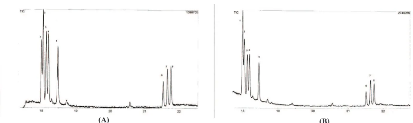
Figure 2. Proile of sugars standards A: 1, rhamnose; 2, ribose; 3, fucose; 4; arabinose; 5, xylose; 6, mannose; 7, glucose, and 8,
galactose. Sugar standards plus sample from hydrolysis of 3, showing an increase in rhamnose (B).
Figure 3. Proile of sugars standards A: 1, rhamnose; 2, ribose; 3, fucose; 4; arabinose; 5, xylose; 6, mannose; 7, glucose, and 8,
galactose. Sugar standards plus sample from hydrolysis of 4, showing an increase in arabinose (B).
(A) (B)
significant cytotoxicity against several tumor cell
lines as HL-60, K562, M4Beu, and HSC-2, with IC50 values ranging from 5.0 to 29.0 μg.mL-1 (Vechia et al.,
2009). It is interesting to note that this triterpenoid
exhibited IC50 values of 7.0 μg.mL-1 and 21 μg.mL-1
against Leishmania donovani (da Silva Filho et al., 2009) and Leishmania major (Takahashi et al., 2004), respectively. In our results it demonstrated IC50 value
of 9.34 μg.mL-1 against L. (L.) amazonensis.
Conclusions
It was isolated five compounds where the chalcone 2 seems be a new one. Although the other four compounds are usual, it was the first time that they were isolated in this genus and species. The chalcones 1 and 2 showed be active against all biological models researched moreover it was the first time that these compounds (1 and 2) were tested on these biological assays. Thus, these two chalcones are promising structures to the development of new drugs, showing the pharmacological potential of the Blepharocalyx salicifolius, a species little studied.
Acknowledgements
The authors thank CNPq-FIOCRUZ PAPES Program and FAPEMIG for all financial support.
References
Arisawa M, Horiuchi T, Hayashi T, Tezuka Y, Kikuchi T,
Morita N 1993. Studies on constituents of Evodia rutaecarpa (Rutaceae). I. Constituents of the leaves.
Chem Pharm Bull 41: 1472-1474.
Belofsky G, Percivill D, Lewis K, Tegos GP, Ekart J 2004.
Phenolic metabolites of Dalea versicolor that enhance antibiotic activity against model pathogenic bacteria. J Nat Prod 67: 481-484.
Cota BB, Magalhães A, Pimenta AMC, Siqueira EP, Alves TMA, Zani CL 2008. Chemical constituents of Habenaria petalodes Lindl. (Orchidaceae). J Braz Chem Soc 19: 1098-1104.
Cunha LCS, Andrade SML, Furtado NAJC, Vinholis AHC, Martins CHG, Silva FAA, Cunha WR 2007.
Antibacterial activity of triterpene acids and semi-synthetic derivatives against oral pathogens. J Biosciences 62: 668-672.
da Silva Filho AA, Resende DO, Fukui MJ, Santos FF, Pauletti
PM, Cunha WR, Silva MLA, Gregorio LE, Bastos JK,
Nanayakkara NPD 2009. In vitro antileishmanial, antiplasmodial and cytotoxic activities of phenolics and triterpenoids from Baccharis dracunculifolia D. C. (Asteraceae). Fitoterapia 80: 478-482.
Espinel-Ingroff A, Boyle K, Sheehan DJ 2001. In vitro
antifungal activities of voriconazole and reference
agents as determined by NCCLS methods: review of Table 1. Results of the biological assays expressed in concentration of the compound (µg.mL-1) suficient to inhibit the growth or to
kill the pathogenic agents.
aConcentration in µg.mL-1 suficient to inhibit 50 % of growth (IC50). Etoposide used as control; bConcentration in µg.mL-1 suficient to kill 50%
of the population of cells (LC50). Amphotericin B used as control; cConcentration in µg.mL-1suficient to inhibit the growth (irst data)/to kill the
culture (second data). Sulfametoxazol-trimetoprim used as control; dConcentration in µg.mL-1 suficient to inhibit 72% of growth of TK-10 strain; eConcentration in µg.mL-1 suficient to inhibit 94% of growth of UACC strain; fConcentration in µg.mL-1 suficient to inhibit 79% of growth of MCF-7
strain; gConcentration in μg.mL-1 suficient to kill 66% of the populations of cells.
Pathogenic agent Compounds
1 2 3 4 5 Control
TK-10a 4.33±0.58 10.83±1.44 Not active Not active 12.83±3.01 16d
UACCa 5.00±0.10 7.00±1.73 Not active Not active 10.83±1.44 16e
MCF-7a 3.33±0.57 3.00±1.80 Not active Not active 11.67±1.44 16f
L.(L.) amazonensisb 5.23±1.84 2.19±1.60 6.25±0.10 20.80±7.21 9.34±6.80 0.20g
Pb-01c 6.20/6.20 3.10/6.20 ≥200/≥200 ≥200/≥200 ≥200/≥200 300/300
Pb-18c 3.10/12.50 3.10/6.20 ≥200/≥200 ≥200/≥200 ≥200/≥200 300/300
Pb-B339c 3.100/12.50 3.10/6.20 ≥200/≥200 ≥200/≥200 ≥200/≥200 75/75
Pb-14c 6.20/6.20 6.20/6.20 ≥200/≥200 ≥200/≥200 ≥200/≥200 75/75
Pb-3c 6.20/6.20 6.20/6.20 ≥200/≥200 ≥200/≥200 ≥200/≥200 300/300
Pb-4c 12.50/12.50 12.50/12.50 ≥200/≥200 ≥200/≥200 ≥200/≥200 150/150
Pb-2c 6.20/6.20 6.20/6.20 ≥200/≥200 ≥200/≥200 ≥200/≥200 150/150
Pb-1578c 3.10/3.10 3.10/6.20 ≥200/≥200 ≥200/≥200 ≥200/≥200 75/75
Pb-ED01c 6.20/6.20 6.20/6.20 ≥200/≥200 ≥200/≥200 ≥200/≥200 75/75
Pb-11c 6.20/6.20 6.20/6.20 ≥200/≥200 ≥200/≥200 ≥200/≥200 150/150
the literature. Mycopathologia 150: 101-115.
Fu L, Zhang S, Li N, Wang J, Zhao M, Sakai J, Hasegawa T, Mitsui T, Kataoka T, Oka S, Kiuchi M, Hirose K, Ando
M 2005. Three new triterpenes from Nerium oleander
and biological activity of the isolated compounds. J Nat Prod 68: 198-206.
Gafner S, Wolfender JL, Mavi S, Hostettmann K 1996.
Antifungal and antibacterial chalcones from Myrica serrata. Planta Med 62: 67-69.
Gonzalez AG, Aguiar ZE, Luis JG, Rivera A, Calle J, Gallo
G 1992. A C-methyl chalcone from Dalea caerullea. Phytochemistry 31: 2565-2566.
Hallett FP, Parks LM 1951. A note on the isolation of
quercitrin from Euphorbia pilulifera. J Am Pharm Assoc 40: 56-57.
Johann S, Cisalpino PS, Watanabe GA, Cota BB, Siqueira EP, Pizzolatti MG, Zani CL, Resende M A 2010.
Antifungal activity of extracts of some plants used in Brazilian traditional medicine against the pathogenic fungus. Pharm Biol 48: 388-396.
Kato H, Li W, Koike M, Wang Y, Koike K 2010. Phenolic
glycosides from Agrimonia pilosa. Phytochemistry 71: 1925-1929.
Leite JPV, Oliveira AB, Lombardi JA, Filho JDS, Chiari E 2006. Trypanocidal activity of triterpenes from Arrabidaea triplinervia and derivatives. Bio Phar Bull 29: 2307-2309.
Limberger RP, Sobral MEG, Zuanazzi JAS, Moreno PRH, Schapoval EES, Henriques AT 2001. Biological
activities and essential oil composition of leaves of
Blepharocalyx salicifolius. Pharm Biol 39: 308-311.
Liu, J 2005. Oleanolic acid and ursolic acid: Research perspectives. J Ethnopharmacol 100: 92-94.
Lopez JA 1982. Isolation of quercitrin from the leaves of Anacardium rhinocarpus D.C., A. excelsum (Bert and Balb.) (Anacardiaceae). Ingen Cienc Quim 6: 148-149.
Lozoya X, Meckes M, Abou-Zaid M, Tortoriello J, Nozzolillo
C, Arnason JT 1994. Quercetin glycosides in Psidium guajava L. leaves and determination of a spasmolytic
principle. Arch Med Res 25: 11-15.
Mair AGR, Pandiyan M, Venkatasubramanian H 1987.
Polyphenolic compounds from flowers of Psidium guajava. Fitoterapia 58: 204-205.
Malterud KE, Anthonsen T, Lorentzen GB 1977. Chemistry
of Myrica gale L. Part 4. Two new C-methylated flavonoids from Myrica gale. Phytochemistry 16: 1805-1809.
Monks A, Scudiero D, Skehan P, Shoemaker R, Paull K,
Vistica D, Hose C, Langley J, Cronise P, Vaigro-Wolff A, Gray-Goodrich M, Campbell H, Mayo J, Boyd M
1991. Feasibility of a high-flux anticancer drug screen using a diverse panel of cultured human tumor cell lines. J Natl Cancer Inst 83: 757-766.
Monteiro AS, Coutinho JOPA, Júnior AC, Rosa CA, Siqueira
EP, Santos VL 2009. Characterization of new biosurfactant produced by CLOA 72 isolated from
dairy industry effluents. J Basic Microb 49: 553-563. Monteiro AS, Bonfim MRQ, Domingues VS, Corrêa JA,
Siqueira EP, Zani CL, Santos VL 2010. Identification
and characterization of bioemulsifier-producing yeasts isolated from effluents of a dairy industry.
Bioresource Technol 101: 5186-5193.
Mors WB, Rizzini CT, Pereira NA 2000. Medicinal Plants of Brazil. 1 ed. Michigan: Reference Publications, p. 501.
Muzitano MF, Tinoco LW, Guette C, Kaiser CR,
Rossi-Bergmann B, Costa SS 2006. The antileishmanial activity assessment of unusual flavonoids from
Kalanchoe pinnata. Phytochemistry 67: 2071-2077.
National Committee for Clinical Laboratory Standards
2002. M27-A2. Method for broth dilution antifungal
susceptibility testing of yeast. 2 ed. Wayne: NCCLS. Pavan FR, Leite CQF, Coelho RG, Coutinho ID, Honda
NK, Cardoso CAL, Vilegas W, Leite SRA, Sato DN 2009. Evaluation of anti-mycobacterium tuberculosis
activity of Campomanesia adamantium (Myrtaceae).
Quim nova 32: 1222-1226.
Pecoul B, Chirac P, Trouiller P, Pinel J 1999. Access to essential drugs in poor countries - a lost battle? J Am Med Ass 281: 361-367.
Portillo A, Vila R, Freixa B, Ferro E, Parella T, Casanova J,
Canigueral S 2005. Antifungal sesquiterpene from the root of Vernonanthura tweedieana. J Ethnopharmacol 97: 49-52.
Seebacher W, Simic N, Weis R, Saf R, Kunert O 2003.
Complete assignments of 1H and 13C NMR resonances
of oleanolic acid, 18a-oleanolic acid, ursolic acid and their 11-oxo derivatives. Magn Reson Chem 41: 636-638.
Silva MGV, Vieira IGP, Mendes FNP, Albuquerque IL,
Santos RN, Silva FO, Morais SM 2008. Variation of ursolic acid content in eight Ocimum species from northeastern Brazil. Molecules 13: 2482-2487.
Siqueira EP, Fagundes EMS, Sobral MEG, Alves TMA, Rabello A, Zani CL 2010. Leishmanicidal activities
of the extract from Blepharocalyx salicifolius (Kunth) O. Berg, Myrtaceae. Rev Bras Farmacogn 20: 416-421.
Takahashi M, Fuchino H, Sekita S, Satake M 2004. In vitro leishmanicidal activity of some scarce natural products. Phytother Res 18: 573-578.
Teixeira MC, Santos RJ, Sampaio RB, Carvalho LLP, Santos WL 2002. A simple and reproducible method to obtain
large numbers of axenic amastigotes of different
Leishmania species. Parasitol Res 88: 963-968.
Torres-Santos EC, Moreira DL, Kaplan MA, Meirelles MN,
Agents Ch 43: 1234-1241.
Vechia LD, Gnoatto SCB, Gosmann G 2009. Derivados
oleananos e ursanos e sua importância na descoberta de novos fármacos com atividade antitumoral, anti-inflamatória e antioxidante. Quim Nova 32: 1245-1252.
Xu F, Matsuda H, Hata H, Sugawara K, Nakamura S, Yoshikawa M 2009. Structures of new flavonoids and
benzofuran-type stilbene and degranulation inhibitors of rat basophilic leukemia cells from the Brazilian herbal medicine Cissus sicyoides. Chem Pharm Bull 57: 1089-1095.
*Correspondence
Ezequias P. Siqueira
Centro de Pesquisa René Rachou
Av. Augusto de Lima, 1715, 30190-002 Belo Horizonte, MG,
Brazil