Patterns of expression of cell wall related genes in sugarcane
D.U. Lima, H.P. Santos, M.A. Tiné, F.R.D. Molle and M.S. Buckeridge*
Abstract
Our search for genes related to cell wall metabolism in the sugarcane expressed sequence tag (SUCEST) database (http://sucest.lbi.dcc.unicamp.br) resulted in 3,283 reads (1% of the total reads) which were grouped into 459 clusters (potential genes) with an average of 7.1 reads per cluster. To more clearly display our correlation coefficients, we constructed surface maps which we used to investigate the relationship between cell wall genes and the sugarcane tissues libraries from which they came. The only significant correlations that we found between cell wall genes and/or their expression within particular libraries were neutral or synergetic. Genes related to cellulose biosynthesis were from theCesAfamily, and were found to be the most abundant cell wall related genes in the SUCEST database. We found that the highest number ofCesAreads came from the root and stem libraries. The genes with the greatest number of reads were those involved in cell wall hydrolases (e.g.β-1,3-glucanases, xyloglucan endo-β-transglycosylase, β-glucosidase and endo-β-mannanase). Correlation analyses by surface mapping revealed that the expression of genes related to biosynthesis seems to be associated with the hydrolysis of hemicelluloses, pectin hydrolases being mainly associated with xyloglucan hydrolases. The patterns of cell wall related gene expression in sugarcane based on the number of reads per cluster reflected quite well the expected physiological characteristics of the tissues. This is the first work to provide a general view on plant cell wall metabolism through the expression of related genes in almost all the tissues of a plant at the same time. For example, developing flowers behaved similarly to both meristematic tissues and leaf-root transition zone tissues. Besides providing a basis for future research on the mechanisms of plant development which involve the cell wall, our findings will provide valuable tools for plant engineering in the near future.
INTRODUCTION
The plant cell wall is a composite of interwoven mers that surround plant cells. Among these polymers, poly-saccharides are the majority, these being usually grouped into three classes: cellulose, hemicelluloses and pectins. Usually, polysaccharides make 80-90% of the wall, the other components being proteins and phenolic compounds such as lignin. Cell wall proteins are either of the structural type (glycine and proline rich proteins) or enzymes (Carpita and Gibeaut, 1993).
The cell wall forms the extracellular matrix of plant tissues and on the basis of what is presently known it can be considered rather as another cell compartment in which several metabolic reactions take place (seePlant Physiol-ogy and Biochemistryv. 38 for an extensive review).
Among the main functions of the wall are control of growth, cell signaling, defense, selective porosity and car-bon storage. As these functions are usually performed si-multaneously, the wall has to be both flexible and tough which requires exact/fine metabolic control. As carbohy-drates are the principal components of the plant cell wall, understanding the chemical changes that take place in dif-ferent tissues at difdif-ferent stages of growth and development is the key to understanding how physiological processes are controlled inside the plant.
On the basis of the discovery that structurally different polymers can perform analogous functions in the walls of plants of different taxonomic groups, Carpita and Gibeaut (1993) proposed the separation of plant cell walls into two groups. The group I type of cell wall is typical of dico-tyledonous plants (and some monocodico-tyledonous plants) hav-ing xyloglucan (one of the major hemicelluloses in higher plants) as the principal hemicellulose and a higher propor-tion of pectins. The group II type cell wall is characteristic of monocotyledonous grasses (thePoaceaeorGramineae) and although xyloglucan is still present the dominant hemicel-luloses are usually arabinoxylans and (1,3)-(1,4)-β-glucan (also known as mixed linkage glucan orβ-glucan) and these walls have a lower proportion of pectins.
The biochemical changes that take place in cell walls are performed mostly by enzymes, principally hydrolases which are of two types, endo- and exo-enzymes (Table I). Endo enzymes attack polymers anywhere in the main chain, producing polymer fragments of low molecular weight while the exo-hydrolases can only attack a polymer by breaking the glycosidic linkage in the non-reducing end. Another family of enzymes which are important in the plant cell wall are the xyloglucan endotransglycosylases (XETs). Be-sides hydrolyzing xyloglucans, these enzymes are also ca-pable of transferring fragments of xyloglucan
inter-molecularly, a phenomenon has been exhaustively studied since about 1991, and it is presently believed that XETs are equally important in the biosynthesis and degra-dation of xyloglucans (Campbell and Braam, 1999).
Except for cellulose and callose, which are synthe-sized at the membrane level, the enzymes related to cell wall polysaccharide biosynthesis can not be strictly consid-ered as cell wall enzymes, since they are active mainly in the cytoplasm on the membranes of the Golgi apparatus. Nevertheless, it is obvious that these enzymes have a direct impact on the events taking place in the cell wall and there-fore for the purpose of this paper they will be considered as being cell wall enzymes.
The plant cell wall is thought to be composed of three independent domains; cellulose-hemicellulose (the micro-fibril and the micromicro-fibril orientating domain) pectin (con-trol of porosity and a source of cell signaling for defense) and protein (cross linking and wall loosening). Clearly all three domains are involved in the maintenance of the me-chanical properties of the wall. Thus, the three of the most relevant features in the control of cell wall function in plants are the topology and specificity of the cell wall re-lated enzymes and the exact timing by which they are pres-ent at the cell wall at the correct stage of developmpres-ent.
All the information necessary for topology, specific-ity and timing to combine and form a pattern of polymers in the wall of a given plant tissue are ‘written’ in the genes that encode the enzymes and other proteins that participate in the process of their production, transport, secretion and action. Therefore, studying gene expression can be very useful to understand how these patterns are formed, main-tained and degraded.
Group II cell walls are of special interest to us be-cause our subject of interest was sugarcane. The composi-tion and chemical structure of the cell walls of sugarcane has not yet been directly studied. However, a few papers have been published that take into account the cell wall polysaccharides in sugarcane, these papers normally con-centrating on the properties of sugarcane sap/juice, the cell
wall polymers usually being called indigenous polysaccharides (IPs) (Robertset al., 1976). The analytic work performed on the composition of IPs clearly indicate that sugarcane has the typical polymers of the group II cell walls.
We studied the expression of cell wall related genes present in the sugarcane expressed sequence tag (SUCEST) database (http://sucest.lbi.dcc.unicamp.br) using digital northern blots constructed by computer analysis of the reads found in the different libraries of the database. Digital northern analysis provided us with an idea of which cell wall proteins were expressed (and at what intensity they were expressed) in different sugarcane tissues. We also constructed correlation surface maps which made possible a general appreciation of the pattern of expression of cell wall related proteins in the entire plant.
MATERIAL AND METHODS
Search of cell wall related proteins
The clusters related to cell wall proteins were ob-tained from the SUCEST database by searching with key-words and the basic local alignment search tool (BLAST) using well known cell wall genes. The keywords used were: arabinosidase, cellulase, cellulose synthase, endopolyga-lacturonase, expansin, extensin, fucosyltransferase, galac-tanase, alfa galactosidase, beta galactosidase, alfa galacto-syltransferase, beta glucanase, beta glucosidase, laccase, lichenase, beta mannanase, beta mannosidase, pectin acety-lesterase pectin methyacety-lesterase, pectinesterase, peroxidase, polygalacturonase, xylanase, xyloglucan endotransglyco-sylase and xylosidase. Keywords that did not show any homology with clusters in the sugarcane database were cellobiohydrolase, galacturonyltransferase, glucoronidase, glycine rich protein, hydroxyproline rich protein, manno-syltransferase, rhamnogalacturonase and xylosyltransfe-rase. The lists of matches (from the NCBI database) related to each cluster, were checked for authenticity as a potential cell wall related gene firstly by association with a plant
Table I- Some features of the principal plant cell wall polysaccharides and the enzymes involved in their degradation.
Polysaccharide Main chain residue Branching residues Enzymes Mannans Mannose Generally none, rarely galactose Endo-β-mannanase
Glucomannan Mannose, Glucose Galactose Endo-β-mannanase, Endo-β-glucanase,α-galactosidase Galactomannans Mannose Galactose Endo-β-mannanase,α-galactosidase, exo-β-mannanase Xyloglucans Glucose Xylose, galactose,
fucose, arabinose
Xyloglucan-endo-β-transglycosilase, Xyloglucan-endo-β-glucanase,
β-galactosidase,α-xylosidase,β-glucosidase,α-fucosidase Galactans Galactose Arabinose Exo-galactanase,α-arabinosidase
Arabinoxylans Xylose Arabinose, Glucuronic acid β-xylosidase, endo-β-xylanase,α-arabinosidase,α-glucuronidase
β-Glucan Glucose None Lichenase,β-(1,3)(1,4)-glucosidase Callose Glucose None β-(1,3)-glucanase
gene, secondly by checking for any existing relevant bibli-ography and thirdly by limiting the e-value to a maximum of 1e-50. Subsequently, the reads within the clusters were grouped by protein and library.
The libraries investigated were: AM1 = apical meris-tem (young leaves and smeris-tem of mature plants), AM2 = api-cal meristem (young leaves and stem of immature plants), CL3, CL4 and CL6 = callus tissue submitted to a 12 hour:12 hour light/dark regime and temperature (4° and 37 °C) stress, FL1 = 1 cm flowers, FL2 = 20 cm flowers with size, FL3 = 5 cm long flower stem, FL4 = 50 cm long flowers stem, FL8 = 10 cm long flower stem, LB1 and LB2 = Lateral buds; LR1 and LR2 = leaf roll, LV1 = etiolated leaves from plantlets, RT1 and RT2 = roots from young plants, RT3 = roots from adult plants, RZ1, RZ2 and RZ3-Leaf-root transition zone, SB1 = Stem bark, SD1 and SD2 = seeds in different stages of development, ST1 = Stem (first internode) and ST3 = Stem (fourth internode).
The data were normalized in order to compare the dif-ferent libraries and proteins. The number of reads obtained for every cell wall related gene found in each library was di-vided by the total number of reads of the respective SUCEST library. Because some values were quite low, we expressed them as the number of reads per 10,000 library reads. To construct the surface maps, the reads were grouped according to the libraries from which they were obtained,e.g.all the root libraries (RT1+RT2+RT3) were grouped together as roots and so forth. The reads from li-brary tests ( 500 reads) were discarded.
Statistical analysis
In order to understand the degree of association in ex-pression (number of reads) for different cell wall proteins, simple linear correlation analysis (r) was used. Using this approach we were able to observe the direction (synergism or antagonism) and the magnitude of each association be-tween two cell wall genes (independently of the library from which they originally came), or between two libraries (independently of the cell wall related gene). The values obtained ranged from -1 to 1: where ‘zero’ means that the variables do not vary together at all, ‘positive’ means that the two variables tend to increase or decrease together (syn-ergism), and ‘negative’ means that the variables are in-versely related (antagonism) (Motulsky, 1995).
The correlation analyses were made using the ‘Tool Analysis’ function of the Excel program (Microsoft Corpo-ration, USA), and the calculated coefficients were statisti-cally tested using the ‘r Table’ of Snedecor (1946) which follows a distribution with η-2 degrees of freedom (η = number of pairs) at the 5% probability level. To view the topographical patterns, the correlation coefficients for li-braries or genes were plotted as surface graphs using the graph tools available in the software. The ‘r’ value was con-sidered as a dependent variable, and only significant statis-tical coefficients were included.
RESULTS AND DISCUSSION
The search for genes related to cell wall metabolism resulted in 459 potential genes (clusters) which corre-sponded to 3,283 reads (7 reads per cluster). The average number of cell wall related reads per library was about 1% of the total number of reads in the database (Figure 1). About 50% of the total number of cell wall related reads oc-curred in the following tissue libraries: RT1+RT2 (roots from young plants), RT3 (roots from adult plants), SB1 (stem bark), FL4 (flower stem) , ST1 (first internode), ST3 (fourth internode), RZ3 (leaf-root transition zone) and AM2 (apical meristem from young plants) (Figure 1A).
Cellulose synthase, peroxidase, expansin,β -glucosi-dases β-1,3 glucanases were among the more highly ex-pressed cell wall genes in sugarcane (Figure 1B). However, the high standard deviation found for each gene suggest
Figure 1- Expression of cell wall related genes shown as percentage of the number of reads. (A) Number of cell wall related reads in relation to the to-tal number of reads in each library. The AD1 (1.2%) and HR1 (1.0) librar-ies were accounted for but are not shown. (B) Total number of reads from a related cell wall gene in relation to the total reads in the sugarcane ex-pressed sequence tag (SUCEST) database. The nomenclature used for li-braries in (A) were the same used in the SUCEST project : AMs = apical meristem, CLs = callus, FLs = flower/stem flower, LBs = lateral buds, LV = etiolated leaves, RTs = roots, RZs = leaf-root transition zone, SB = stem bark, SDs = seeds, STs = stems. Libraries from equivalent tissues were placed together,e.g.CL3+CL4+CL6, RT1+RT2, RZ1+RZ2, SD1+SD2. In (B) the proteins are represented by numbers: 1 = cellulose synthase; 2 = peroxidase; 3 = expansin, 4 =β-glucosidase, 5 =β-(1,3) glucanase, 6 = XET, 7 = Endopolygalacturonase; 8 = Pectin(acetyl)(methyl)esterase; 9 = Galactanases; 10 = Lichenase; 11 = Cellulase; 12 = Mannanase/manno-sidase; 13 = Laccase; 14 =α-galactosidase; 15 =β-1,4 xylosidase; 16 =
that expression can be highly dependent on the tissue (li-brary) investigated.
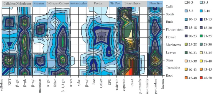
This survey, containing the frequency distribution of reads (per 10,000 reads) of each cell wall related gene per library, generated a highly complex pattern of cell wall gene expression and in order to better visualize the tenden-cies, surface maps were constructed (Figures 2, 3 and 4).
The proportion of reads in each library varied greatly and the pattern of expression (Figure 2) revealed that cellu-lose synthase is highly expressed in root and stem tissue as well as in tissues from the transition zone. Some other genes appeared to be expressed simultaneously in these li-braries such as β-glucosidase, β-1,3-glucanase (callose), pectin esterase and peroxidase (phenolic compounds). XET and mannanase were expressed in stems but not in roots (Figure 2).
Pectins presented a differential pattern of expression with pectin esterase present in stem and roots, endo-poly-galacturonase mainly in leaves and galactanase mainly in flowers (Figure 2). Mannans seem to have been associated almost exclusively with stems, since a higher number of reads was found in normal stem and flower stem.
The CesAgene family, related to cellulose biosyn-thesis (Hollandet al. 2000), deserves a special mention. It is by far the largest cell wall related gene family found in sug-arcane. Before this study we had already found 18 different CesAclusters (genes) and their distribution within the cane
as well as comparisons with theCesAfamilies from maize, Arabidopsis, rice and cotton will be published elsewhere. Since the studies on polysaccharide synthases are not as de-veloped as for hydrolases, this high number of reads should be viewed with some caution because other glycosyltrans-ferases (such as otherβ-glucan, xylan and mannan syntha-ses) may have been included among the CesA genes, artificially increasing the number of reads of this gene found in the libraries.
Peroxidase is an important cell wall enzyme thought to be related to wall tightening (Boudet, 2000) and genes for this enzyme were found at a high frequency in root and stem tissues, including flower-stem tissues (Figure 2). This pattern of expression is consistent with tissues where the formation of vascular bundles is more intense.
Correlation between genes that encode the action of enzymes in the cell wall
One of the most important events during development is the orientation of cellulose microfibrils in the cell wall (McCann and Roberts, 1991; Talbott and Ray, 1992; Car-pita and Gibeaut, 1993) and two steps, division and exten-sion, are thought to be key phases in wall development. During cell division, the formation of the phragmoplast (the equatorial region of the spindle during anaphase from which the cell plate develops at telophase from which it is believed the middle lamella is formed) involves the
synthesis of cellulose and the assembly of cellulose mole-cules into microfibrils simultaneously with the orientation of these microfibrils by hemicelluloses synthesized in the Golgi apparatus at practically the same time. On the other hand, the formation of the phragmoplast has also to involve polysaccharide hydrolysis, since the newly synthesized wall has to fuse with an existent one. After division, plant cells continue to grow by extension and this process also
in-volves microfibril orientation since important changes in
the mechanical properties of the wall have to be performed in order to allow elongation in a specific direction
(Cosgrove, 2000; Whitneyet al., 1995). In order to stop growth, cross links between polymers can be made and this is thought to be performed by phenolic compounds and
structural proteins such as extensin (Fry, 1988).
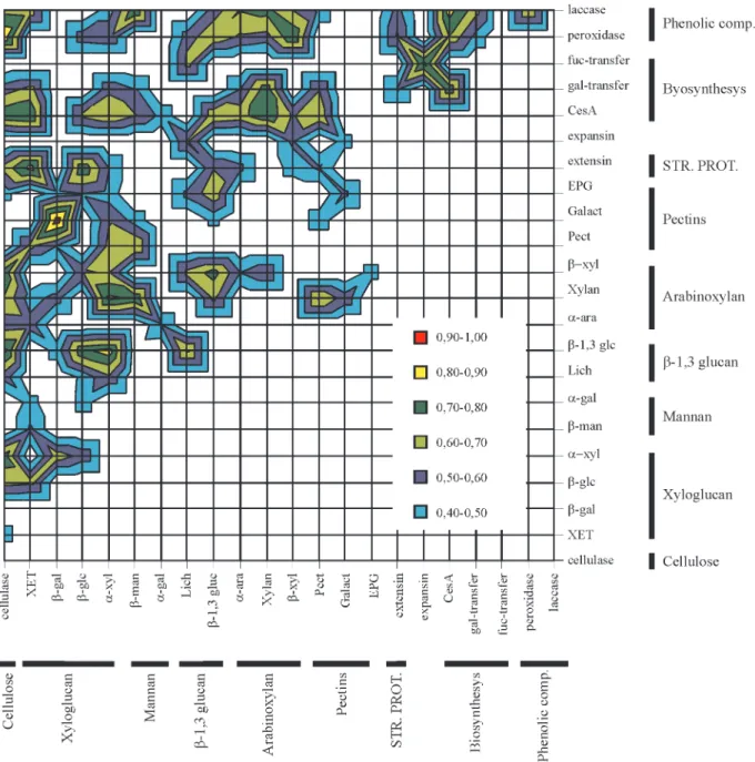
Figure 3- Correlation between sugarcane cell wall related genes. Calculations were performed with the number of reads obtained for each gene by a basic local alignment search tool (BLAST) search using keywords and the amino acid sequences of genes known to be related to cell wall metabolism. The cor-relation analyses were made using the ‘Tool Analysis’ function contained in the Excel® software package (Microsoft Corporation®, USA), and the cal-culated coefficients were statistically tested using the ‘r Table’ of Snedecor (1946) based on a distribution withη-2 degrees of freedom (η= number of pairs) at the 5% probability level. (r = 0.423) XET = xyloglucan endo-β-transglycosylase;β-gal =β-galactosidase;β-glc =β-glucosidase;α-xyl =
Figure 3 shows the correlation between the groups of genes that work either to hydrolyze or synthesize a given sugarcane cell wall polymer. As only significant correla-tion coefficients are shown, one outstanding feature of this map is that there is no significant antagonic expression be-tween the groups of cell wall genes analyzed, indicating that ‘biochemical operations’ on cell wall polymers can be
lulose microfibrils (McCann and Roberts, 1991, Carpita and Gibeaut, 1993). The correlation we observed probably means that any modification in microfibril orientation has to have a concomitant change in the size of pores in the wall so that the xyloglucan-degrading enzymes can reach their substrate.
Another situation where there was a strong correla-tion of gene expression was between the genes that encode xyloglucan and callose/β-glucan exo-hydrolases. As pro-posed for xyloglucan,β-glucan is thought to be related to microfibril orientation, although this proposition is based solely on the fact thatβ-glucan binds specifically to cellu-lose (Mishimaet al., 1998). Another relevant feature re-gardingβ-glucans is that, in grasses, their appearance is thought to be transitory and related to wall development (Mishimaet al., 1998). If these two polymers are really re-lated to microfibril orientation (an important event in wall development) we should indeed expect correlation between genes coding for enzymes involved with these polymers.
Another striking set of correlations was between he-micellulose hydrolysis (cellulases, xyloglucanases,β -glu-canases and arabinoxylanases) and cellulose biosynthesis. There are two ways to explain this correlation. One is through cell wall turnover, during which both hydrolysis and synthesis occur simultaneously and with approxi-mately the same intensity (Gorshkovaet al., 1997). An-other, non-exclusive, possibility is that the correlation between hydrolytic and synthetic events is reflecting other developmental processes occurring at the same time. This possibility is quite plausible in sugarcane because this plant has a very high rate of growth. Figure 3 shows that lignin hydrolysis (laccase genes) and biosynthesis (peroxidase genes) correlate with almost exactly the same pattern, and this supports the hypothesis that some genes are linked to hydrolysis/biosynthetic events. The situation is still un-clear, however, since Boudet (2000) has suggested that laccase also has some role in lignification.
Expression of cell wall related genes in different sugarcane tissues
Another instructive way to look at the expression of cell wall related genes in sugarcane is to construct surface correlation maps of the libraries which show more clearly the relationships between genes.
For the groups of cell wall genes, only positive corre-lation coefficients were found (Figure 3), but in Figure 4 the absence of correlation can be used as a measure of the uniqueness of certain libraries. For example, the seed li-braries correlated weakly with only a few other lili-braries, re-lated to root, young flowers and leaf tissues.
The flower libraries are also very interesting in that we detected a high correlation (0.6) between some flower libraries (FL1, FL3 and FL5) and leaf and apical meristem libraries. It is interesting to note that during flower
develop-ment the flower libraries tended to reduce their association with apical meristem (AM) libraries and increase their as-sociation with leaf roll (LR) libraries. Libraries obtained from the leaf-root transition zone (RZ) and the first inter-node of stems (ST1) showed a lower, but still significant, correlation with developing flowers. This is probably re-lated to the fact that the leaf-root transition zone correre-lated highly with apical meristem. Indeed, leaf-root transition zone is typical of a region with high meristematic activity. However, it is important to note that the cDNA from which the apical meristem libraries were constructed was in fact prepared from a mixture containing some leaf and some stem tissues. On the other hand, flower stem library FL8 presented only a little correlation with the young flower braries FL1 and FL3 while the 50 cm flower stem FL4 li-brary presented some correlation with stem and root libraries.
Other important correlations were between the bud and flower, bud and apical meristem, bud and root, and stem and root libraries. Again, this is also to be expected from our knowledge of plant physiology.
Topology, specificity and timing, the three features cited as being the keys to our understanding of enzyme ac-tion in the cell wall, could not be directly evaluated using gene expression data (ESTs) but a reasonable picture of the co-ordination of events relevant to the whole sugarcane plant as a whole could be distinguished. The fairly high number of reads which we found for theCesAgene family is not surprising, since cellulose synthesis is probably one of the most important anabolic processes in plants. Holland et al. (2000) reminds us that this is probably the reason why considerable redundancy is found for this gene family.
Most of the correlations that we found in sugarcane were quite ‘logical’ and fit in well with the classical views of plant anatomy and physiology. To our knowledge this is the first time that this approach has been applied to plant cell walls and it would be interesting to see this technique applied to species such asArabidopsis thaliana andZea mays,which are among the most studied genomes to date. We are now starting biochemical studies to confirm our ob-servations, and we hope to combine these studies with cor-relation studies ofArabidopsisand maize to develop other powerful tools for use in plant bioengineering.
RESUMO
expressão esteja associada à raiz e colmo. Entre as hidro-lases, β-1,3-glucanases, xiloglucano endo-β -transglicosi-lase, β-glucosidase e endo-β-mananase foram os genes com o maior número de clones. Análise de correlação (por mapas de superfície) revelou que a expressão dos genes relacionados à biossíntese parece estar associada à hidrólise de hemicelulose, enquanto as hidrolases de pectina estão relacionadas principalmente às hidrolases de xiloglucano. O padrão de expressão de genes relacionados à parede celular, baseado no número de “reads” por “cluster” refletiu bem as características fisiológicas esperadas para cada tecido. Este é o primeiro trabalho que fornece uma visão geral do metabolismo de parede celular através da expressão dos genes em vários tecidos ao mesmo tempo. Por exemplo, inflorescências em desenvolvimento se comportaram de forma semelhante a tecidos meristemáticos e à região de transição folha-raiz. Estes da-dos servirão tanto para pesquisas em fisiologia e desenvolvimento de cana, como também para o desenvolvimento de processos industriais.
ACKNOWLEDGEMENTS
Authors acknowledge Dr Marcia Regina Braga for a critical review of the manuscript and FAPESP-SUCEST for financial support (grant number 00/07436-9).
REFERENCES
Boudet, A.M.(2000). Lignins and lignification: Selected issues. Plant Physiol. Biochem. 38: 81-96
Campbell, P.andBraam, J.(1999). Xyloglucan endotransglyco-sylases: diversity of genes, enzymes and potential wall-mo-difying functions.Trends Plant Sci. 9(4): 361-366.
Carpita, N.C.andGibeaut, D.M.(1993). Structural models of primary cell walls in flowering plants: consistency of molec-ular structure with the physical properties of the walls during growth.Plant J. 3: 1-30.
Cosgrove, D.J. (2000). Expansive growth of plant cell walls. Plant Physiol. Biochem.38: 109-124.
Fry, S.C.(1988). The growing plant cell wall: chemical and meta-bolic analysis. Longman, Harlow, Essex.
Gorshkova, T.A., Chemikosova, S.B., Lozovaya, V.V. and Carpita, N.C.(1997). Turnover of galactans and other cell wall polysaccharide in developing flax plants.Plant Physiol. 114: 723-729.
Holland, N., Holland, D., Helentjaris, T., Dhugga, K.S., Ca-zares, B.X.andDelmer, D.P.(2000). A comparative analy-sis of the plant cellulose synthase (CesA) gene family. Plant Physiol. 123: 1313-1323.
McCann, M.C.andRoberts, K.(1991). Architecture of the pri-mary cell wall. In: The Cytoskeletal Basis of Plant Growth and Form(Lloyd, C.W. ed.). Academic Press, London, pp. 109-129.
Mishima, T. Hisamatsu, M., York, W.S., Teranishi, K.and Yamada, T.(1998). Adhesion ofβ-D-glucans to cellulose. Carbohydr. Res. 308: 389-395.
Motulsky, H.(1995). Intuitive Biostatistics.Oxford University Press, New York, 386p.
Roberts, E.J., Godshall, M.A., Carpenter, F.G.andClarke, M.A. (1976). Composition of soluble indigenous polysa-ccharide from sugarcane.Int. Sugar J.78: 163-165. Snedecor, G.W.(1946).Statistical Methods. The Iowa State
Col-lege Press,USA, 4 ed.
Talbott, L.D.andRay, P.M.(1992). Molecular size and separa-bility features of pea cell wall polysaccharides. Implications for primary wall structure.Plant Physiol. 98: 357-368. Whitney, S.E.C., Brigham, J.E., Darke, A.H., Reid, J.S.G.and