Polymorphism in the promoter region of von Willebrand factor gene and von
Willebrand disease type 1
Daniel Simon
1,2, Eliane Bandinelli
1and Israel Roisenberg
11
Universidade Federal do Rio Grande do Sul, Instituto de Biociências, Departamento de Genética,
Porto Alegre, RS, Brazil.
2
Universidade Luterana do Brasil, Canoas, RS, Brazil.
Abstract
The -1185A/G polymorphism in the 5’-regulatory region of the von Willebrand factor (VWF) gene was associated with VWF plasma levels in a normal population. This study was undertaken to evaluate whether there is a relationship between this polymorphism and type 1 von Willebrand disease (VWD), a disorder characterized by a quantitative deficiency of VWF. The association between this polymorphism and plasma VWF levels in normal Brazilian individuals was also analyzed. Control subjects (n = 460) and type 1 VWD patients (n = 41) were studied. Polymerase chain reaction (PCR) amplification of the 864-bp VWF promoter region followed by AccII restriction-digestion was used to identify the -1185A/G genotypes. The -1185G allele frequency was 57% in normal individuals and 63% in type 1 VWD patients, this difference was not significant (p = 0.29). No significant association was observed between -1185A/G genotypes and VWF plasma levels in normal individuals, although VWF levels were in the same direction as those reported by another study, with subjects carrying the G allele having the lower levels. These results suggest that -1185A/G polymorphism is not associated with the partial deficiency of VWF in type 1 VWD patients.
Key words:von Willebrand factor, promoter polymorphisms, genetics, von Willebrand disease. Received: June 26, 2003; Accepted: October 21, 2003.
Introduction
Von Willebrand disease (VWD) is the most frequent inherited human bleeding disorder, with a prevalence of up to 1% in the general population (Rodeghieroet al., 1987; Werneret al., 1993). It is caused by quantitative or qualita-tive defects in the von Willebrand factor (VWF), a multimeric glycoprotein present in megakaryocytes, plate-lets, endothelial cells, and plasma. VWF plays two main hemostatic roles: (a) binding of platelets to subendothelium at sites of vascular injury, and (b) stabilization and protec-tion of coagulaprotec-tion factor VIII (FVIII).
VWD exhibits a wide heterogeneity of phenotypes, which are classified into three main groups (Sadler, 1994). Type 1 VWD refers to a partial, quantitative deficiency of VWF, the most frequent form of VWD, accounting for 70-80% of the diagnosed cases, and is considered as an autosomal dominant disease with incomplete penetrance and variable expressivity (Sadler et al., 1995). Type 2
VWD refers to all qualitative variants of VWF and is gener-ally autosomal dominant with regard to inheritance, except for type 2N VWD which is autosomal recessive. Type 3 VWD, the severe form of the disorder, is characterized by very low levels of VWF and is associated with profound, life-threatening bleeding. The mode of inheritance in type 3 VWD is autosomal recessive, associated with homo-zygosity or compound heterohomo-zygosity for a defective VWF allele.
The molecular basis of VWD has been established principally for the types 2 and 3 (Nichols and Ginsburg, 1997), but the genetic mechanisms underlying type 1 VWD remain unexplained for the majority of the cases. However in addition to the VWF gene mutations, it is likely that ge-netic and environmental modifying factors contribute to the incomplete penetrance and variable expressivity of type 1 VWD (Levy and Ginsburg, 2001).
Recently, four single nucleotide polymorphisms (SNPs) in the 5’-regulatory region of the VWF gene have been associated with plasma VWF levels in a normal popu-lation (Keightleyet al., 1999; Harveyet al., 2000). These polymorphisms were in strong linkage disequilibrium and segregated in two distinct haplotypes. In one of these SNPs,
Send correspondence to Israel Roisenberg. Universidade Federal do Rio Grande do Sul, Instituto de Biociências, Departamento de Genética, Caixa Postal 15053, 91501-970 Porto Alegre, RS, Brazil. E-mail: israberg@ufrgs.br.
the -1185A/G polymorphism, AA homozygotes were sig-nificantly associated with the highest levels of VWF, GG homozygotes showed the lowest levels, and heterozygotes intermediate levels (Keightleyet al., 1999).
The present study examined the hypothesis that the -1185G allele may be contributing to the phenotype of type 1 VWD. The association between the -1185A/G polymor-phism and VWF levels in normal Brazilian individuals was also investigated.
Subjects and Methods
Subjects. The control group was comprised of 460 unrelated healthy Caucasian volunteers (220 males, 240 fe-males), with no personal or familiar reference of bleeding disease. They were of European ancestry, mainly from Portugual, Spain, Italy and Germany, and consisted of 212 blood donors and 238 students or members of the Univer-sity staff, living in Porto Alegre, the capital of Brazil’s southernmost state. The mean age was 34.1 years (± 15.7 years).
The patient group consisted of 41 unrelated Cauca-sian individuals (21 males, 20 females), with a mean age of 24.6 years (± 9.8 years), diagnosed with type 1 VWD by Hemostasis Laboratory at the Department of Genetics of the Federal University of Rio Grande do Sul (UFRGS). The diagnosis of VWD was based on clinical history (mucocutaneous bleeding such as epistaxis, bleeding after dental extractions and menorrhagia) and in laboratory find-ings (decreased plasma VWF levels, correlated decreased plasma FVIII coagulant activity and prolonged bleeding time) (Favaloro and Koutts, 1997). All individuals who agreed to participate in the study were evaluated through a detailed questionnaire which provided information about personal and family bleeding history. Twenty seven pa-tients (65.9%) referred other affected individuals in the family and in twelve the pattern was compatible with autosomal dominant inheritance. In the other individuals with familiar recurrence the references were not compatible with definitive pattern of inheritance. In the other families where recurrence occurred, the inheritance could not be de-fined. There was no reference of consanguineous marriages among the parents of the affected individuals.
Hemostatic Analyses. Plasma VWF antigen (VWF:Ag) was quantified by immunoelectrophoresis us-ing a polyclonal rabbit antihuman VWF antibody (Fischer
et al., 1996), the assays being performed at two different plasma dilutions. FVIII procoagulant activity (FVIII:C) was determined by a one-stage clotting assay, using a hu-man FVIII deficient plasma. Bleeding time was measured by the Ivy method.
DNA Analyses. High molecular weight DNA was ex-tracted from whole blood using a non-enzymatic technique (Lahiri and Nurnberger, 1991), and an 864-bp fragment was amplified by polymerase chain reaction (PCR), as pre-viously reported (Simonet al., 2002). DNA fragments were
cut with theAccII restriction enzyme, separated by electro-phoresis on 1% agarose gel containing ethidium bromide, and visualized under ultraviolet light. In the absence of a cleavage siteAccII digestion yielded one 864-bp band (the A allele) while the presence of a cleavage site resulted in a 668-bp band and a 196-bp band (the G allele).
Statistical Analyses. Allele frequencies were deter-mined by the direct count of the alleles. Departures from the Hardy-Weinberg equilibrium were evaluated by the chi-square (χ2) test. The skewness of the plasma VWF:Ag dis-tribution was normalized by logarithmic (log) transforma-tion, although for convenience, back-transformed means and 95% confidence intervals are reported in the text and tables. The Student’s t-test was used to compare VWF lev-els between individuals of O and non-O blood groups and between males and females. Allele, genotype and ABO blood group frequencies of the patients and controls were compared by theχ2 test. Linear regression analysis was used to adjust plasma VWF levels for the age effect. Two-way ANOVA was used to compare age-adjusted VWF lev-els among genotype groups, with the ABO blood group as a correction factor. In addition, subjects were divided into two age groups (≤40 years and > 40 years); subgroup anal-ysis was performed using two-way ANOVA, as cited above. In the patients, for analysis of the VWF levels by ge-notype, we combined the AA and AG genotypes and com-pared them with GG genotype because the frequency of AA was very low.
Results
The mean plasma VWF level in type 1 VWD patients was 25.3 U/dL (95% CI: 21.3-30.1) while in the control group it was 116.3 U/dL (95% CI: 112.0-120.7). As ex-pected, there were significant differences in plasma VWF levels between subjects of O (104.9 U/dL) and non-O blood groups (128.3 U/dL) in the control group (t(458) = -5.92,
p < 0.001). In patients, no significant differences were ob-served in VWF levels between O (27.4 U/dL) and non-O subjects (22.8 U/dL) (t(39)= 0.542, p = 0.466). Frequencies
of ABO blood groups were significantly different between patients and the control group (χ2= 13.79, p < 0.001), with type 1 VWD patients showing a higher blood group O fre-quency (80%) than normal subjects (49%).
al. (1999) for predominantly Caucasian Canadian subjects (G allele = 0.64;χ2= 6.02, p = 0.014), and from those de-scribed for Dutch subjects (G allele = 0.64; χ2 = 6.52, p = 0.011) (Kamphuisenet al., 2001).
Table 2 gives the plasma VWF levels by -1185A/G genotypes of patients and control subjects. There was no significant association between the -1185A/G genotypes and the VWF levels in controls. The two-factor ANOVA analysis of the effect of the -1185A/G polymorphism and ABO blood group on plasma VWF levels showed no signif-icant interaction between these factors (-1185A/G effect, F = 0.62, p = 0.54; ABO effect, F = 25.35, p < 0.001; -1185A/G-ABO interaction, F = 0.73, p = 0.48). In the pa-tients no significant differences were observed in plasma VWF levels between the GG genotype and AA and AG pooled genotypes (t(39)= 0.096, p = 0.758).
After adjustment for ABO blood group, a significant age effect in regard to VWF levels was observed in control subjects, with a rise ~10.1 U/dL for each 10 year increase in age (p < 0.001). There were no significant differences in plasma VWF levels with regard to the sex either in control subjects (p = 0.107) or in patients (p = 0.741). When normal individuals were subgrouped according to age no signifi-cant differences were observed in the VWF levels with re-gard to the different genotypes (≤ 40 years: F = 1.21, p = 0.48; > 40 years: F = 0.98, p = 0.65). There were no sig-nificant differences in the -1185A/G genotype frequencies when the patients were subgrouped as to the family history (p = 0.423) and the clinical symptoms (p = 0.372).
Discussion
Zhanget al.(1994) were the first to describe SNPs in the promoter region of the VWF gene. These authors de-scribed four SNPs and studied the association between plasma VWF levels and three of these SNPs but found no association, although this study did not take into account the influence of ABO blood groups when evaluating plasma VWF levels. Keightley et al. (1999) studied the same three SNPs, which included the -1185A/G polymor-phism, in O group blood donors and found an association between these SNPs and plasma VWF levels. But, when the sample of 261 Canadian subjects was subgrouped
accord-ing to age, the significant association between SNPs and VWF levels was maintained only for subjects > 40 years of age. Similar results were seen in the same sample when an-other promoter SNP (at nucleotide -1793) was studied (Harveyet al., 2000). Kamphuisenet al. (2001) analyzed the same SNPs studied by Zhanget al. (1994) and did not find any association between them and VWF levels in 301 thrombotic patients and 301 matched healthy controls.
Apart from promoter polymorphisms, data on genetic variation and inter-individual differences in VWF are scarce. Heywoodet al. (1996) studied the Ile471Val poly-morphism in the VWF gene and found no association be-tween it and the plasma VWF levels in type 2 diabetic patients. Lacquemantet al. (2000) examined three VWF gene polymorphisms in type 1 diabetic patients and found association between the Thr789Ala polymorphism and plasma VWF levels, but these authors suggest that the Ala789 allele may not be the real functional variant associ-ated with the high VWF levels but a marker of this variant. Although different polymorphisms were studied and the re-sults of these studies differ, they have in common the fact that the subjects were diabetic patients, and it is well known that elevated plasma VWF levels are found in diabetic pa-tients (Vischeret al., 1998). Studies on normal individuals have involved only promoter polymorphisms (Zhanget al., 1994; Keightley et al., 1999; Harvey et al., 2000; Kamphuisenet al., 2001), and, as noted above, controver-sial association results were obtained. Interestingly, Souto
et al. (2003) in a genome-wide linkage analysis reported that the structural VWF gene itself had a very low influence on the variability of plasma levels of VWF in a Spanish population.
Our results show that -1185A/G genotypes, adjusted for ABO blood group and age effects, were not associated with plasma VWF levels either in normal individuals or in patients. In spite of this, VWF levels were in the same di-rection as reported by Keightleyet al. (1999), with subjects carrying the G allele having the lower levels. The lack of association in our study could be the result of some specific genotype-environment effect. In this context, age might be considered as an index of changes in a number of environ-mental factors, such as diet, smoking, and alcohol intake during the lifetime of an individual. These last two factors were reported as affecting the plasma VWF levels (Meena
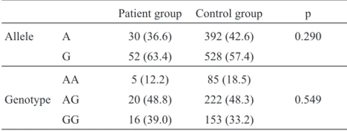
et al., 2000), and may be contributing to the age-associated Table 1- Allele and genotype frequencies of the -1185A/G polymorphism
of the VWF gene in type 1 VWD patient group (n = 41) and control group (n = 460).
Patient group Control group p
Allele A 30 (36.6) 392 (42.6) 0.290
G 52 (63.4) 528 (57.4)
AA 5 (12.2) 85 (18.5)
Genotype AG 20 (48.8) 222 (48.3) 0.549
GG 16 (39.0) 153 (33.2)
Values in parentheses are percentages.
Table 2- Mean of plasma VWF levels (U/dL) with regard to the -1185A/G genotypes.
Genotype Patient group Control group
Mean 95% CI Mean 95% CI
AA 28.4 22.1-34.7 122.1 112.7-132.5
AG 27.1 21.9-33.2 115.8 110.0-122.5
results of Keightleyet al. (1999). In our study we found a significant age effect in regard to plasma VWF levels, as has been reported in other studies (Colan et al., 1993; Kamphuisenet al., 1998; Kadiret al., 1999). Although the plasma VWF levels were adjusted for this effect no signifi-cant association between the –1185A/G polymorphism and plasma VWF levels was found.
Mohlke and Ginsburg (1997) have stated that the overlapping distribution of VWF levels observed between normal individuals and type 1 VWD patients suggest that VWD should be viewed more appropriately as a complex trait. As in other complex traits, it may not be possible to define a clear threshold plasma VWF level that distin-guishes type 1 VWD patients from normal subjects or that accurately predicts the development of symptoms. The ABO blood group is the main genetic variable known to be involved in the plasma VWF levels, and our data showed that there were significantly more O blood group individu-als among type 1 VWD patients, as previously reported (Gillet al., 1987). Several studies have shown that O blood group individuals have, on average, lower VWF levels than non-O blood group individuals (Gillet al., 1987; Shimaet al., 1995; Kamphuisenet al., 1998; Soutoet al., 2000), and perhaps, the O blood group trait and some SNPs in VWF gene would act in an addictive way to decrease VWF levels. This would account for some of the type 1 VWD patients, mainly those nearest to the lower limit of the normal distri-bution.
Our study was, for the first time, concerned with the relationship between VWD and the -1185A/G polymor-phism, the data showing that type 1 VWD patients do not have a higher G allele frequency than control subjects. Few mutations have been detected in type 1 VWD families, de-spite the efforts in this direction. The great majority of mu-tations described in type 1 VWD refer to null alleles segregating in type 3 VWD genealogies (Zhang et al., 1992; Eikenboomet al., 1993, 1998). Heterozygous rela-tives of type 3 VWD patients with a single null allele can exhibit a considerable variability, either being asymptom-atic or exhibiting a type 1 VWD phenotype (Zhanget al., 1995). This variability shows that the normal allele may have a significant influence on VWD expression. Further-more, only two mutations, both disrupting cysteine pairing in the D3 domain of the VWF gene, have been associated with classical dominant type 1 VWD (Eikenboomet al., 1996; Castamanet al., 2000), but, as with the null alleles, this does not appear a common event in type 1 VWD (Coughlanet al., 1999; Keeneyet al., 1999).
In conclusion, our data does not support the hypothe-sis that the –1185A/G polymorphism plays a role in the de-velopment of type 1 VWD disease, and apart from this, polymorphism was not associated with levels of VWF in normal Brazilian individuals. The role of other SNPs in the VWF gene should be investigated further.
Acknowledgments
This research was supported by the Programa de Apoio a Núcleos de Excelência (PRONEX), Financiadora de Estudos e Projetos (FINEP), and Conselho Nacional de Desenvolvimento Científico e Tecnológico (CNPq).
References
Castaman G, Eikenboom JCJ, Missiaglia E and Rodeghiero F (2000) Autossomal dominant type 1 von Willebrand disease due to G3639T mutation (C1130F) in exon 26 of von Willebrand factor gene: description of five Italian families and evidence for a founder effect. Br J Haematol 108:876-879.
Conlan MG, Folsom AR, Finch A, Davis CE, Sorlie P, Marcucci G and Wu KK (1993) Associations of factor VIII and von Willebrand factor with age, race, sex, and risk factors for atherosclerosis. The Atherosclerosis Risk in Communities (ARIC) study. Thromb Haemost 70:380-385.
Coughlan TC, Blagg JL, Abulola M, Daly ME, Hampton KK, Makris M, Peake IR and Goodeve AC (1999) Null alleles are not a common cause of type 1 von Willebrand disease in the British population. Thromb Haemost 82:1373-1375. Eikenboom JCJ, Reistma PH, Peerlinck KMJ and Briët E (1993)
Recessive inheritance of von Willebrand’s disease type 1. Lancet 341:982-986.
Eikenboom JCJ, Matsushita T, Reistma PH, Tuley EA, Castaman G, Briet E and Sadler JE (1996) Dominant type 1 von Willebrand disease caused by mutated cysteine residues in the D3 domain of von Willebrand factor. Blood 88:2433-2442.
Eikenboom JCJ, Castaman G, Vos HL, Bertina RM and Rode-ghiero F (1998) Characterization of the genetic defects in re-cessive type 1 and type 3 von Willebrand disease patients of Italian origin. Thromb Haemost 79:709-717.
Favaloro EJ and Koutts J (1997) Laboratory assays for von Willebrand factor: relative contribution to the diagnosis of von Willebrand’s disease. Pathology 29:385-391.
Fischer RR, Lucas EM, Pereira AMB and Roisenberg I (1996) Preparation of a heterologous antiserum for the determina-tion of von Willebrand factor in human plasma. Braz J Med Biol Res 29:1641-1644.
Gill JC, Endres-Brooks J, Bauer PJ, Marks Jr WJ and Montgom-ery RR (1987) The effect of ABO blood group on the diag-nosis of von Willebrand disease. Blood 69:1691-1695. Harvey PJ, Keightley AM, Lam YM, Cameron C and Lillicrap D
(2000) A single nucleotide polymorphism at nucleotide -1793 in the von Willebrand factor (VWF) regulatory region is associated with plasma VWF:Ag levels. Br J Haematol 109:349-353.
Heywood DM, Mansfield MW and Grant PJ (1996) Levels of von Willebrand factor, insulin resistance syndrome, and a com-mon VWF gene polymorphism in non-insulin-dependent (type 2) diabetes mellitus. Diabetic Med 13:720-725. Kadir RA, Economides DL, Sabin CA, Owens D and Lee C
(1999) Variations in coagulation factors in women: effects of age, ethnicity, menstrual cycle and combined oral contra-ceptive. Thromb Haemost 82:1456-1461.
Familial clustering of factor VIII and von Willebrand factor levels. Thromb Haemost 79:323-327.
Kamphuisen PW, Eikenboom JCJ, Rosendaal FR, Koster T, Blann AD, Vos HL and Bertina RM (2001) High factor VIII antigen levels increase the risk of venous thrombosis but are not associated with polymorphisms in the von Willebrand factor and factor VIII gene. Br J Haematol 115:156-158. Keeney S, Cumming A and Hay C (1999) Mutations in von
Willebrand factor multimerization domains are not a com-mon cause of classical type 1 von Willebrand disease. Thromb Haemost 82:1446-1450.
Keightley AM, Lam YM, Brady JN, Cameron CL and Lillicrap D (1999) Variation at the von Willebrand factor (VWF) gene locus is associated with plasma VWF:Ag levels: identifica-tion of three novel single nucleotide polymorphisms in the VWF gene promoter. Blood 93:4277-4283.
Lacquemant C, Gaucher C, Delorme C, Chatellier G, Gallois Y, Rodier M, Passa P, Balkau B, Mazurier C, Marre M and Froguel P (2000) Association between high von Willebrand factor levels and the Thr789Ala VWF gene polymorphism but not with nephropathy in type I diabetes. Kidney Int 57:1437-1443.
Lahiri DK and Nurnberger JI Jr (1991) A rapid non-enzimatic method for preparation of HMW DNA from blood for RFLP studies. Nucleic Acids Res 19:5444.
Levy G and Ginsburg D (2001) Getting at the variable expressivity of von Willebrand disease. Thromb Haemost 86:144-148.
Meena K, Marmot M and Brunner E (2000) Social determinants of von Willebrand factor: The Whitehall II study. Arterioscler Thromb Vasc Biol 20:1842-1847.
Mohlke KL and Ginsburg D (1997) von Willebrand disease and quantitative variation in von Willebrand factor. J Lab Clin Med 130:252-261.
Nichols WC and Ginsburg D (1997) von Willebrand disease. Medicine 76:1-20.
Rodeghiero F, Castaman G and Dini E (1987) Epidemiological in-vestigation of the prevalence of von Willebrand’s disease. Blood 69:454-459.
Sadler JE (1994) A revised classification of von Willebrand dis-ease. Thromb Haemost 71:520-525.
Sadler JE, Matsushita T, Dong Z, Tuley EA and Westfield LA (1995) Molecular mechanism and classification of von Willebrand disease. Thromb Haemost 74:161-166. Shima M, Fujimura Y, Nishiyama T, Tsujiuchi T, Narita N,
Matsui T, Titani K, Katayama M, Yamamoto F and Yoshioka A (1995) ABO blood group genotype and plasma von Willebrand factor in normal individuals. Vox Sang 68:236-240.
Simon D, Palatnik M and Roisenberg I (2002) Analysis of the -1185A/G von Willebrand factor (VWF) gene polymor-phism in two Brazilian ethnic groups and its effect on the plasma VWF levels. Thromb Res 105:519-522.
Souto JC, Almasy L, Muñiz-Diaz E, Soria JM, Borrell M, Bayen L, Mateo J, Madoz P, Stone W, Blangero J and Fontcuberta J (2000) Functional effects of the ABO locus polymorphism on plasma levels of von Willebrand factor, factor VIII, and activated partial thromboplastin time. Arterioscler Thromb Vasc Biol 20:2024-2028.
Souto JC, Almasy L, Soria JM, Buil A, Stone W, Lathrop M, Blangero J and Fontcuberta J (2003) Genome-wide linkage analysis of von Willebrand factor plasma levels: results from the GAIT project. Thromb Haemost 89:468-474. Vischer UM, Emeis JJ, Bilo HJG, Stehouwer CDA, Thomsen C,
Rasmussen O, Hermansen K, Wollheim CB and Ingerslev J (1998) von Willebrand factor (vWf) as a plasma marker of endothelial activation in diabetes: improved reliability with parallel determination of the vWf propeptide (vWf:AgII). Thromb Haemost 80:1002-1007.
Werner EJ, Broxson EH, Tucker EL, Giroux DS, Shults J and Abshire TC (1993) Prevalence of von Willebrand disease in children: A multiethnic study. J Pediatric 123:893-898. Zhang ZP, Lindstedt M, Falk G, Blombäck M, Egberg N and
Anvret M (1992) Nonsense mutations of the von Willebrand factor gene in patients with von Willebrand disease type III and type I. Am J Hum Genet 51:850-858.
Zhang ZP, Blombäck M, Egberg N, Falk G and Anvret M (1994) Characterization of the von Willebrand factor gene (VWF) in von Willebrand disease type III patients from 24 families of Swedish and Finnish origin. Genomics 21:188-193. Zhang ZP, Lindstedt M, Blombäck M and Anvret M (1995)
Ef-fects of the mutant von Willebrand factor gene in von Willebrand disease. Hum Genet 96:388-394.