online | memorias.ioc.fiocruz.br
Pseudomonas aeruginosa is a major agent of sepsis, a clinical syndrome characterised by the infection-trig-gered activation of the inflammatory and coagulation systems and by endothelial dysfunction (Esmon 2005).
The endothelium usually functions as an antithrom-botic surface and is critical for maintaining normal vas-cular homeostasis (Schouten et al.2008). However, under conditions of sepsis, endothelial cells are activated to se-crete and express surface procoagulant molecules such as tissue factor (TF) and von Willebrand factor (vWF) (Val-let & Wiel 2001). Activated endothelial cells also release increased amounts of procoagulant microparticles, which are small membrane-derived vesicles shed by cells fol-lowing activation or during apoptosis under a variety of pathophysiological circumstances (Martinez et al.2005). Microparticle membranes exhibit an antigen repertoire that is representative of the cell membranes from which they are derived, except for the loss of phospholipid asym-metry. Consequently, microparticle membranes contain anionic phosphatidylserine in their exoplasmic leaflet,
enabling the assembly of clotting enzyme complexes and the generation of thrombin. Hardly detectable in the pe-ripheral blood of healthy individuals, elevated levels of circulating procoagulant microparticlesare often associ-ated with thromboticpotential. The expression of TF (del Conde et al. 2005) and vWF (Jimenez et al.2003) on en-dothelial microparticle membranes further contributes to their potential role in hemostasis dysfunction.
VWF is a large multimeric glycoprotein that is syn-thesised in endothelial cells and megakaryocytes and plays a pivotal role in hemostatic plug formation and thrombosis (Ruggeri 2007). The main functions of vWF are to mediate the adhesion of platelets to sites of vas-cular injury and to act as a bridge molecule for plate-let aggregation, promoting the interaction of specific binding sites with the platelet surface receptors GP Iba and aIIbb-3 integrin (Ruggeri 2007, Varga-Szabo et al. 2008). High-molecular-weight vWF multimers, the most important factors for physiological platelet function, are released from endothelial cells towards both the lumen and the subendothelial matrix (Ruggeri 2003, 2007). In-creased concentrations of circulating vWF are detected in individuals with many pathologic conditions, includ-ing acute infectious diseases (Pottinclud-inger et al.1989,,Kayal et al.1998), and are generally accepted as a marker of en-dothelium activation/dysfunction (Vischer et al.2000).
One of the most important P. aeruginosa virulence factors is ExoU, a cytotoxin with phospholipase A2 activ-ity (Sato et al. 2003, Engel & Balachandran 2009). Pre-Financial support: CNPq, FAPERJ
CF was supported by a scholarship from CNPq. + Corresponding author: [email protected] Received 8 November 2011
Accepted 9 April 2012
The infection of microvascular endothelial cells
with ExoU-producing
Pseudomonas aeruginosa
triggers
the release of von Willebrand factor and platelet adhesion
Carla Freitas1, Maria-Cristina Assis2, Alessandra Mattos Saliba1, Veronica Maria Morandi3,
Camila Castro Figueiredo3, Mirian Pereira4, Maria-Cristina Plotkowski1/+
1Departamento de Microbiologia e Imunologia 3Departamento de Biologia Celular, Universidade do Estado do Rio de Janeiro, Rio de Janeiro, RJ, Brasil 2Departamento de Ciências Biológicas e da Saúde, Centro Universitário Estadual da Zona Oeste,
Rio de Janeiro, RJ, Brasil 4Departamento de Ultra-Estrutura e Biologia Celular-Fiocruz, Rio de Janeiro, RJ, Brasil
An increased plasma concentration of von Willebrand factor (vWF) is detected in individuals with many infec-tious diseases and is accepted as a marker of endothelium activation and prothrombotic condition. To determine whether ExoU, a Pseudomonas aeruginosa cytotoxin with proinflammatory activity, enhances the release of vWF, microvascular endothelial cells were infected with the ExoU-producing PA103 P. aeruginosastrain or an exoU -defi-cient mutant. Significantly increased vWF concentrations were detected in conditioned medium and subendothelial extracellular matrix from cultures infected with the wild-type bacteria, as determined by enzyme-linked immunoas-says. PA103-infected cells also released higher concentrations of procoagulant microparticles containing increased amounts of membrane-associated vWF, as determined by flow cytometric analyses of cell culture supernatants. Both flow cytometry and confocal microscopy showed that increased amounts of vWF were associated with cytoplasmic membranes from cells infected with the ExoU-producing bacteria. PA103-infected cultures exposed to platelet sus-pensions exhibited increased percentages of cells with platelet adhesion. Because no modulation of the vWF mRNA levels was detected by reverse transcription-polymerase chain reaction assays in PA103-infected cells, ExoU is likely to have induced the release of vWF from cytoplasmic stores rather than vWF gene transcription. Such release is likely to modify the thromboresistance of microvascular endothelial cells.
vious studies from our group have highlighted the abil-ity of ExoU to elicit a potent inflammatory response in microvascular endothelial cells (Saliba et al. 2005) and to enhance the release of soluble ICAM-1 from P. aeru- ginosa-infected endothelial cells (Lins et al. 2010), dem-onstrating the toxin’s ability to induce endothelial activa-tion/dysfunction (Gando et al. 2005). In the present study, we investigated whether ExoU would promote the release of vWF from endothelial cells, thereby contributing to enhanced platelet adhesion/aggregation and potentially to the generation of prothrombotic conditions.
SUBJECTS, MATERIALS AND METHODS
Bacterial strains and culture conditions - P. aerugi-nosa PA103 and the ExoU-deficient PA103ΔexoU mu-tant (Saliba et al. 2005) were used throughout this study. Bacteria weregrown in Luria-Bertani broth at 37ºC for 16-18 h, harvested by centrifugation and resuspended in MCDB 131 cell culture medium (Sigma-Aldrich) supplemented with 10% foetal calf serum, 10 ng mL-1
EGF (Sigma-Aldrich), 1 μg mL-1hydrocortisone (Sigma-Aldrich) and glutamine (complete culture medium).
Cell culture and infection - Cells from thehuman mi-crovascular endothelial cell line-1 (HMEC-1) (Ades et al. 1992) were cultured in complete MCDB-131 culture medium for 48 h. Bacterial suspensions were added to the cell cultures at a multiplicity of infection of approxi-mately 100 bacteria per cell and centrifuged (1,000 g for 10 min) to promote close contact between the bacteria and host cells (Saliba et al.2005). Control cultures were exposed to culture medium alone. After incubation for 1 h at 37ºC, cells were incubated with culture medium
containing 300 μg/mL gentamicin for different lengths
of time to reduce the bacterial challenge and to assess the host response at later time points (Saliba et al. 2005, Lins et al.2010).
Detection of released vWF - To detect vWF released into the cell supernatant, cells cultured in 6-well plates (4.5 x 10⁵ cells/well) were infected for 1 h, rinsed and incubat-ed with gentamicin-containing culture mincubat-edium for 72 h (Ribeiro et al. 1995). The conditioned media from control and infected cultures were then concentrated 20-fold with a Speed Vac Plus (Savant) and the vWF concentration was assayed in duplicate by ELISA using purified rabbit anti-human vWF and a horseradish peroxidase-conjugat-ed anti-vWF secondary antibody (Dako), as previously described (Bonnefoy et al. 2001). A standard curve was constructed from serial dilutions of normal pooled human plasma, assuming a vWF concentration of 1 unit/mL.
To detect the released vWF that was incorporated into the subendothelial matrix, cells were cultured in 96-well dishes (0.3 x 10⁴ cells/well), infected for 1 h (or not in-fected) and treated with gentamicin-containing medium for 72 h. Cells were then washed with phosphate buffered saline (PBS) and treated for 20 min with PBS containing 0.1 M NH4OH and 0.1% Triton X-100 to expose the na-tive subendothelial matrix (Bonnefoy et al. 2001). After washing, the wells of the microtitre plate were fixed with 4% paraformaldehyde, treated with 1% bovine serum al-bumin (BSA) in PBS and exposed to the
anti-vWF-horse-radish peroxidase conjugate. The concentrations of vWF in the supernatant and the subendothelial matrix were normalised and reported in U/mL released by 105 cells.
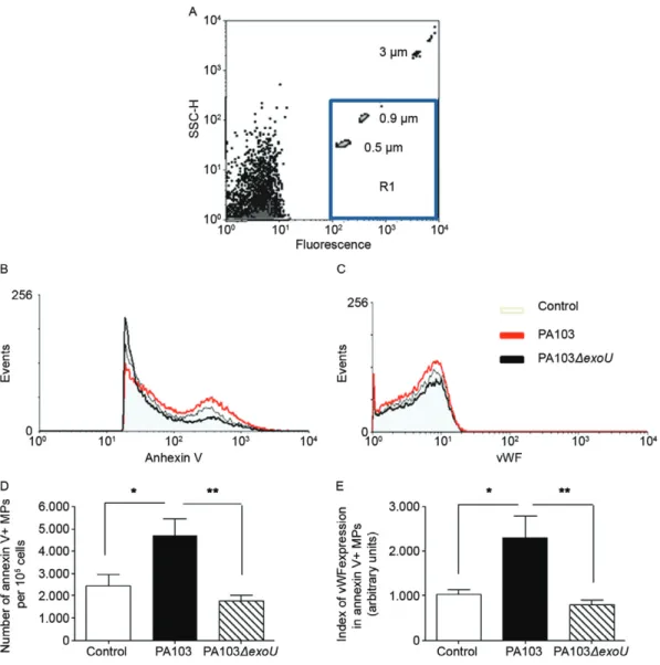
Detection of vWF-expressing microparticles in cell supernatants - Cells were infected for 1 h and then treated with gentamicin-containing medium. Control cells were treated similarly with antibiotic-containing medium. After incubation at 37ºC for 19 h, the incubation media were cleared of cell debris by centrifugation (5,000 g for 15 min) and then centrifuged at 17,500 g for 30 min at 15ºC (Plotkowski et al. 2008). Sedimented microparticles were washed and treated for 30 min at room temperature (RT) with annexin V-Alexa Fluor 647 complexes (Invitro-gen) to identify surface phosphatidylserine residues. After washing, microparticles were suspended in PBS contain-ing 1% BSA and analysed by flow cytometry. The region corresponding to shed microparticles was defined in side-angle light scatter vs. fluorescent intensity dot plot
rep-resentations using 0.5 μm and 0.9 μm fluorescent beads
(Megamix) as a reference, according to the manufacturer’s instructions. Because ExoU cytotoxicity leads to the par-tial death of PA103-infected cultured cells, the numbers
of viable cells in the PA103 and PA103ΔexoU-infected culture wells and in the control uninfected culture wells were determined in parallel after the 1 h infection period. The microparticle concentrations detected in the cell cul-ture supernatants were then normalised and reported as microparticles shed by 105 cells. In other assays, before treatment with the annexin V complex, sedimented mi-croparticles were incubated with rabbit anti-human vWF for 1 h and then incubated for 30 min at RT with goat anti-rabbit IgG-FITC. After washing, microparticles were analysed by flow cytometry. The results were expressed as a “vWF-expression index” as described (Leytin et al. 2000) and this value was obtained by the following for-mula: percentage of vWF-expressing microparticles x median microparticle fluorescence intensity.
Detection of cell-associated vWF - Two different ap-proaches were used to determine whether any portion of the secreted vWF remained associated with the cultured cells: flow cytometry and confocal microscopy analy-sis of nonpermeabilized cells. For the flow cytometry analysis, cells were infected for 1 h, treated with anti-biotic-containing medium for an additional 5 h, washed with PBS to remove all microparticles that were associ-ated with cell membranes, detached from the wells by trypsinisation and fixed in 4% paraformaldehyde in PBS containing 4% sucrose for 20 min. After washing with PBS, cells were treated with a rabbit anti-human vWF antibody for 1 h at 4ºC, washed and incubated with a goat anti-rabbit IgG-FITC complex (Santa Cruz Biotechnol-ogy). The cells were then washed again and suspended in PBS containing 1% BSA. At least 5,000 cells were analysed by flow cytometry. Control uninfected cells were processed similarly. The results were expressed as a “vWF-expression index”, calculated as described above.
membranes, fixed with 4% paraformaldehyde and stained with anti-vWF antibody and goat anti-rabbit IgG-Cy3 complex (Zymed). In some assays, cells were subsequent-ly stained with phalloidin-FITC (Sigma-Aldrich), washed and incubated with Topro (Invitrogen) for 5 min. Cover-slips were mounted on glass slides and observed under a Zeiss LSM 510 Meta confocal microscope.
Detection of vWF mRNA by reverse transcription-polymerase chain reaction (RT-PCR) - Total RNA was isolated from uninfected (control) HMEC-1 cells or cells infected for 1 or 3 h using the Qiagen RNeasy kit. cDNA was synthesised from the total RNA by RT with the SuperScript™ First-Strand Synthesis System for RT-PCR (Invitrogen). The isolated cDNA was subjected to PCR under the following conditions: denaturation at 95ºC for 2 min, 33 (vWF) or 23 (ß-actin) cycles of de-naturation at 95ºC for 45 sec, annealing at 50ºC for 45 sec and extension at 72ºC for 45 sec, and an addition-al extension step of 5 min at 72ºC after the last cycle. The primers used in the reactions were 5’-AGTTCAT-GGAGGAGGTGATTCAGC-3’ (vWF sense), 5’-AGC-CATCCAGGAGAAGGATCACG-3’ (vWF antisense), 5’-CCTCGCCTTTGCCGATCC-3’ (ß-actin sense) and 5’-GGATCTTCATGAGGTAGTCAGTC-3’ (ß-actin an-tisense). PCR products were subjected to electrophoresis in a 1% agarose gel and densitometry was performed us-ing Lab Image software (Kaplan GmbH, Germany).
Platelet adhesion to P. aeruginosa-infected cells - To investigate the relationship between increased vWF re-lease by PA103-infected cells and enhanced platelet ad-hesion, cells cultured on glass coverslips were infected for 1 h and then treated with gentamicin-containing cul-ture medium for additional 5 h. In parallel, human blood was collected in ACD anticoagulant solution (130 mM citric acid, 153 mM citrate trisodium and 111 mM glu-cose; pH 6.5) containing PGE1 at 1 U/mL and centrifuged at 250 g for 8 min. The resulting platelet-rich plasma was centrifuged at 1,500 g for 10 min. Sedimented platelets were washed in platelet buffer (36 mM citric acid, 5 mM glucose, 5 mM KCl, 2 mM CaCl2, 1 mM MgCl2 and 103 mM NaCl; pH 6.4) and resuspended at a concentration of 1.0 x 10⁷ platelets/mL. Infected and control uninfected
endothelial cells were then incubated with the platelet suspension for 30 min at RT. In other assays, platelet sus-pensions were perfused over endothelial cells for 5 min (10 mL/min) with a peristaltic pump (Pump-1, Pharmacia Biotech). Cells were then washed, fixed for 20 min with 4% paraformaldehyde in PBS containing 4% sucrose and stained with May-Grunwald-Giemsa stain. Coverslips were mounted on glass slides and examined with a Zeiss Axioplan optical microscope. The percentage of cells with adherent platelets present in at least 10 different mi-croscopic fields was determined in two different assays, both of which were carried out in duplicate.
Statistical analysis - The results were expressed as the means ± standard error of the means of data obtained in at least three different experiments. Significant dif-ferences between groups were determined with one-way analysis of variance followed by Dunnett’s or Bonfer-roni’s multiple comparisons post-test. Statistical signifi-cance was set at a pvalue of < 0.05.
RESULTS
ExoU enhanced the release of vWF and vWF-bear-ing microparticles from microvascular endothelial cells - As shown in Fig. 1, the concentrations of vWF in both the cell culture supernatant (Fig. 1A) and subendothe-lial matrix (Fig. 1B) of endothesubendothe-lial cells infected with the ExoU-producing bacterial strain were significantly higher than those in control cultures or in cultures in-fected with the exoU-deficient mutant.
ExoU similarly enhanced the release of microparti-cles containing anionic phosphatidylserine in their exo-plasmic leaflet, as revealed by their reactivity with the annexin V-FITC complex (Fig. 2B, D). More important-ly, the vWF expression indices for microparticles shed after PA103 infection were significantly higher than those for microparticles shed by control cells or by cells
infected with the PA103ΔexoU mutant (Fig. 2C, E). PA103-infected cells exhibited increased vWF expres-sion - Studies have shown that vWF multimers released from endothelial cells are anchored to the endothelial sur-face in the form of structures that are capableof binding platelets before being cleaved by plasma metalloproteases
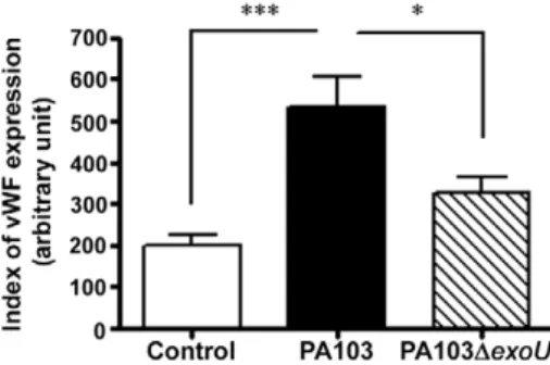
(Dong et al. 2002). To investigate whether PA103-infect-ed cells harbour increasPA103-infect-ed amounts of surface-associatPA103-infect-ed vWF, nonpermeabilized control and infected cells were treated with an anti-vWF antibody and submitted to flow cytometry and immunocytochemistry analyses.
As shown in Fig. 3, the vWF-expression index of cells infected with the wild-type bacteria was significantly higher than those of uninfected control cells and cells infected with the mutant bacteria. Confocal microscopy analyses confirmed the increased expression of vWF on the surface of PA103-infected cells (Fig. 4).
Because ExoU is a phospholipase, we wondered whether the increased staining of PA103-infected cells with the anti-vWF antibody, detected by both flow cytometry and confocal microscopy, may have been
caused by the permeabilization of cell membranes by the toxin; this permeabilization would enable the anti-vWF antibody to penetrate infected cells and stain anti-vWF stored in Weibel Palade bodies. To address this question, nonpermeabilized fixed control and infected cells were treated with phalloidin-FITC, which labels cytoplasmic actin and with the anti-vWF antibody and then examined by confocal microscopy. Control and infected cells were equally poorly labelled with phalloidin-FITC (data not shown), thereby ruling out the false-positive staining of PA103-infected cells.
ExoU did not modulate the level of VWF mRNA ex-pressed by infected cells - To determine whether the increased vWF expression and release from PA103-in-fected cells depended on the modulation of vWF mRNA
Fig. 2: release of microparticles (MPs) by control and infected endothelial cells detected by flow cytometry analysis. A: dot plot histogram show-ing the distribution of fluorescent beads used for the flow cytometer settshow-ing before analysis of MPs in region defined as R1; B: representative his-togram showing increased number of annexin V-labelled MPs released by PA103-infected cells; C: representative hishis-togram showing increased number of annexin V positive MPs expressing von Willebrand factor (vWF) released by PA103-infected cells; D: mean number and standard error of the means (SEM) of MPs released by 105 cells, as determined in three assays carried out in triplicate; E: index of vWF expression by
levels, we performed RT-PCR assays. No difference in mRNA levels was detected between control and P. aeruginosa-infected cells (data not shown).
ExoU enhanced platelet adhesion to infected en-dothelial cells - To evaluate whether the ExoU-induced release of vWF favoured platelet adhesion to endothelial cells, platelet suspensions were either incubated with or perfused over HMEC-1 cells cultured on glass cover-slips. In PA103-infected cultures incubated with plate-lets under static conditions, the percentage of cells with adherent platelets was significantly higher than that in control cultures or in cultures infected with the exoU mutant (Fig. 5A). More interestingly, PA103-infected cultures exposed to platelets under flow conditions for only 5 min also exhibited a significantly higher percent-age of cells with adherent platelets (Fig. 5B). No signifi-cant difference in the percentage of cells with adherent platelets was observed between cultures infected with the bacterial mutant and control cultures.
DISCUSSION
Endothelial cells usually provide an ideal surface for blood flow by inhibiting platelet adhesion and the initiation of blood clotting. In contrast, platelets adhere avidly to activated or injured endothelium via several adhesion molecules that are expressed on these cells (Wu & Thiagarajan 1996, Varga-Szabo et al. 2008). Because P. aeruginosa ExoU maydirectly damage endothelial cells (Saliba et al. 2006), we investigated whether this toxin might alter the thromboresistant phenotype of the microvascular endothelium and pro-mote platelet adhesion.
We demonstrated that ExoU enhanced the release of vWF into the endothelial cell culture supernatant and subendothelial matrix. One explanation for this phenomenon might be that arachidonic acid released by PA103-infected cells, as previously reported by our group (Saliba et al. 2005), enhances the fusion of Weibel Palade bodies with the endothelial plasma membrane, resulting in vWF exocytosis (Connell et al. 2007, Darios et al. 2007). A second potential explanation is that vWF release may have resulted from ExoU-induced endothe-lial cell lysis. Because a similar injury of endotheendothe-lial
cells is likely to occur in vivo, the vWF release reported here is likely relevant; the immobilisation of vWF re-leased towards the lumen of blood vessels on exposed subendothelial matrix is considered to be the predomi-nant mechanism that initiates platelet response in areas of vascular injury (Ruggeri 2003).
A proposed model for the regulation of vWF release is that upon stimulation, ultra-large vWF multimers are released and immediately anchored to the surface of endothelial cells via binding to P-selectin. These mul-timers are then stretched by fluid shear stress into an open conformation and cleaved by the metalloproteinase ADAMTS-13 into fragments of various sizes that are de-tected in the plasma of healthy individuals (Dong et al. 2002, Padilla et al. 2004). Because cultured endothelial cells are not exposed to fluid shear, uncleaved ultra-large vWF is likely to remain anchored to cell membranes. Such anchorage would explain both the increased ex-pression of surface-associated vWF, as detected by confocal microscopy and the increased adhesion of non-stimulated platelets to PA103-infected cells.
The physiological relevance of vWF-mediated plate-let adhesion to endothelial cells, rather than to sites of endothelial injury, has been highlighted by in vivo studies showing that resting platelets adhere to intact mouse mesenteric venules treated with a secretagogue of Weibel Palade bodies through a vWF-dependent and P-selectin-independent mechanism (Sporn et al. 1986, Andre et al. 2000). Although platelets recruited to the endothelium may release proinflammatory cytokines and modulate leukocytefunction, thereby contributing to local defence mechanisms, they can also initiate del-eterious thrombotic events.
Another finding from our study that may have in vivo relevance is the enhanced release of vWF-expressing mi-croparticles from PA103-infected cells. Mimi-croparticles released from activated or apoptotic endothelial cells participate in platelet aggregation owing to the pres-ence of functional adhesive glycoproteins in the plasma membranes. These glycoproteins bind to the A1 domain of immobilised and soluble vWF (Reininger 2008). In addition, high percentages of endothelial-derived mi-croparticles (EMP) express ultra-large vWF multimers (Jimenez et al. 2003, Jy et al. 2005, Othman et al. 2007). The multimeric size of vWF is important for its function because lower molecular weight multimersshow less binding to platelets and to collagen (Doucet-de Bruine et al. 1978, Santoro et al.1983). In a study on the interac-tion between EMP and platelets, Jy et al. (2005) high-lighted the importance of the large molecular weight vWF expressed by EMP. In the presence of ristocetin, EMP induced the aggregation of up to 95% of platelets and EMP-induced platelet aggregates were more stable than those induced by free soluble vWF. These results further support the procoagulant properties of endothe-lial microparticles.
In conclusion, this report describes the effects of en-dothelial cell infection with the ExoU-producing PA103 P. aeruginosa, showing that ExoU enhanced vWF release and that at least a portion of the released vWF remained associated with the cell surface. ExoU also enhanced the Fig. 3: index of cell-associated von Willebrand factor (vWF) in control
release of prothrombotic vWF-bearing microparticles and platelet adhesion to infected cells. Although it is tempting to conclude that there is a cause-effect relation-ship between the increased expression of cell-associated vWF and increased platelet adhesion to PA103-infected cells, further studies are necessary to prove that vWF is involved in platelet-endothelial cell association.
In recent years, our laboratory has made progress in understanding the contribution of ExoU to the pathogen-esis of P. aeruginosa infections. We first demonstrated that ExoU may induce a procoagulant activity in airway epithelial cells, resulting from the upregulated expres-sion of TF and the increased release of tissue factor-positive microparticles (Plotkowski et al. 2008). More recently, Machado et al. (2010) demonstrated that ExoU can induce vascular hyper-permeability, platelet
acti-vation and thrombus formation in the lungs and renal microvasculature of mice with experimental P. aerugi-nosa pneumosepsis; these authors also observed an anti-fibrinolytic environment in mice airways secondary to the induction of enhanced plasminogen activator inhib-itor-1 production (Machado et al. 2011). These results, as well as those from the present report, demonstrate the potential of ExoU to induce a prothrombotic state in host organisms.
ACKNOWLEDGEMENTS
To Maria Angélica P da Silva and Pedro Paulo de Abreu Manso, for her technical assistance, Dr Marcelo Santiago (Instituto de Biofísica Carlos Chagas Filho, UFRJ), for his support with confocal microscopy, and to PDTIS, for use of its facilities.
Fig. 4: confocal microscopy detection of von Willebrand factor (labelled in red) at the surface of nonpermeabilized paraformaldehyde-fixed
control (A), PA103 (B) and PA103ΔexoU-infected endothelial cells (C). Cell nuclei appear labelled in blue.
REFERENCES
Ades EW, Candal FJ, Swerlick RA, George VG, Summers S, Bosse DC, Lawley TJ 1992. Establishment of an immortalized human micro-vascular endothelial cell line. J Invest Dermatol99: 683-690.
Andre P, Denis CV, Ware J, Saffaripour S, Hynes RO, Ruggeri ZM, Wagner DD 2000. Platelets adhere to and translocate on von Willebrand factor presented by endothelium in stimulated veins.
Blood 96: 3322-3328.
Bonnefoy A, Harsfalvi J, Pfliegler G, Fauvel-Lafève F, Legrand C 2001. The subendothelium of the HMEC-1 cell line supports thrombus formation in the absence of von Willebrand factor and collagen types I, III and VI. Thromb Haemos85: 552-559.
Connell E, Darios F, Broersen K, Gatsby N, Peak-Chew S, Rickman C, Davletov B 2007.Mechanism of arachidonic acid action on syntaxin-Munc18. EMBO Rep8: 414-419.
Darios F, Connell E, Davletov B 2007. Phospholipases and fatty acid signalling in exocytosis. J Physiol585: 699-704.
del Conde I, Shrimpton CN, Thiagarajan P, López JA 2005. Tissue-factor-bearing microvesicles arise from rafts and fuse with acti-vated platelets to initiate coagulation. Blood106: 1604-1611.
Dong J, Moake JL, Nolasco L, Bernardo A, Arceneaux W, Shrimpton CN, Schade AJ, McIntire LV, Fujikawa K, Lopez JA 2002. AD-AMTS-13 rapidly cleaves newly secreted ultralarge von Wille-brand factor multimers on the endothelial surface under flowing conditions. Blood100: 4033-4039.
Doucet-de Bruine MHM, Sixma JJ, Over J, Beeser-Visser NH 1978. Heterogeneity of factor VIII: characterization of forms of factor VIII binding to platelets in the presence of ristocetin. J Lab Clin Med92: 96-107.
Engel J, Balachandran P 2009. Role of Pseudomonas aeruginosa type III effectors in disease. Curr Opin Microbiol12: 61-66.
Esmon CT 2005. The interactions between inflammation and coagu-lation. Br J Haematol131: 417-430.
Gando S, Kameue T, Matsuda N, Hayakawa M, Hoshino H, Kato H 2005. Serial changes in neutrophil-endothelial activation markers during the course of sepsis associated with disseminated intra-vascular coagulation. Thromb Res116: 91-100.
Jimenez JJ, Jy W, Mauro LM, Horstman LL, Soderland C, Ahn YS 2003. Endothelial microparticles released in thrombotic throm-bocytopenic purpura express von Willebrand factor and markers of endothelial activation. Br J Haematol123: 896-902.
Jy W, Jimenez JJ, Mauro LM, Horstman LL, Cheng P, Ahn ER, Bidot CJ, Ahn YS 2005. Endothelial microparticles induce formation of platelet aggregates via a von Willebrand factor/ristocetin depen-dent pathway, rendering them resistant to dissociation. J Thromb Haemost3: 1301-1308.
Kayal S, Jais JP, Aguini N, Chaudiere J, Labrousse J 1998. Elevated circulating E-selectin, intercellular adhesion molecule 1 and von Willebrand factor in patients with severe infection. Am J Respir Crit Care Med157: 776-784.
Leytin V, Mody M, Semple JW, Freedman J 2000. Flow cytometric parameters for characterizing platelet activation. Biochem Bioph Res Comm269:85-90.
Lins RX, de Assis MC, Mallet de Lima CD, Freitas C, Plotkowski MC, Saliba AM 2010. ExoU modulates soluble and membrane-bound ICAM-1 in Pseudomonas aeruginosa-infected endothelial cells. Microb Infect12: 154-161.
Machado GB, de Assis MC, Leão R, Saliba AM, Silva MC, Suas-suna JH, de Oliveira AV, Plotkowski MC 2010. Exou-induced vascular hyperpermeability and platelet activation in the course of experimental Pseudomonas aeruginosa pneumosepsis. Shock 33: 315-321.
Machado GB, de Oliveira AV, Saliba AM, de Lima CD, Suassuna JH, Plotkowski MC 2011. Pseudomonas aeruginosa toxin ExoU
in-duces a PAF-dependent impairment of alveolar fibrin turnover secondary to enhanced activation of coagulation and increased expression of plasminogen activator inhibitor-1 in the course of mice pneumosepsis. Respir Res 5: 104.
Martinez MC, Tesse A, Zobairi F, Andriantsitohaina R 2005. Shed mi-croparticles from circulating and vascular cells in regulating vascu-lar function. Am J Physiol Heart Circ Physiol288: H1004-H1009.
Othman M, Labelle A, Mazzetti I, Elbatarny HS, Lillicrap D 2007. Adenovirus-induced thrombocytopenia: the role of von Wil-lebrand factor and P-selectin in mediating accelerated platelet clearance. Blood109: 2832-2839.
Padilla A, Moake JL, Bernardo A 2004. P-selectin anchors newly re-leased ultralarge von Willebrand factor multimers to the endothe-lial cell surface. Blood103: 2150-2156.
Plotkowski MC, Feliciano LFP, Machado GBS, Cunha Jr LG, Frei-tas C, Saliba AM, Assis MC 2008. ExoU-induced procoagulant activity in Pseudomonas aeruginosa-infected airway cells. Eur Respir J32: 1591-1598.
Pottinger BE, Read CR, Paleolog EM, Higgins PG, Pearson JD 1989. von Willebrand factor is an acute phase reactant in man. Thromb Res15: 387-394.
Reininger AJ 2008. VWF attributes impact on thrombus formation.
Thromb Res122 (Suppl. 4): S9-S13.
Ribeiro MJA, Phillips DJ, Benson JM, Evatt BL, Ades EW, Hooper WC 1995. Hemostatic properties of the SV-40 transfected human micro-vascular endothelial cell line (HMEC-1). Thromb Res79: 153-161.
Ruggeri ZM 2003. von Willebrand factor, platelets and endothelial cell interactions. J Throm Haemost1: 1335-1342.
Ruggeri ZM 2007. The role of von Willebrand factor in thrombus for-mation. Thromb Res120 (Suppl. 1): S5-S9.
Saliba AM, Assis MC, Nishi R, Raymond B, Marques EA, Lopes UG, Touqui L, Plotkowski MC 2006. Implications of oxidative stress in the cytotoxicity of Pseudomonas aeruginosa ExoU. Microb Infect8: 450-459.
Saliba AM, Nascimento DO, Silva MCA, Assis MC, Gayer CRM, Raymond B, Coelho MGP, Marques EA, Touqui L, Albano RM, Lopes UG, Paiva DD, Bozza PT, Plotkowski MC 2005. Eicosa- Eicosa-noid-mediated proinflammatory activity of Pseudomonas aeru-ginosa ExoU. Cell Microbiol7: 1811-1822.
Santoro SA 1983. Preferential binding of high molecular weight forms of von Willebrand factor to fibrillar collagen. Biochim Biophys Acta756: 123-126.
Sato H, Frank DW, Hillard CJ, Feix JB, Pankhaniya RR, Moriyama K, Finck-Barbançon V, Buchaklian A, Lei M, Long RM, Wie-ner-Kronish J, Sawa T 2003. The mechanism of action of the
Pseudomonas aeruginosa-encoded type III cytotoxin, ExoU.
EMBO J22: 2959-2969.
Schouten M, Wiersinga WJ, Levi M, van der Poll T 2008. Inflammation, endothelium and coagulation in sepsis. J Leukoc Biol83: 1-10.
Sporn LA, Marder VJ, Wagner DD 1986. Inducible secretion of large biologically potent von Willebrand factor multimers. Cell46: 185-190.
Vallet B, Wiel E 2001. Endothelial cell dysfunction and coagulation.
Crit Care Med29 (Suppl. 7): S36-S41.
Varga-Szabo D, Pleines I, Nieswandt B 2008. Cell adhesion mecha-nisms in platelet. Arterioscler Thromb Vasc Biol28: 403-412.
Vischer UM, Barthm H, Wollheim CB 2000. Patterns of cytoskeletal remodeling in cultured endothelial cells regulated von Willebrand factor secretion is associated with agonist-specific. Arterioscler Thromb Vasc Biol 20: 883-891.