MEETING REVIEW
Trends in tissue repair and regeneration
Brigitte Galliot1,*, Marco Crescenzi2, Antonio Jacinto3and Shahragim Tajbakhsh4
ABSTRACT
The 6th EMBO conference on the Molecular and Cellular Basis of Regeneration and Tissue Repair took place in Paestum (Italy) on the 17th-21st September, 2016. The 160 scientists who attended discussed the importance of cellular and tissue plasticity, biophysical aspects of regeneration, the diverse roles of injury-induced immune responses, strategies to reactivate regeneration in mammals, links between regeneration and ageing, and the impact of non-mammalian models on regenerative medicine.
KEY WORDS: Cell plasticity and cell reprogramming, Blastema dynamics, Model systems, Immune response, Transcriptional control of regeneration, Wound healing, Injury-induced signaling, Senescence and ageing
Introduction
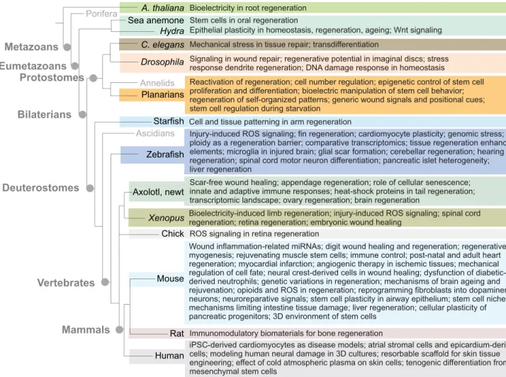
Since 2002, a series of conferences dedicated to the mechanisms underlying tissue repair and regeneration has taken place in Europe every other year, and under the EMBO umbrella since 2006. This year, the conference was organized by Patrizia Ferretti (University College London), James Godwin (The Jackson Laboratory), Sophie Jarriault (Strasbourg University), Nadia Mercader (Bern University), Henry Roehl (Sheffield University) and Andras Simon (Karolinska Institute). This conference series has become the key international meeting for scientists investigating basic biological questions in regeneration and repair using a whole animal or an organ system approach. The aim is to provide a multidisciplinary and integrative platform where scientists using a wide variety of models, including plants, cnidarians, planarians, insects, nematodes, echinoderms, fish, amphibians, birds, rodents, and human organoids, and also those developing innovative biomaterials (Fig. 1) can meet to promote creative thinking and foster new collaborations. A key goal is to identify the mechanisms that underlie injury-induced regeneration as evolutionarily conserved processes and/or as taxon-specific innovations, as well as the barriers that prevent regeneration in mammals and the factors that lead to the deterioration of tissue repair with age.
In their seminal review on comparative aspects of animal regeneration, Brockes and Kumar (2008) wrote: “regenerative phenomena present a continuum in relation to mechanisms, and exact definitions can be difficult to justify”.However, there is an emerging view to split this continuum into three major processes: wound healing, tissue repair, and regeneration. The first, which corresponds to the healing of injury with full or partial functional
restoration (typically the skin), is often replaced by fibrotic scarring in mammals; the second involves functional restoration of the injured organ with no patterned 3D reconstruction (e.g. heart, muscles, liver, lung, bone); the third involves regrowth and 3D patterning of a complex structure, such as appendages or body parts, and relies on blastema formation. Both stem and differentiated cells can contribute to blastema formation and this process involves various forms of cellular plasticity triggered by injury, namely cell cycle re-entry, changes in cell proliferation, cell dedifferentiation and cell transdifferentiation (Galliot and Ghila, 2010; Stocum and Cameron, 2011; Tanaka and Reddien, 2011).
To understand the regenerative responses to injury and to develop therapeutic approaches, it is crucial to understand how homeostatic tissues initiate the regeneration program by triggering a coherent immune response, appropriate cell plasticity, as well as stem and stromal cell responses following injury. We discuss here the strategies and key advances reported at this conference.
Wound repair and injury-induced signaling
Among the first responses to injury is the production of reactive oxygen species (ROS), which appear as universal injury-induced signals, although their sources and activities are diverse. Several questions were debated during the meeting: what makes ROS signals sufficient to trigger regeneration? How do ROS signals initiate regeneration? When do ROS signals impede regeneration? In Xenopustadpoles, where a localized and sustained ROS production is necessary for tail regeneration (Love et al., 2013), Enrique Amaya (Manchester University, UK) showed that NADPH oxidases from wounded tissues, but not from inflammatory cells, produce ROS. Interestingly, during early embryonic development, mitochondrial ROS production is triggered by Ca2+bursts, resulting in the regulation of TGFβ/Nodal and Wnt signaling and impacting on the cell cycle. Provocatively, Amaya proposed that, upon wounding, adult tissues return to an embryonic-like oxidative state favoring regeneration, and that fertilization acts as an injury-like signal.
Henry Roehl (Sheffield University, UK) showed that during larval tail regeneration in zebrafish, ROS directly activate the expression of Hedgehog ligands, which in turn activate other known regenerative pathways, possibly Fgf10a and Wnt10a signals, the latter of which is essential for regeneration. Interestingly, Hedgehog signaling is not required for embryonic tail development, providing a clear example of divergence between development and regeneration. However, ROS signaling can also be deleterious for regeneration. The chick embryo provides a classical model to understand how the retina regenerates, either via transdifferentiation of the retinal pigment epithelium or through the proliferation of progenitors in the ciliary marginal zone (CMZ). Nancy Echeverri (Del Rio-Tsonis laboratory, Miami University, Oxford, USA) showed that the antioxidant N-acetylcysteine (NAC) induces regeneration by promoting progenitor proliferation from the CMZ. NAC activates the MAPK pathway and counteracts the ROS-promoted cell differentiation. These findings indicate that tissue sensitivity to redox changes constrains the regeneration process.
1Department of Genetics and Evolution, Institute of Genetics and Genomics in
Geneva (iGE3), University of Geneva, CH-1211 Geneva 04, Switzerland.
2Department of Cell Biology and Neurosciences, National Institute of Health, I-00161
Roma, Italy.3CEDOC, NOVA Medical School, NOVA University of Lisbon, Lisboa
1169-056, Portugal.4Department of Developmental & Stem Cell Biology, Stem Cells
& Development Unit, CNRS UMR 3738, Institut Pasteur, 75015 Paris, France.
*Author for correspondence ([email protected])
B.G., 0000-0001-7596-8284; A.J., 0000-0002-4193-6089; S.T.,
0000-0003-1809-7202
DEVEL
O
To identify regulators of scarless wound healing, Karen Echeverri (Minneapolis University, USA) compared the expression profiles of axolotl and human wounds and identified the transcription factor Sall4 as upregulated exclusively in axolotl. Sall4 appears to prevent scarring: Sall4-depleted axolotls deposit higher levels of collagen I and collagen XII after injury and display a scar-like phenotype. Cutaneous wound healing is compromised in peripheral neuropathies. Investigating this phenomenon in mouse, Vadims Parfejevs (Sommer laboratory, Zürich University, Switzerland) reported that, upon injury, peripheral nerve-associated glia appear to dedifferentiate and acquire neural crest stem cell (NCSC) properties, contributing to the regenerating tissue. This process is compromised inSox10conditional mutants and is associated with reduced TGFβ
signaling. Hence, NCSCs originating from peripheral nerve glia might widely contribute to skin repair.
Unlike the commonly used C57BL/6 mouse, the MRL inbred mouse strain exhibits a remarkable ability to completely regenerate ear hole punches. Ellen Heber-Katz (LIMR, Wynnewood, USA) reported that HIF1αplays a crucial role in this process, with its knockdown blocking healing. In non-regenerative mice, pharmacological inhibition of HIF1αdegradation leads to MRL-type ear hole wound closure (Zhang et al., 2015). Cécile Dromard (Casteilla laboratory, EFS, Toulouse, France) uses lipectomy (excision of adipose tissue) to monitor regeneration in adult MRL and C57BL/6 mice. Unlike C57BL/6 mice, MRL mice display high pain sensitivity and transient ROS activity upon lipectomy. Exposing MRL mice to an opioid receptor agonist abolishes ROS
activity and compromises regeneration, whereas blocking the opioid receptor in C57BL/6 mice induces ROS production from granulocytes and promotes regeneration. These comparative mechanistic studies provide a valuable framework to identify factors that control tissue regeneration.
Not all wounds initiate regeneration, and the signals that convert a generic wound response to a regenerative process are unknown (Wurtzel et al., 2015; Owlarn and Bartscherer, 2016). Kerstin Bartscherer (MPI, Muenster, Germany) showed that by blocking wound-induced ERK activity in planarians, amputated animals close their wound but do not regenerate until ERK activity is restored, thereby dissociating these two processes. She terms this delayed permissive statusʻdormant’, a status that can also be evidenced in zebrafish (in collaboration with the Weidinger laboratory). Finally, Antonio Jacinto (CEDOC, Lisbon, Portugal) reported that the Toll pathway is required for wound healing in Drosophila epithelia, independently of its known role in the innate immune response. Toll controls the tissue repair response through the regulation of E-cadherin remodeling and cell proliferation (Carvalho et al., 2014).
Anti- and pro-regenerative immune responses
Inflammation is generally thought to promote fibrosis, an irreversible barrier to regenerative processes. However, macrophage depletion produces significant fibrotic scarring in salamanders, pointing to a balance between innate and adaptive immune cells that promote regeneration (Godwin et al., 2013). To trace these anti-fibrotic macrophages, James Godwin (The Jackson Laboratory, Bar Harbor, A. thaliana
Porifera
Hydra C. elegans
Drosophila
Zebrafish
Axolotl, newt
Chick
Mouse Eumetazoans
Metazoans
Vertebrates Deuterostomes
Bilaterians Protostomes
Mammals
Starfish Ascidians
Mechanical stress in tissue repair; transdifferentiation
Epithelial plasticity in homeostasis, regeneration, ageing; Wnt signaling
Planarians
Cell and tissue patterning in arm regeneration Bioelectricity in root regeneration
Signaling in wound repair; regenerative potential in imaginal discs; stress response dendrite regeneration; DNA damage response in homeostasis Stem cells in oral regeneration
Injury-induced ROS signaling; fin regeneration; cardiomyocyte plasticity; genomic stress; ploidy as a regeneration barrier; comparative transcriptomics; tissue regeneration enhancer elements; microglia in injured brain; glial scar formation; cerebellar regeneration; hearing regeneration; spinal cord motor neuron differentiation; pancreatic islet heterogeneity; liver regeneration
Scar-free wound healing; appendage regeneration; role of cellular senescence; innate and adaptive immune responses; heat-shock proteins in tail regeneration; transcriptomic landscape; ovary regeneration; brain regeneration
Reactivation of regeneration; cell number regulation; epigenetic control of stem cell proliferation and differentiation; bioelectric manipulation of stem cell behavior; regeneration of self-organized patterns; generic wound signals and positional cues; stem cell regulation during starvation
Bioelectricity-induced limb regeneration; injury-induced ROS signaling; spinal cord regeneration; retina regeneration; embryonic wound healing
Human
Wound inflammation-related miRNAs; digit wound healing and regeneration; regenerative myogenesis; rejuvenating muscle stem cells; immune control; post-natal and adult heart regeneration; myocardial infarction; angiogenic therapy in ischemic tissues; mechanical regulation of cell fate; neural crest-derived cells in wound healing; dysfunction of diabetic- derived neutrophils; genetic variations in regeneration; mechanisms of brain ageing and rejuvenation; opioids and ROS in regeneration; reprogramming fibroblasts into dopaminergic neurons; neuroreparative signals; stem cell plasticity in airway epithelium; stem cell niches; mechanisms limiting intestine tissue damage; liver regeneration; cellular plasticity of pancreatic progenitors; 3D environment of stem cells
Xenopus
ROS signaling in retina regeneration
iPSC-derived cardiomyocytes as disease models; atrial stromal cells and epicardium-derived cells; modeling human neural damage in 3D cultures; resorbable scaffold for skin tissue engineering; effect of cold atmospheric plasma on skin cells; tenogenic differentiation from mesenchymal stem cells
Rat Immunomodulatory biomaterials for bone regeneration
Annelids
Sea anemone
Fig. 1. Tree depicting the regenerative‘zoo’and the issues discussed at Paestum in talks and during the poster sessions.
DEVEL
O
USA) used flow cytometry and live imaging strategies to investigate the homing potential of immune cells. He found that salamanders with amputated limbs predominantly recruit myeloid cells of liver origin. This myeloid-based immune system is also active in mammalian fetuses that heal wounds without scars, thus providing a pro-regenerative signature. Nadia Rosenthal (Imperial College London, UK and The Jackson Laboratory, Bar Harbor, USA), who is interested in identifying common regulators of regeneration, reported that insulin growth factor 1 (IGF1), which is essential for macrophage polarization (Gallego-Colon et al., 2016), links cardiac regeneration and immune tolerance–maintaining a balance between T-regulatory cells (anti-inflammatory) and T-effector cells. As discussed above, the genetic background can significantly influence regenerative potential. C57BL/6 mice regenerate less efficiently than A/J and NOD mouse strains, and the Rosenthal laboratory is using crosses between multiple strains to study the links between immune response and regeneration. Francesca Peri (EMBL, Heidelberg, Germany) showed that the injury-induced immune response in the zebrafish brain contributes to maintaining homeostasis as microglial cells, a subset of macrophages, clear debris. This phagocytic function is performed by a v0-ATPase, which mediates vesicular fusion in microglia (Casano and Peri, 2015). However, the persistence of the microglia in damaged areas contributes to neurodegeneration. The sigma-1 receptor is responsible for microglial attraction towards and departure from the damaged area, offering an entry point for pharmacological control.
To help identify and test immune modulators of regeneration, Wuhong Pei (Burgess laboratory, NIH, Bethesda, USA) has developed a CRISPR/Cas9-based high-throughput mutagenesis and phenotyping pipeline in zebrafish. Short guide RNAs (sgRNAs) can be evaluated in three days and mutations that transmit to the germline identified within three months. This screen has identified three classes of genes involved in both hearing and caudal fin regeneration in zebrafish larvae: heat-shock proteins, such as Hsp60, which acts extracellularly to recruit leukocytes and to promote an inflammation-resolving phenotype of macrophages (Pei et al., 2016); glycosylation enzymes that negatively impact regeneration; and RNA splicing factors. Using transgenic zebrafish lines, Beryl Laplace-Builhe (Djouad laboratory, IRMB, Montpellier, France) described how macrophages can be visualized in vivo at inflammation sites and their inflammation-promoting phenotype identified using a combination of fluorescent markers driven by thempeg1andtnfapromoters (Nguyen-Chi et al., 2015). Using a pro-regenerative artificial microenvironment to model immune responses, Susana Santos (Barbosa laboratory, INEB, Porto, Portugal) reported progress in developing biomaterials with immune-modulatory properties for regenerative medicine. Fibrinogen 3D scaffolds show some promise, stimulating bone repair in rat and modulating the inflammatory and immune responses at local and systemic levels (Vasconcelos et al., 2016).
Biomechanical and bioelectrical control of regeneration
In addition to chemical signals, changes in mechanical forces at the time of injury can activate genetic cascades that impact regeneration. The embryo of the nematodeC. elegansprovides a useful model with which to dissect the role of mechanical forces in elongating tissues and also to study their effects on muscle homeostasis, repair and degeneration. Michel Labouesse (IBPS, Paris, France) showed that during embryonic elongation driven by muscle contraction, the apical extracellular matrix contributes to maintaining tissue integrity and distributing stress. He also provided insights into the degeneration of muscle cells induced by physical exercise in dystrophin mutants (Brouilly et al., 2015). Sara Wickström (MPI, Cologne, Germany)
showed how extrinsic forces lead to attenuation of transcription, chromatin changes and silencing of Polycomb target genes in human epidermal progenitor cells (Le et al., 2016). Her laboratory identified a mechanosensory complex comprising emerin, non-muscle myosin IIA and actin that, upon strain, accumulates at the outer nuclear membrane, thus reducing the amount of actin within the nucleus. This reduction attenuates the transcription of differentiation genes, providing one mechanism by which mechanical forces can impinge on morphogenesis.
Most cells, not just excitable neurons and muscle, communicate via bioelectricity. By genetically and pharmacologically manipulating ion channels and electrical synapses in Xenopus and planarians, Michael Levin (Tufts University, Boston, USA) and colleagues modulate bioelectrical circuitsin vivo. He showed that such manipulations lead to large-scale changes in growth and form, altering organ-level anatomy and axial patterning in development and regeneration (Sullivan et al., 2016). Many human-approved ion channel drugs exist, potentially serving as therapeutics for regenerative medicine. Nestor Oviedo (UC Merced, USA) showed that by applying external electric fields to regenerating planarians, one can obtain predictable modifications in the cell cycle, inducing cellular proliferation and changes in shape in specific body regions, notably the brain (Barghouth et al., 2015). By exposing a small external area of the planarian for a few seconds to a detergent, leading to ion leakage, stem cell proliferation was triggered. Electric fields also affect plant regeneration: Giovanni Sena (Imperial College London, UK) showed that weak electric fields applied parallel to the stump of cut Arabidopsis roots increase the probability of regeneration from 60% to 80% (Kral et al., 2016).
Blastema dynamics and tissue remodeling
David Stocum’s keynote talk (IUPUI, Indianapolis, USA) covered the vast panorama of events that characterize appendage regeneration, demonstrating the value of the axolotl model to interrogate the dynamics of the regenerative blastema. These include cellular origins, interactions between the apical epidermal cap and mesenchymal cells, nerve dependence for blastema growth, and positional identities along the limb and within the blastema (Stocum and Cameron, 2011). InXenopustadpoles, the progressive loss of appendage regeneration with development does not result from loss of signaling, but rather from an inflammatory response that becomes inappropriate with time (Mescher et al., 2013). Gufa Lin (Tongji University, Shanghai, China and Minneapolis University, USA) showed that heterochronic progenitor cell transplants can efficiently counteract this barrier (Lin et al., 2013). He is now developing transplantation strategies that combine growth factors on biomaterials together with limb progenitors or induced pluripotent stem cell (iPSC)-derived progenitors to enhance appendage regeneration in mammals, which is currently restricted to the most distal digit phalange. Ken Muneoka (Texas A&M University, USA) found that digit regeneration in mice, which is enhanced when the wound remains open, is preceded by histolysis and bone resorption in the stump. Bone resorption is a crucial initiator of the regenerative process: it regulates blastema size and thus the extent of regeneration (Simkin et al., 2015). His laboratory has also established a new experimental model in which a simple wound of the nail triggers bone resorption and a regenerative response.
In most regenerative contexts, cells that migrate towards the wound after amputation contribute to blastema formation. To further understand this migratory behavior, Joshua Currie (Tanaka laboratory, CRT, Dresden, Germany) performed live imaging on
DEVEL
O
amputated digits in axolotl, when cells are randomly marked with fluorescent reporters using a multicolorʻbrainbow’recombination system (Currie et al., 2016). Chondrocytes and pericytes contribute poorly, whereas periskeletal and dermal cells enter by waves into the blastema, building the regenerate skeleton and soft connective tissue. PDGF signaling appears to be a major positive regulator of this migratory behavior. Embryonic limb connective tissues specifically express the paired-related gene Prrx1. Tatiana Sandoval-Guzmán (Tanaka laboratory, CRT, Dresden, Germany) used the mousePrrx1enhancer to inducibly label cells before or immediately after wounding. These cells contribute to multiple tissue compartments beyond their original labeling, even more widely when labeled post wounding, indicating that the Prrx1 enhancer is activated upon injury, and marking connective tissue progenitors that participate in wound healing and tissue formation in mammals. The adult zebrafish also provides a model for appendage regeneration. The fin skeleton includes a fish-specific structure, the actinotrichia, which rely on evolutionarily conserved mechanisms to regulate their regeneration. Désirée König (Jazwinska laboratory, Fribourg University, Switzerland) showed that a complex mixture of growth factors (TGFβ, FGF, BMP) is required to regrow actinotrichia (Thorimbert et al., 2015).
In Drosophila larvae, the imaginal discs have a remarkable potential for regeneration and cells from the anterior and posterior compartments can reprogram after massive damage (Herrera and Morata, 2014). However, Gines Morata (Madrid University, Spain) showed that, within the wing disc, the wing regenerates very efficiently and the notum very poorly. Furthermore, the wing cells never regenerate a notum and the notum cells, pushed to proliferate, never regenerate a wing. JNK activity, which is essential to promote cell proliferation through caspase-independent Wg/Dpp-dependent signaling, significantly differs between these two regions, demonstrating the existence of barriers to regeneration and reprogramming in this tissue.
Comparing the regenerative potential of planarian species worldwide, Jochen Rink (MPI-CPG, Dresden, Germany) reported that many are regeneration deficient. In non-regenerating species, silencingβ-catenin suffices to rescue head regeneration (Liu et al., 2013; Sikes and Newmark, 2013; Umesono et al., 2013). A low-to-highβ-catenin gradient from head to tail is an important element of planarian regeneration. Gradient formation involves feedback loops reminiscent of the local interplays between activators and inhibitors as initially proposed by Turing and later by Gierer and Meinhardt (1972). During both development and regeneration, morphogenetic signals must be switched off when the proper size is reached, and the activity gradients essential for regeneration must scale with animal size. As a theoretical model of scalable pattern formation, Benjamin Friedrich (TU-Dresden, Germany) proposed a mechanism by which feedback between the activator and repressor, mediated by an
‘expander’molecule, can always produce a gradient that correctly sets relative positional information, independent of animal size (Werner et al., 2015). However, the regulators that control cell number are largely unknown. Emili Salo (Barcelona University, Spain) reported on a peptide termed Blitzschnell (Smedbs) that modulates cell proliferation and death in planarians. When inhibited, animals regenerate faster and increase cell number, ending with epidermal overgrowths.
Model systems to study stemness and cellular plasticity
Invertebrate systems have proved valuable models for the study of regeneration and cell plasticity. The sea anemoneNematostellais emerging as a novel model of body regeneration (Amiel et al.,
2015). Aldine Amiel (Röttinger laboratory, IRCAN, Nice, France) showed that tissue crosstalk between the mesenteries and the surrounding epithelia is required to induce a stem cell-based regenerative response, offering a new system to study cellular cooperation in tissue reprogramming. Models for regenerative studies also serve as models for stem cell-related human diseases (Mangel et al., 2016). Aziz Aboobaker (Oxford University, UK) showed that planarians knocked down for the H3K4 histone methyltransferase ortholog Smed-LTP exhibit changes in histone modifications, deficient stem cell differentiation, hyperproliferation and ectopic growth. These functions are reminiscent of the mammalian Mll3/4 (Kmt2c/d) tumor suppressor phenotype, suggesting that planarians can develop cancer-like phenotypes driven by epigenetic changes.C. elegansprovides a valuable model for natural plasticity: one particular rectal cell stereotypically transdifferentiates into a neuronal cell in vivo. Sophie Jarriault (IGBMC, Strasbourg, France) reported on the mechanistic details of this multifactorial process, which involves the reprogramming factors OCT4/CEH-6 and SOX-2, a modified histone methylation state (Zuryn et al., 2014), and Notch activity. Whereas a pulse of Notch activity promotes the competence to change identity, sustained Notch activation prevents transdifferentiation, underscoring the importance of timing and dosage of Notch activity in this process.
The zebrafish pancreas is a highly plastic organ, containing
β-cells and β-cell progenitors that respond to metabolic and regenerative cues (Ninov et al., 2013). Using multicolor lineage tracing, Sumeet Singh (Ninov laboratory, CRT, Dresden, Germany) showed that the developmental origin, replicative history and functionality of β-cells within zebrafish islets are highly heterogeneous. The next step will be to determine the regenerative competence of these subpopulations. Moving into mammals, Emma Rawlins (Cambridge University, UK) showed how quiescence is actively maintained in the mouse tracheal epithelium. Using clonal lineage analysis of basal and secretory cells, she showed that–under steady-state conditions – basal cells display heterogeneity and appear to undergo asymmetric cell divisions to yield two basal cells, one of which generates columnar secretory and ciliated cells in consecutive divisions. Signaling via FGFR1 that is stabilized by the inhibitor sprouty 2 is required to restrain basal cell proliferation and maintain quiescence (Balasooriya et al., 2016). In a regeneration model in the mouse airway, Purushothama Rao Tata (Duke University, Durham, USA) showed that differentiated secretory cells respond to stem cell ablation by proliferating and converting into functional basal stem cells (Tata et al., 2013). This dedifferentiation ability allows for multiple cellular reservoirs of regenerative capacity and provides an effective repair response when stem cells are damaged.
Experimental manipulation of stem cells is a key issue in regenerative medicine. Massimiliano Caiazzo (Lutolf laboratory, EPFL, Lausanne, Switzerland) illustrated the properties of home-made polyethylene glycol (PEG)-based hydrogels with a stiffness, degradability and biochemical composition that can be manipulated at will. When cultured in such gels, mouse embryonic stem cells proliferate and retain pluripotency even in the absence of LIF (Caiazzo et al., 2016). As an interestingin vivo approach, Marco Crescenzi (NIH, Rome, Italy) showed the rapid positive impact on muscle trophism, strength and endurance of transient suppression of the cyclin-dependent kinase inhibitor p21 (Cdkn1a) (Biferi et al., 2015). This strategy, which accelerates cell proliferation with no obvious deleterious effects, might be applicable to the repair of
other organs.
DEVEL
O
Cell plasticity in heart regeneration
Myocardial infarction imposes a heavy burden on human communities. Zebrafish regenerate their heart throughout life, whereas cardiac regeneration terminates soon after birth in mammals, and so interrogating this evolutionary change is an active area of research. Upon injury, zebrafish cardiomyocytes (CMs) can re-enter the cell cycle and proliferate, thus helping to regenerate the lost myocardium. The extent to which CMs from different regions of the cardiac ventricle contribute to heart regeneration remains largely unknown. Using lineage-tracing approaches, Nadia Mercader (Bern University, Switzerland and CNIC, Madrid, Spain) reported that CMs can contribute to new domains and adopt a different gene expression profile, thus revealing high plasticity in cell fate. Mohan Dalvoy (Weidinger laboratory, Ulm University, Germany) found DNA damage-response genes to be upregulated in zebrafish proliferating CMs and manyγ-H2AX-positive cells at the wound. Suppressing the Atr or Atm kinases (which are involved in detecting and repairing DNA damage) reduces PCNA-positive CMs by 25%, suggesting that resolving genomic stress is necessary for heart regeneration. Indeed, Anna Jazwinska (Fribourg University, Switzerland) showed that acute stress exerts a suppressive effect on heart regeneration in zebrafish, whereas preconditioning (thoracotomy, noxious stimulus) enhances heart regeneration (de Preux Charles et al., 2016). Ken Poss (Duke University, Durham, USA) recently identified tissue regeneration enhancer elements (TREEs) that are active during heart regeneration (Kang et al., 2016). To explore this chromatin regulatory landscape, his laboratory has used a transgenic zebrafish strain that expresses biotinylated H3.3, a histone variant that is deposited outside of S-phase and identifies open chromatin. This resource provides a rich repertoire of TREEs for investigating heart regeneration, some of which are functional in a mouse neonatal heart injury model.
In adult mice, 95% of CMs are polyploid, whereas very few are in zebrafish. Juan Manuel Gonzalez-Rosa (Burns laboratory, Harvard, Boston, USA) asked whether polyploidy explains the inability of mammals to regenerate their hearts. By expressing a dominant-negative mutant of the RhoA activator Ect2 in zebrafish, cytokinesis was impaired and high proportions of polyploid CMs were generated.
Upon induction of regeneration, diploid CMs outcompeted polyploid CMs, and regeneration was definitively lost when at least 50% of the CMs were polyploid, suggesting that polyploidization blocks heart regeneration. Thomas Braun (MPI-HLR, Bad Nauheim, Germany) showed how, after myocardial infarction, oncostatin M (OSM) signaling drives the dedifferentiation of CMs via a feedback loop that involves inflammatory cells. Dedifferentiating CMs release the chemokine Reg3β, which recruits macrophages and leukocytes; the latter release OSM, thus closing the loop (Lörchner et al., 2015). Christine Mummery (Leiden University, The Netherlands) discussed recent breakthroughs generating and utilizing human iPSC-derived CMs. Although initially restricted to ventricular-like cells, specific culture conditions and markers now identify pacemaker-, atrial- and epicardial-like cells. To overcome their limited numbers, a doxycycline-inducible Myc model can produce greater numbers of CMs, endothelial and smooth muscle cells in vitro (Birket et al., 2015) and also after transplantation in mouse heart. A major objective is to identify pharmacological agents that regulate the physiological readouts that can be mimicked in culture and that show differences between human and mouse. This includes cardiac arrhythmias and K-channel functions.
Regeneration of the CNS
CNS regeneration is efficient in non-mammalian species but is limited in mammals, including humans, where late-onset neurodegenerative diseases cast a shadow over ageing populations. Catherina Becker (Edinburgh University, UK) showed that during spinal cord regeneration in larval and adult zebrafish,olig2-positive spinal progenitor cells generate differentiating motor neurons after injury, a process that requires the injury-induced cellular response of the innate immune system (Ohnmacht et al., 2016). Adult zebrafish can also regenerate most cell types in the brain. Two main types of resident stem cells, namely radial glia and neuroepithelial stem cells, give rise to the cerebellum in development (Kaslin et al., 2013). Michael Brand (CRT, Dresden, Germany) showed that during adult cerebellar regeneration only the neuroepithelial stem cells, but not the glial cells, remain active and thus account for a partial regeneration. Retinal regeneration also occurs in fish and chick, but not in humans, where retinal degeneration is a major health problem in ageing
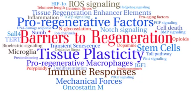
Fig. 2. Word cloud summary of this Meeting Review.The word cloud was randomly generated with TAGUL from keywords selected from this Meeting Review, showing barriers to regeneration (red), pro-regenerative factors (blue) and modulators (brown) that can positively or negatively impact regeneration as discussed
in the text.
DEVEL
O
populations. Muriel Perron (University Paris-Saclay, France) showed that both CMZ and Müller glial cells regenerate the retina inXenopus, where the Hippo pathway effector YAP regulates S-phase progression of CMZ retinal stem cells (Cabochette et al., 2015). In mouse Müller cells, YAP appears to control the balance between the beneficial and detrimental effects of glial reactivation, positively regulating cell cycle genes and neurotrophic factors, and negatively controlling gliosis genes that prevent retina regeneration in mammals. In salamanders, a feedback mechanism exists whereby dopamine inhibits the proliferation of midbrain dopaminergic progenitors and hence controls the formation of new dopaminergic neurons when mature ones disappear. Andras Simon (Karolinska Institute, Stockholm, Sweden) has now identified a population of Lmx1a-expressing ventricular cells in the adult mouse midbrain that express dopamine receptors, are found in a dopaminergic-innervated region and become activated upon depletion of dopaminergic neurons. During embryogenesis, Lmx1a+progenitor proliferation appears to be repressed by dopaminergic signaling (Hedlund et al., 2016), supporting the conservation of the feedback mechanism discovered in salamanders and opening new paths for treating Parkinson’s disease (in which dopaminergic neurons degenerate). Using human neural stem cells, Patrizia Ferretti (University College London, UK) reported recent progress on the development of 3D culture systems to study neural damage. She also showed that in developing human brains major histocompatibility complex class II antigens (MHC-II) are unexpectedly expressed in the germinal zone in a subset of SOX2-positive neural progenitors (Vagaska et al., 2016). This expression, which is seemingly regulated independently of inflammatory stimuli, challenges the accepted notion that microglia are the only MHC-II-expressing cells in the developing CNS. It also rekindles the issue of a possible neuroectodermal contribution to microglia during development.
Senescence, regeneration and ageing
The Hydra polyp provides a unique system with which to investigate the mechanisms that maintain homeostasis, regeneration and slow ageing over many years. Brigitte Galliot (Geneva University, Switzerland) showed thatH. vulgarisepithelial cells adapt to the loss of neurogenesis by overexpressing genes with potential neurogenic, neurotransmission or reprogramming functions (Wenger et al., 2016). By contrast, some H. oligactis strains lack this property, and animals rapidly lose regenerative capacity and develop an ageing phenotype. A positive role for senescent cells in regeneration has recently emerged as a new direction in the field (Demaria et al., 2014). Maximina Yun (University College London, UK) reported that, in salamander, endogenous senescent cells are detected during early phases of limb regeneration and are subsequently cleared by a ROS-dependent surveillance mechanism mediated by macrophages (Yun et al., 2015). Interestingly, transplantation of senescent cells into the regenerating blastema stimulates regeneration, whereas their elimination hinders it, highlighting their pro-regenerative nature. Similarly, Shahragim Tajbakhsh (Institut Pasteur, Paris, France) reported on a potentially positive role for cellular senescence in muscle regeneration in adult mice. UsingNumb;Numblconditional mutants in muscle stem cells, he showed that myogenic cells undergo cellular senescence following acute muscle injury, a process that is dependent on p53 (Trp53) and oxidative stress (Le Roux et al., 2015). Senescence of endothelial cells also occurs during early regeneration in wild-type mice, although this is independent of p53 and oxidative stress. These findings point to different modes by which senescent cells influence regeneration.
Ageing also affects skeletal muscles, with stem cell contribution to muscle fibers dramatically decreasing with age. Helen Blau (Stanford University, USA) underscored the importance of telomere length in this process. Indeed, in the Mdx dystrophic mouse, telomerase deficiency exacerbates skeletal and cardiac muscle phenotypes. Remarkably, telomere shortening occurs in the heart independently of cell division (Chang et al., 2016). Blau’s laboratory has developed a transient modified mRNA approach to deliver TERT for only 48 h, which overcomes problems associated with constitutive retroviral delivery, and leads to a marked increase in telomere length and rescue of cell proliferative defects (Ramunas et al., 2015). Finally, Saul Villeda (UCSF, USA), who explores the mechanisms of brain ageing, used mouse parabiosis experiments to show that β2-microglobulin (B2M) is a brain ageing factor, the action of which is mediated by the MHC (Smith et al., 2015). Indeed, mice missing the H2-Dband H2-HbMHC alleles perform better in hippocampus-mediated spatial tasks, and decreasing B2M levels improves cognition in ageing mice.
Emerging trends of regeneration research
This conference underscored the diverse nature of injury responses not only across species, but also across structures, organs and tissues within a given species (Fig. 2). Intriguingly, some factors–such as ROS and immune responses–act positively in some conditions and negatively in others. As barriers to regeneration, we learned about genomic stress, telomere attrition, polyploidy, opioids, neurotransmitters such as dopamine, and B2M. Positive regulators of regeneration tend to attract more attention and an increasingly long list is now available. We heard about mechanical forces, bioelectric control, transient senescence, telomere maintenance, cellular plasticity, anti-scarring pathways and pro-regenerative networks. We also learned how evolutionarily conserved pathways can regulate taxon-specific genes – highlighting regeneration as a process in which robust ancestral modules are combined with more recent innovations. Another new direction in the field is understanding how homeostatic conditions impact regenerative potential. Indeed, evidence is accumulating to show that ageing or environmental conditions can modulate the permissive status of a given organ to achieve regeneration. Finally, efforts to develop biomaterials forex vivoandin vivostudies will help bridge the gap between the biology of regeneration and regenerative medicine.
Acknowledgements
We thank all participants who agreed to share data for discussion in this report, and Muriel Perron (Paris-Saclay) Jochen Rink (Dresden) and Ryan Debuque (Melbourne) for their comments in the manuscript. This Meeting Review is dedicated to the memory of Panagiotis (Takis) Tsonis, a passionate scientist who devoted his life to the study of regeneration, and who sadly passed away a few days before this conference.
Competing interests
The authors declare no competing or financial interests.
Funding
B.G. is funded by the Canton of Geneva, the Swiss National Foundation (SNF 31003A_169930), and the National Institutes of Health (R01AG037962); A.J. is funded by the Fundação para a Ciência e Tecnologia (PTDC/BIM-MED/0659/2014); S.T. is funded by Institut Pasteur, Centre National de la Recherche Scientifique (CNRS), Agence Nationale de la Recherche (ANR-10-LABX-73), and a European Research Council (ERC) Advanced Research Grant 332893.
References
Amiel, A. R., Johnston, H. T., Nedoncelle, K., Warner, J. F., Ferreira, S. and Röttinger, E. (2015). Characterization of morphological and cellular events underlying oral regeneration in the sea anemone, Nematostella vectensis.
Int. J. Mol. Sci.16, 28449-28471.
DEVEL
O
Balasooriya, G. I., Johnson, J.-A., Basson, M. A. and Rawlins, E. L.(2016). An FGFR1-SPRY2 signaling axis limits basal cell proliferation in the steady-state airway epithelium.Dev. Cell37, 85-97.
Barghouth, P. G., Thiruvalluvan, M. and Oviedo, N. J. (2015). Bioelectrical regulation of cell cycle and the planarian model system.Biochim. Biophys. Acta
1848, 2629-2637.
Biferi, M. G., Nicoletti, C., Falcone, G., Puggioni, E. M. R., Passaro, N., Mazzola, A., Pajalunga, D., Zaccagnini, G., Rizzuto, E., Auricchio, A. et al.(2015). Proliferation of multiple cell types in the skeletal muscle tissue elicited by acute p21 suppression.Mol. Ther.23, 885-895.
Birket, M. J., Ribeiro, M. C., Verkerk, A. O., Ward, D., Leitoguinho, A. R., den Hartogh, S. C., Orlova, V. V., Devalla, H. D., Schwach, V., Bellin, M. et al. (2015). Expansion and patterning of cardiovascular progenitors derived from human pluripotent stem cells.Nat. Biotechnol.33, 970-979.
Brockes, J. P. and Kumar, A.(2008). Comparative aspects of animal regeneration. Annu. Rev. Cell Dev. Biol.24, 525-549.
Brouilly, N., Lecroisey, C., Martin, E., Pierson, L., Mariol, M.-C., Qadota, H., Labouesse, M., Streichenberger, N., Mounier, N. and Gieseler, K.(2015). Ultra-structural time-course study in theC. elegansmodel for Duchenne muscular dystrophy highlights a crucial role for sarcomere-anchoring structures and sarcolemma integrity in the earliest steps of the muscle degeneration process. Hum. Mol. Genet.24, 6428-6445.
Cabochette, P., Vega-Lopez, G., Bitard, J., Parain, K., Chemouny, R., Masson, C., Borday, C., Hedderich, M., Henningfeld, K. A., Locker, M. et al.(2015). YAP controls retinal stem cell DNA replication timing and genomic stability.eLife4, e08488.
Caiazzo, M., Okawa, Y., Ranga, A., Piersigilli, A., Tabata, Y. and Lutolf, M. P. (2016). Defined three-dimensional microenvironments boost induction of pluripotency.Nat. Mater.15, 344-352.
Carvalho, L., Jacinto, A. and Matova, N.(2014). The Toll/NF-kappaB signaling pathway is required for epidermal wound repair in Drosophila.Proc. Natl. Acad. Sci. USA111, E5373-E5382.
Casano, A. M. and Peri, F.(2015). Microglia: multitasking specialists of the brain. Dev. Cell32, 469-477.
Chang, A. C. Y., Ong, S.-G., LaGory, E. L., Kraft, P. E., Giaccia, A. J., Wu, J. C. and Blau, H. M. (2016). Telomere shortening and metabolic compromise underlie dystrophic cardiomyopathy. Proc. Natl. Acad. Sci. USA 113, 13120-13125.
Currie, J. D., Kawaguchi, A., Traspas, R. M., Schuez, M., Chara, O. and Tanaka, E. M.(2016). Live imaging of axolotl digit regeneration reveals spatiotemporal choreography of diverse connective tissue progenitor pools. Dev. Cell 39, 411-423.
de Preux Charles, A.-S., Bise, T., Baier, F., Sallin, P. and Jaźwińska, A.(2016). Preconditioning boosts regenerative programmes in the adult zebrafish heart. Open Biol.6, pii:160101.
Demaria, M., Ohtani, N., Youssef, S. A., Rodier, F., Toussaint, W., Mitchell, J. R., Laberge, R.-M., Vijg, J., Van Steeg, H., Dollé, M. E. T. et al.(2014). An essential role for senescent cells in optimal wound healing through secretion of PDGF-AA. Dev. Cell31, 722-733.
Gallego-Colon, E., Villalba, M., Tonkin, J., Cruz, F., Antonio Bernal, J., Jimenez-Borregureo, L. J., Schneider, M. D., Lara-Pezzi, E. and Rosenthal, N.(2016). Intravenous delivery of adeno-associated virus 9-encoded IGF-1Ea propeptide improves post-infarct cardiac remodelling.Regen. Med.1, 16001.
Galliot, B. and Ghila, L.(2010). Cell plasticity in homeostasis and regeneration. Mol. Reprod. Dev.77, 837-855.
Gierer, A. and Meinhardt, H.(1972). A theory of biological pattern formation. Kybernetik12, 30-39.
Godwin, J. W., Pinto, A. R. and Rosenthal, N. A.(2013). Macrophages are required for adult salamander limb regeneration.Proc. Natl. Acad. Sci. USA110, 9415-9420.
Hedlund, E., Belnoue, L., Theofilopoulos, S., Salto, C., Bye, C., Parish, C., Deng, Q., Kadkhodaei, B., Ericson, J., Arenas, E. et al.(2016). Dopamine receptor antagonists enhance proliferation and neurogenesis of midbrain Lmx1a-expressing progenitors.Sci. Rep.6, 26448.
Herrera, S. C. and Morata, G. (2014). Transgressions of compartment boundaries and cell reprogramming during regeneration in Drosophila.eLife
3, e01831.
Kang, J., Hu, J., Karra, R., Dickson, A. L., Tornini, V. A., Nachtrab, G., Gemberling, M., Goldman, J. A., Black, B. L. and Poss, K. D. (2016). Modulation of tissue repair by regeneration enhancer elements. Nature532, 201-206.
Kaslin, J., Kroehne, V., Benato, F., Argenton, F. and Brand, M. (2013). Development and specification of cerebellar stem and progenitor cells in zebrafish: from embryo to adult.Neural Dev.8, 9.
Kral, N., Hanna Ougolnikova, A. and Sena, G.(2016). Externally imposed electric field enhances plant root tip regeneration.Regeneration3, 156-167.
Le, H. Q., Ghatak, S., Yeung, C.-Y. C., Tellkamp, F., Günschmann, C., Dieterich, C., Yeroslaviz, A., Habermann, B., Pombo, A., Niessen, C. M. et al.(2016). Mechanical regulation of transcription controls Polycomb-mediated gene silencing during lineage commitment.Nat. Cell Biol.18, 864-875.
Le Roux, I., Konge, J., Le Cam, L., Flamant, P. and Tajbakhsh, S.(2015). Numb is required to prevent p53-dependent senescence following skeletal muscle injury. Nat. Commun.6, 8528.
Lin, G., Chen, Y. and Slack, J. M. W.(2013). Imparting regenerative capacity to limbs by progenitor cell transplantation.Dev. Cell24, 41-51.
Liu, S.-Y., Selck, C., Friedrich, B., Lutz, R., Vila-Farré, M., Dahl, A., Brandl, H., Lakshmanaperumal, N., Henry, I. and Rink, J. C.(2013). Reactivating head regrowth in a regeneration-deficient planarian species.Nature 500, 81-84.
Lörchner, H., Pöling, J., Gajawada, P., Hou, Y., Polyakova, V., Kostin, S., Adrian-Segarra, J. M., Boettger, T., Wietelmann, A., Warnecke, H. et al.(2015). Myocardial healing requires Reg3beta-dependent accumulation of macrophages in the ischemic heart.Nat. Med.21, 353-362.
Love, N. R., Chen, Y., Ishibashi, S., Kritsiligkou, P., Lea, R., Koh, Y., Gallop, J. L., Dorey, K. and Amaya, E.(2013). Amputation-induced reactive oxygen species are required for successful Xenopus tadpole tail regeneration.Nat. Cell Biol.15, 222-228.
Mangel, M., Bonsall, M. B. and Aboobaker, A.(2016). Feedback control in planarian stem cell systems.BMC Syst. Biol.10, 17.
Mescher, A. L., Neff, A. W. and King, M. W.(2013). Changes in the inflammatory response to injury and its resolution during the loss of regenerative capacity in developing Xenopus limbs.PLoS ONE8, e80477.
Nguyen-Chi, M., Laplace-Builhe, B., Travnickova, J., Luz-Crawford, P., Tejedor, G., Phan, Q. T., Duroux-Richard, I., Levraud, J.-P., Kissa, K., Lutfalla, G. et al. (2015). Identification of polarized macrophage subsets in zebrafish.eLife4, e07288.
Ninov, N., Hesselson, D., Gut, P., Zhou, A., Fidelin, K. and Stainier, D. Y. R. (2013). Metabolic regulation of cellular plasticity in the pancreas.Curr. Biol.23, 1242-1250.
Ohnmacht, J., Yang, Y., Maurer, G. W., Barreiro-Iglesias, A., Tsarouchas, T. M., Wehner, D., Sieger, D., Becker, C. G. and Becker, T.(2016). Spinal motor neurons are regenerated after mechanical lesion and genetic ablation in larval zebrafish.Development143, 1464-1474.
Owlarn, S. and Bartscherer, K.(2016). Go ahead, grow a head! A planarian’s guide to anterior regeneration.Regeneration3, 139-155.
Pei, W., Tanaka, K., Huang, S. C., Xu, L., Liu, B., Sinclair, J., Idol, J., Varshney, G. K., Huang, H., Lin, S. et al.(2016). Extracellular HSP60 triggers tissue regeneration and wound healing by regulating inflammation and cell proliferation. Regen. Med.1, 16013.
Ramunas, J., Yakubov, E., Brady, J. J., Corbel, S. Y., Holbrook, C., Brandt, M., Stein, J., Santiago, J. G., Cooke, J. P. and Blau, H. M.(2015). Transient delivery of modified mRNA encoding TERT rapidly extends telomeres in human cells. FASEB J.29, 1930-1939.
Sikes, J. M. and Newmark, P. A.(2013). Restoration of anterior regeneration in a planarian with limited regenerative ability.Nature500, 77-80.
Simkin, J., Sammarco, M. C., Dawson, L. A., Tucker, C., Taylor, L. J., Van Meter, K. and Muneoka, K. (2015). Epidermal closure regulates histolysis during mammalian (Mus) digit regeneration.Regeneration2, 106-119.
Smith, L. K., He, Y., Park, J.-S., Bieri, G., Snethlage, C. E., Lin, K., Gontier, G., Wabl, R., Plambeck, K. E., Udeochu, J. et al.(2015). beta2-microglobulin is a systemic pro-aging factor that impairs cognitive function and neurogenesis.Nat. Med.21, 932-937.
Stocum, D. L. and Cameron, J. A.(2011). Looking proximally and distally: 100 years of limb regeneration and beyond.Dev. Dyn.240, 943-968.
Sullivan, K. G., Emmons-Bell, M. and Levin, M.(2016). Physiological inputs regulate species-specific anatomy during embryogenesis and regeneration. Commun. Integr. Biol.9, e1192733.
Tanaka, E. M. and Reddien, P. W. (2011). The cellular basis for animal regeneration.Dev. Cell21, 172-185.
Tata, P. R., Mou, H., Pardo-Saganta, A., Zhao, R., Prabhu, M., Law, B. M., Vinarsky, V., Cho, J. L., Breton, S., Sahay, A. et al.(2013). Dedifferentiation of committed epithelial cells into stem cells in vivo.Nature503, 218-223. Thorimbert, V., Konig, D., Marro, J., Ruggiero, F. and Jazwinska, A.(2015).
Bone morphogenetic protein signaling promotes morphogenesis of blood vessels, wound epidermis, and actinotrichia during fin regeneration in zebrafish.FASEB J.
29, 4299-4312.
Umesono, Y., Tasaki, J., Nishimura, Y., Hrouda, M., Kawaguchi, E., Yazawa, S., Nishimura, O., Hosoda, K., Inoue, T. and Agata, K.(2013). The molecular logic for planarian regeneration along the anterior–posterior axis.Nature500, 73-76.
Vagaska, B., New, S. E. P., Alvarez-Gonzalez, C., D’Acquisto, F., Gomez, S. G., Bulstrode, N. W., Madrigal, A. and Ferretti, P. (2016). MHC-class-II are expressed in a subpopulation of human neural stem cells in vitro in an IFNgamma-independent fashion and during development.Sci. Rep.6, 24251.
Vasconcelos, D. M., Gonçalves, R. M., Almeida, C. R., Pereira, I. O., Oliveira, M. I., Neves, N., Silva, A. M., Ribeiro, A. C., Cunha, C., Almeida, A. R. et al. (2016). Fibrinogen scaffolds with immunomodulatory properties promote in vivo bone regeneration.Biomaterials111, 163-178.
DEVEL
O
Wenger, Y., Buzgariu, W. and Galliot, B.(2016). Loss of neurogenesis in Hydra leads to compensatory regulation of neurogenic and neurotransmission genes in epithelial cells.Philos. Trans. R. Soc. B Biol. Sci.371, 20150040.
Werner, S., Stückemann, T., Beirán Amigo, M., Rink, J. C., Jülicher, F. and Friedrich, B. M. (2015). Scaling and regeneration of self-organized patterns. Phys. Rev. Lett.114, 138101.
Wurtzel, O., Cote, L. E., Poirier, A., Satija, R., Regev, A. and Reddien, P. W. (2015). A generic and cell-type-specific wound response precedes regeneration in planarians.Dev. Cell35, 632-645.
Yun, M. H., Davaapil, H. and Brockes, J. P. (2015). Recurrent turnover of senescent cells during regeneration of a complex structure.eLife4, e05505. Zhang, Y., Strehin, I., Bedelbaeva, K., Gourevitch, D., Clark, L., Leferovich, J.,
Messersmith, P. B. and Heber-Katz, E.(2015). Drug-induced regeneration in adult mice.Sci. Transl. Med.7, 290ra292.
Zuryn, S., Ahier, A., Portoso, M., White, E. R., Morin, M.-C., Margueron, R. and Jarriault, S. (2014). Sequential histone-modifying activities determine the robustness of transdifferentiation.Science345, 826-829.