Transforming Growth Factor-
β
1 T869C Gene
Polymorphism Is Associated with Acquired
Sick Sinus Syndrome via Linking a Higher
Serum Protein Level
Jan-Yow Chen1,2,3, Jiung-Hsiun Liu3, Hong-Dar Isaac Wu4, Kuo-Hung Lin1,3, Kuan-Cheng Chang1,3, Ying-Ming Liou2*
1Division of Cardiology, Department of Medicine, China Medical University Hospital, Taichung, Taiwan,
2Department of Life Sciences, National Chung Hsing University, Taichung, Taiwan,3School of Medicine, China Medical University, Taichung, Taiwan,4Department of Applied Mathematics and Institute of Statistics, National Chung Hsing University, Taichung, Taiwan
Abstract
Background
Familial sick sinus syndrome is associated with gene mutations and dysfunction of ion channels. In contrast, degenerative fibrosis of the sinus node tissue plays an important role in the pathogenesis of acquired sick sinus syndrome. There is a close relationship between transforming growth factor-β1 mediated cardiac fibrosis and acquired arrhythmia. It is of interest to examine whether transforming growth factor-β1 is involved in the pathogenesis of acquired sick sinus syndrome.
Methods
Overall, 110 patients with acquired SSS and 137 age/gender-matched controls were screened for transforming growth factor-β1 and cardiac sodium channel gene polymor-phisms using gene sequencing or restriction fragment length polymorphism methods. An enzyme-linked immunosorbent assay was used to determine the serum level of transform-ing growth factor-β1.
Results
Two transforming growth factor-β1 gene polymorphisms (C-509T and T+869C) and one cardiac sodium channel gene polymorphism (H588R) have been identified. The C-dominant CC/CT genotype frequency of T869C was significantly higher in acquired sick sinus syn-drome patients than in controls (OR 2.09, 95% CI 1.16–3.75, P = 0.01). Consistently, the level of serum transforming growth factor-β1 was also significantly greater in acquired sick sinus syndrome group than in controls (5.3±3.4 ng/ml vs. 3.7±2.4 ng/ml, P = 0.01). In addi-tion, the CC/CT genotypes showed a higher transforming growth factor-β1 serum level than the TT genotype (4.25±2.50 ng/ml vs. 2.71±1.76 ng/ml, P = 0.028) in controls.
a11111
OPEN ACCESS
Citation:Chen J-Y, Liu J-H, Wu H-DI, Lin K-H, Chang K-C, Liou Y-M (2016) Transforming Growth Factor-β1 T869C Gene Polymorphism Is Associated with Acquired Sick Sinus Syndrome via Linking a Higher Serum Protein Level. PLoS ONE 11(7): e0158676. doi:10.1371/journal.pone.0158676
Editor:Qiang Ding, University of Alabama at Birmingham, UNITED STATES
Received:December 22, 2015
Accepted:June 20, 2016
Published:July 5, 2016
Copyright:© 2016 Chen et al. This is an open access article distributed under the terms of the
Creative Commons Attribution License, which permits unrestricted use, distribution, and reproduction in any medium, provided the original author and source are credited.
Data Availability Statement:All relevant data are within the paper and its Supporting Information files.
Funding:This study was supported by funding (NSC 101-2314-B-039-042) from Taiwan National Science Council (Ministry of Science and Technology) (https:// www.most.gov.tw/en/public). JYC received the funding.
Conclusion
Transforming growth factor-β1 T869C polymorphism, correlated with high serum transform-ing growth factor-β1 levels, is associated with susceptibility to acquired sick sinus
syndrome.
Introduction
Sick sinus syndrome (SSS) is a group of abnormal heart rhythm disorders resulting from mal-functions of the sinus node. The prevalence of SSS is about 1/600 in the population with age over 65 years. Over half of the global pacemaker implantations were performed for the SSS
patients [1,2]. The electrophysiological abnormalities of SSS patients include sinus node arrest,
sinus node exit block, profound sinus bradycardia and bradycardia-tachycardia syndrome [1, 2]. Evidence also indicated that genetic mutations in ion channels might lead to familial SSS [3–6]; however, limited information is available regarding the pathophysiologic mechanism of
age-related acquired SSS [7,8]. Acquired SSS, most commonly observed in the elderly, results
from degenerative fibrosis and aging sinus node tissue [2,6]. Apparently, variations in the
genes encoding proteins involved in fibrosis and aging would affect the susceptibility to acquired SSS [8]. The spontaneous depolarization of the sinus node has been attributed to the
inward current driven through sodium (INa), calcium (L-type Ca2+current, ICa,L; and T-type
Ca2+current, ICa,T) and funny currents (If). In addition, the voltage-gated sodium channel,
type V alpha subunit (SCN5A) gene encoding the cardiac sodium channel has been shown to
trigger the action potential of the sinus node [6]. INaplays an important role in pacemaking
and action potential conduction, particularly in the periphery of the sinus node [6]. In addition, SCN5A mutation or variation has also been associated with cardiac arrhythmia, sinus node
dysfunction, and conduction disorders [9,10]. However, it remains unclear whether alterations
in SCN5A gene expression contribute to acquired SSS.
Transforming growth factorβ1(TGF-β1), a member of the transforming growth factor
fam-ily, has been associated with cell growth, proliferation and differentiation, tissue fibrosis, tumor
progression and metastasis, wound healing, embryogenesis and immune modulation [10–13].
In addition, TGF-β1 involved in the renin-angiotensin system (RAS) plays a role in cardiac
fibrosis [12]. A study using the SCN5A knockout mouse model showed that TGF-β1-mediated
fibrosis and ion channel remodeling could lead to sinus node dysfunction [10]. Clinical studies
also reported that the TGF-β1 serum level is highly associated with the risk of atrial arrhythmia
[14,15]. However, a role for TGF-β1 in the pathogenesis of age-related acquired SSS has not
been determined. Nevertheless, studies have shown that TGF-β1 gene polymorphisms
modu-lating TGF-β1 serum levels [13,16,17]. Thus, we hypothesized that TGF-β1 gene
polymor-phisms might exert a significant effect on sinus node function and the susceptibility to
acquired SSS via linking TGF-β1 serum levels.
Materials and Methods
Ethics statement
The protocol of present study was approved by the Institutional Review Board of China Medi-cal University Hospital (DMR-IRB2-158) in Taiwan. Written informed consent was obtained
from each participant or each participant’s guardian after a full explanation for the present
Subjects
During the period from August 1, 2012 to July 31, 2013, a total of 110 consecutively eligible patients with documented SSS and a control group of 137 subjects matched for age and gender in frequency were included for the present study. All of the SSS patients and controls are Tai-wanese population. Medication such as digitalis and antiarrhythmic drugs that can modify the heart rate had been discontinued for at least five half-lives before observing the heart rate. Acquired SSS was diagnosed based on evidence of irreversible symptomatic bradycardia with a sinus long pause of greater than 3 seconds or profound sinus bradycardia of less than 40 beats/
min for more than 1 min while awake [18,19]. The sinus pauses and bradycardia were
recorded by a series of resting 12-lead electrocardiogram (ECG) and ambulatory ECG record-ers. All SSS patients met the indications for permanent pacemaker implantation.
All control subjects were randomly selected from cardiovascular outpatient department and ward and were frequency-matched to SSS patients by gender and age at the same hospital during the same period. Patients with history of reversible bradycardia, familial SSS, serious systemic dis-ease, unstable angina, acute myocardial infarction or acute myocarditis were excluded.
Genotyping and association study
Blood samples from 137 controls and 110 SSS patients were prepared to isolate genomic DNA
using a commercially available DNA extraction kit (IllustraTM, GE Healthcare). A final reaction
of 50μL mixture including 100 ng genomic DNA, 2–6 pmol of selected primers, 1X Taq
poly-merase buffer, and 0.25 units of AmpliTaq GoldTMpolymerase (Roche) was prepared for
Poly-merase chain reactions (PCRs). A programmable thermal cycler (GeneAmp PCR system 2700, Applied Biosystems, CA) was used for PCRs. The PCR cycling conditions were set with 1 cycle at 94°C for 5 minutes, followed by 35 cycles at 94°C for 30 seconds, 56°C for 30 seconds, and 72°C for 60 seconds, with a final extension cycle at 72°C for 7 minutes. The primers for the
TGF-β1 C-509T (rs1800469) polymorphism were 5'-CGCAACTTCGACCGCTAC -3'
(for-ward) and 5'-CTCGGCGACTCCTTCCTC-3' (reverse). The primers for TGF-β1 T+869C
poly-morphism (rs1800470) were 5’-TTCCCTCGAGGCCCTCCTA-3 (forward) and 5’-GCCGCA
GCTTGGACAGGATC-3’(reverse). The primers were designed according to the sequences of
the human TGF-β1 gene (GenBank accession number: J04431) and TGF-β1 precursor gene
(exon 1; X05839) [13]. The primers for the SCN5A polymorphism A1673G (H558R, rs1805124)
were 5’-GCGAGACCTGGGTTCTGA-3’(forward) and 5’-GTGGCTTCCTGGGGATGTT-3’
(reverse). The quality of the PCR products was assessed through the electrophoresis of 4μL of
the PCR reactions on a 3% agarose gel containing ethidium bromide. Genotyping of the TGF-β1
gene C-509T and T+869C polymorphisms was determined through DNA sequencing. A gene sequencing analyzer (ABI 3730 XL DNA Analyzer, Applied Biosystems) was used to determine the gene sequences of the PCR products. Genotyping of the H558R polymorphism was per-formed through restriction fragment length polymorphism (RFLP) analysis as described in detail previously [20]. The corresponding A to G nucleotide transition creates the sequence -CCGC-,
which is the recognition site for the restriction enzymeAciI. This variant, located in exon 12 of
SCN5A, was first amplified within a 251-base pair (bp) PCR product. Restriction digestion of the
PCR product usingAciIresulted in the following fragment sizes: H allele(-CCAC-): 201 and 50
bp; and R allele (-CCGC-): 181, 50 and 20 bp. The fragments were separated using agarose gel electrophoresis. Hence, the H and R alleles could be distinguished based on the presence of 201 and 181bp fragments, respectively.
An association study between the gene polymorphisms and acquired SSS was performed via
measuring the frequency of the alleles, genotypes and haplotypes of the TGFβ1and SCN5A
Measurement of the serum TGF-
β
1 concentration
Serum samples were randomly obtained from 44 of the 110 acquired SSS patients and 49 of the 137
control individuals. The samples were stored at -20°C until further analysis. Serum TGF-β1 levels
were detected using the enzyme-linked immunosorbent assay kit (ELISA) (Invitrogen, CA, USA). The detection minimum of this assay was 15.6 pg/mL, and the intra-assay and inter-assay coeffi-cients of variation (CVs) were 5.5 and 7.5%, respectively. The sera were activated through acidifica-tion/neutralizing and tested at a 1:40 dilution. The optical density (OD) was measured using an ELISA Microplate Reader set to 450 nm (VersaMax, Molecular Devices, Sunnyvale, CA, US). The
samples were run in duplicate technical repeats for the serum protein levels. The TGF-β1
concen-tration was determined using a standard curve according to the manufacturer’s instructions.
Quantification of TGF
β
1 mRNA expression
A centrifuge (Kubota) was used to isolate the peripheral blood mononuclear cells (PBMC) for
TGF-β1 mRNA isolation. mRNA samples were randomly obtained from 15 of the 110 acquired
SSS patients and 18 of the 137 control individuals. Real-time quantitative polymerase chain
reaction (qPCR) was used for quantitative measurement of mRNA expression of TGF-β1.
TRI-zol1Reagent (Invitrogen) was used to isolate total RNA based on the manufacturer’s protocol.
Reverse transcriptase (M-MLV-RT, Genemark), oligo-dT and 2μg of total RNA was used for
reverse transcription. SYBR qPCR Kit (Genemark) and an ABI Prism 7300 sequence detection system (Applied Biosystems) were used for quantitative analysis of mRNA level. qPCR was run in duplicate for each sample. SYBR Green dye was used as a real-time reporter of the presence
of double-stranded DNA. The primers specific for TGF-β1 were listed as followings: forward,
5’- CCCAGCATCTGCAAAGCTC-3’, reverse, 5’- GTCAATGTACAGCTGCCGCA-3.
Statistical Analysis
Continuous data were expressed as the means ± standard deviation (SD). Student’s t test was
used for the continuous data with normal distribution. Mann-Whitney U test was used for the continuous data without normal distribution. Conventional chi-squire test was used for com-parison of categorical data if the observation numbers in all categories were larger than 5; oth-erwise, the Fisher exact test was replaced for data analysis. One-way analysis of variance
(ANOVA) was used for comparison among numeric variables for the TGF-β1 and SCN5A
genotypes. Theχ2 goodness-of-fit test with 1 degree of freedom was utilized to exam the
devia-tion of the genotype distribudevia-tion from Hardy-Weinberg equilibrium (HWE) for each TGF-β1
or SCN5A polymorphism. Haploview software was used to analyze the haplotype frequency of the gene polymorphisms [21]. The gene polymorphisms were likely not separated through recombination process and linkage disequilibrium (LD) might be developed. The LD between
the polymorphisms of TGF-β1 was analyzed via pairwise measurement of LD. In addition,
owing to the D’magnitude varies according to the sample size; ther2-value was used to confirm
the LD. We also utilized expectation-maximization (EM)-based haplotype frequency estima-tion with a permutaestima-tion test to confirm whether a specific haplotype was associated with
acquired SSS [22,23]. D’>0.7 and.r2>1/3 were defined as statistic significance of the LD [23,
24].Pvalues less than 0.05 were considered statistically significant.
Results
Patient characteristics
The clinical characteristics of the SSS patients and controls were summarized inTable 1.
HWE test and LD analysis
In the present study, two polymorphisms at positions -509and +869 in the TGF-β1 gene (Fig
1) and the polymorphism H558R (A+1673C) in the SCN5A gene were observed. HWE test was utilized to exam the deviations of the genotype counts of each polymorphism. The P-values were 0.09, 0.22 and 0.18 for C-509T, T869C and A1673G gene polymorphisms, respectively.
We also used the HWE test to exam the deviations of TGF-β1and SCN5A genotype
distribu-tion in the SSS and control groups respectively. In control group, the P-values were 0.36, 0.75 and 0.76 for C-509T, T869C and A1673G gene polymorphisms, respectively. In SSS patients, the P-values were 0.15, 0.085 and 0.087 for C-509T, T869C and A1673G gene polymorphisms, respectively.
We used the LD test with D’and r2to assess the pair-wise linkage between the two
polymor-phic sites of TGF-β1 gene. The D’value of the -509/+869 loci pairs was 0.87 and the
corre-sponding r2value was 0.556. The D’and r2values indicated a significant but not complete
linkage at the loci pair -509/+869, reflecting the distribution of haplotypes for the TGF-β1
gene.
Table 1. Clinical characteristics of study patients.
SSS Control
(N = 110) (N = 137) p
Age (years) 70.3±11.1 70.2±9.1 0.898*
Gender (male/female) 34/76 46/91 0.656§
Heart rate (beats/min) 45.9±11.5 75.2±13.6 <0.0001
PR interval (ms) 175.3±33.0 161.6±25.3 0.002
HT (n, %) 47 (42.7%) 62 (45.3%) 0.691§
DM (n, %) 25 (22.7%) 27 (19.7%) 0.563§
CAD (n, %) 11 (10.0%) 13 (9.5%) 0.893§
AF (n, %) 22 (20.0%) 24 (17.5%) 0.62§
*Mann-Whitney U test
§χ2test; SSS = sick sinus syndrome; HT = hypertension; DM = diabetes mellitus; CAD = coronary artery
disease; AF = atrialfibrillation
doi:10.1371/journal.pone.0158676.t001
Fig 1. Gene sequencing for the different genotypes of the TGF-β1 T869C polymorphism.The polymorphic sites of CC, TT and CT genotypes were indicated by the arrows.
Gene association studies
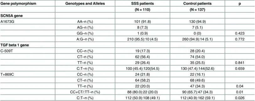
The genotype distribution at position +869 was significant different between SSS and controls (Table 2, P = 0.04). The C-dominant CC/CT genotype frequency of T869C was significantly
higher in acquired SSS patients than in control individuals (OR 2.09, 95% CI 1.16–3.75,
P = 0.01). There was also a significant difference in the distribution of allele frequency at posi-tion +869 between subjects in the SSS and control groups (P = 0.026). The C allele frequency of
TGF-β1 was significantly higher in the SSS group than in the control group (OR = 1.5, 95% CI:
1.049–2.45, P = 0.026) (Table 2). No significant difference in the genotype and allele frequency
distribution was observed in TGF-β1 C-509T and SCN5A H558R polymorphisms between the
subjects in the SSS and control groups (P>0.05).
Haplotype analysis
Four haplotypes in the TGF-β1 gene were identified to examine the relationship of these
varia-tions with SSS (Table 3). The TT haplotype (-509T,+869T) occurred at a significantly lower fre-quency in the SSS group than in the control group (haplotype frefre-quency: 0.0627 vs. 0.1380; OR
0.418, P = 0.0066;Table 3). The TC haplotype (-509T, +869C) showed a significantly higher
Table 2. Genotype and allele distribution of TGFβ1 and SCN5A in SSS patients and controls,
Gene polymorphism Genotypes and Alleles SSS patients Control patients p
(N = 110) (N = 137)
SCN5A gene
A1673G AA–n (%) 101 (91.8) 130 (94.9)
AG–n (%) 8 (7.3) 7 (5.1)
GG–n (%) 1 (0.9) 0 (0) 0.423
A:G–n (%) 210 (95.5):10 (4.5) 260 (94.9):14 (5.1) 0.772
TGF beta 1 gene
C-509T CC–n (%) 19 (17.3) 28 (20.4)
CT–n (%) 62 (56.4) 74 (54.0)
TT–n (%) 29 (26.4) 35 (25.5) 0.841
C:T–n (%) 100 (45.4):120(54.5) 130 (47.4):144(52.6) 0.659
T+869C CC–n (%) 24 (21.8) 22 (16.1)
CT–n (%) 64 (58.2) 68 (49.6)
TT–n (%) 22 (20.0) 47 (34.3) 0.04
CC+CT/:TT–n (%) 88 (80.0):22 (20.0) 90 (65.7):47 (34.3) 0.01
C:T–n (%) 112 (50.9):108 (49.1) 112 (40.9):162 (59.1) 0.026
SSS = sick sinus syndrome. The upperPvalue is for comparison of genotype frequencies and the lower is for allele frequencies.
doi:10.1371/journal.pone.0158676.t002
Table 3. Haplotype frequency of the TGFβ1 gene polymorphisms in SSS patients and controls.
Haplotype
Overall SSS Controls
-509 +869 (N = 247) (N = 110) (N = 137) OR P*
C T 0.442 0.4282 0.4533 0.903 0.5761
T C 0.426 0.4827 0.3803 1.521 0.0221
T T 0.105 0.0627 0.1380 0.418 0.0066
C C 0.028 0.0263 0.0285 0.924 0.8889
*There results were confirmed through permutation test. TT is the only significant candidate haplotype (P = 0.0226).
frequency in the SSS group than in the control group (haplotype frequency: 0.4827 vs. 0.3803; OR 1.521, P = 0.0221).
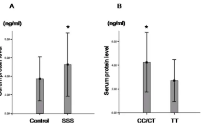
Serum TGF-
β
1 protein levels in the various TGF-
β
1 polymorphisms
ELISA revealed that the level of TGF-β1 in the serum was significantly greater in the SSS group
(N = 44) than in the control group (N = 49) (5.3 ± 3.4 ng/ml vs. 3.7 ± 2.4 ng/ml, P = 0.01) among randomly selected subjects. The 44 SSS patients included 35 patients with CC/CT genotypes and 9 patients with TT genotypes. The 49 controls included 32 subjects with CC/CT genotypes and 17 subjects with TT genotype. In addition, the CC/CT genotype (N = 32) showed a higher
TGF-β1 serum level than the TT genotype (N = 17) (CC/CT vs. TT = 4.25 ± 2.50 ng/ml vs. 2.71 ± 1.76
ng/ml, P = 0.028) in the control group (N = 49) (Fig 2).
TGF-
β
1 mRNA expression levels of the various TGF-
β
1 gene
polymorphisms in SSS patients and controls
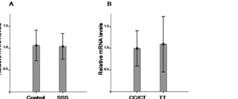
The samples were randomly collected from 18 controls and 15 SSS patients. Quantitative
analy-sis of TGF-β1 mRNA expression revealed there is no significant difference in the TGF-β1
mRNA expression levels between SSS patients (N = 15) and control individuals (N = 18) (rela-tive mRNA expression levels, SSS vs. control = 1.05 ± 0.27 vs. 1.06 ± 0.35 P = 0.976) (Fig 3A). The 15 SSS patients included 10 patients with CC/CT genotypes and 5 patients with TT geno-types. The 18 controls included 11 subjects with CC/CT genotypes and 7 subjects with TT
genotype. There is also no significant difference in the TGF-β1 mRNA expression levels
between CC/CT and TT genotypes in the controls (relative mRNA expression levels, CC/CT
vs. TT = 0.98 ± 0.40 vs. 1.07 ± 0.63, P = 0.899) (Fig 3B). The results indicates that the TGF-β1
T869C gene polymorphism could not significantly affect the TGF-β1 mRNA expression.
Discussion
The present study demonstrated the C-dominant CC/CT genotype frequency of TGF-β1T
+869C polymorphism is significantly higher in the SSS group. The TGF-β1 serum protein level
was significantly higher in the CC/CT genotypes than in the TT genotypes in the control
group. The serum TGF-β1 protein levels were also higher in SSS patients than in the controls.
These data suggested that CC/CT genotypes associated with higher TGF-β1 protein levels in
serum might be associated with disease susceptibility to acquired SSS.
Fig 2. Serum TGF-β1 protein levels.(A) The samples were randomly collected from 49 controls and 44 SSS patients. The serum TGF-β1 protein level in SSS patients (N = 44) is significantly higher than that in control individuals (N = 49). (B) The samples were all from control group (N = 49) including 32 cases with CC/ CT genotypes and 17 cases with TT genotypes. The serum TGF-β1 protein level in controls with a CC/CT genotype is significantly higher than in those with a TT genotype.*<0.05.
Aging and acquired sick sinus syndrome
Studies in animal models have shown that TGF-β1-mediated fibrosis and ion channel
remodel-ing play key roles in the sinus node dysfunction linked to SCN5A-deficiency and agremodel-ing.[10]
The loss of connexin-43 expression and the down-regulation of L-type calcium channels (Ica,L)
have been associated with sinus node dysfunction in aging [25,26]. The down regulation of
sodium and potassium channels has also been linked to sinus node aging process [27,28].
These findings suggested that atrial remodeling with fibrosis and the down regulation of car-diac ion channels and gap junction proteins are important factors in the pathophysiological mechanism of aging-dependent acquired SSS.
TGF-
β
1 affecting sinus node function via linking the RAS molecules
Acquired SSS has been attributed to cardiac fibrosis during aging process and the modificationof ion channels through RAS [8,29]. The overexpression of RAS downregulates the expression
of sodium channel and gap junction proteins, leading to low-voltage electrical activity and con-duction in the heart [30]. Studies using transgenic mice demonstrated that the overexpression of angiotensin II type I (AT1) receptor in myocardium is associated with sinus bradycardia [31]. These results indicate that overexpression of RAS molecules might result in fibrosis, ion
channel modification and sinus node dysfunction. TGF-β1 has been associated with the RAS
and cardiac fibrosis [12]. In vitro studies have shown that angiotensin II might upregulate
TGF-β1 expression through the mitogen-activated protein kinase (MAPK) signaling pathway
via the AT1 receptor in cardiac fibroblasts, myofibroblasts, and myocytes [12]. A TGF-β1
auto-crine/paracrine cellular response induced through the binding of angiotensin II to the AT1 receptor has been observed in cardiac fibroblasts [12]. These studies indicate a critical role of
TGF-β1 in the activation of the RAS in fibrosis and the modulation of ion channels, resulting
in the dysfunction of the pacemaking activity in fibrotic heart tissue.
Gene polymorphisms and susceptibility to acquired SSS
Information concerning the influence of gene polymorphisms on the pathophysiological
mech-anisms of acquired SSS is limited [7,8]. A recent genome-wide association study reported that
a rare variant in the MYH6 gene, which encodes the alpha heavy chain subunit of cardiac myo-sin, is associated with a high risk of acquired SSS based on the population of Iceland [7]. This
Fig 3. TGF-β1 mRNA expression levels in different genotypes of SSS patients and controls.(A) Quantitative analysis of TGF-β1 mRNA expression using GAPDH mRNA as inner control revealed there is no significant difference in the TGF-β1 mRNA expression levels between SSS patients (N = 15) and control individuals (N = 18) (relative mRNA expression levels, SSS vs. control = 1.05±0.27 vs. 1.06±0.35 P = 0.976). (B) The samples were all from control group (N = 18) including 11 cases with CC/CT genotypes and 7 cases with TT genotypes. There is no significant difference in the TGF-β1 mRNA expression levels between CC/CT and TT genotypes (relative mRNA expression levels, CC/CT vs. TT = 0.98±0.40 vs. 1.07±0.63, P = 0.899). In (A) and (B), each value represents the mean±SEM.
study showed that a rare missense variant, C2161T, results in the conceptual amino acid substi-tution Arg721Trp, associated with acquired SSS. The variant C2161T is considered to be linked to cardiac-specific microRNA and gap junction protein expression, and subsequently affects sinus node function [7]. This study proposed differential mechanisms for acquired and familial SSS. However, acquired SSS is also a complex disease with a collection of manifestations of
sinus node dysfunction rather than a single etiology and pathogenesis [32,33]. Notably, the
complex susceptibility of acquired SSS might be determined through multiple susceptibility loci, ethnic population, environment factors and age differences.
In a previous study, we demonstrated that the angiotensinogen (AGT) promoter polymorphism G-6A is highly associated with susceptibility to acquired SSS.[8] Based on functional studies, including electrophoretic mobility shift assay (EMSA) and luciferase assay results, we confirmed that nucleotide G at position -6 of the AGT gene modulates binding affinity with nuclear factors and yields lower transcriptional activity than nucleotide A. The G-6A AGT promoter polymor-phism modulates AGT expression and therefore associated with acquired SSS. The results indicate that the pathophysiologic mechanism of acquired SSS is associated with the RAS [8].
In the present study, we fist reported that TGF-β1 T869C gene polymorphism is associated
with the disease susceptibility to acquired SSS. It is likely that TGF-β1 T869C gene
polymor-phism could affect the sinus node function via linking to the modulation of TGF-β1 protein
levels in serum.
Gene association study of TGF-
β
1 T869C gene polymorphism with SSS
associated cardiovascular diseases
AF, CAD and HT have been reported to be associated with acquired SSS [2,32,34]. The results
of the present association study between the TGF-β1 T869C gene polymorphism and disease
sus-ceptibility of acquired SSS might be influenced by these cardiovascular diseases. We have per-formed analysis by doing the same association study for AF, CAD and HT before/after excluded known SSS patients. Before excluded SSS patients, no significant difference in genotype
distribu-tion (CC+CT/TT) of TGF-β1 T869C gene polymorphism was observed between the subjects
with and without AF (34/12 vs. 144/57, OR = 1.122, 95% CI 0.543–2.318, p = 0.757), CAD (19/5
vs. 159/64, OR = 1.530, 95% CI 0.548–4.271, p = 0.417) or HT (74/35 vs. 104/34, OR = 0.691,
95% CI 0.396–1.208, p = 0.195). After excluded SSS patients, there was also no significant
differ-ence in the genotype distribution (CC+CT/TT) between the subjects with and without AF (14/10
vs. 76/37, OR = 0.682, 95% CI 0.277–1.679, p = 0.405), CAD (9/4 vs. 81/43, OR = 1.194, 95% CI
0.348–4.105, p = 1.000) or HT(39/23 vs. 81/43, OR = 0.789, 95% CI 0.393–1.6915, p = 0.532).
The results of the association study indicate that TGFβ1 T869C gene polymorphism is not
asso-ciated with AF, CAD or HT. The results also support that TGFβ1 T869C gene polymorphism
might be independently associated with the disease susceptibility of acquired SSS
The role of TGF-
β
1 gene polymorphisms in the pathogenesis of acquired
SSS
T869C gene polymorphism is located in exon 1 of the TGF-β1 gene. With the T to C transition,
the T869C gene polymorphism has been associated with elevated TGF-β1 serum
concentra-tions.[17,35] In the present study, there was no significant difference in the TGF-β1 mRNA
expression levels between SSS patients and control individuals. There was also no significant
difference in the TGF-β1 mRNA expression levels between CC/CT and TT genotypes of
TGF-β1 gene polymorphism in the control group. The results indicated the TGF-β1 T869C gene
polymorphism could not significantly affect the TGF-β1 mRNA expression. However, TGF-β1
exportation efficiency of the synthesized protein to the endoplasmic reticulum. TGF-β1 T869C
gene polymorphism might make serum TGF-β1 protein level increased via affecting the
pro-tein trafficking and promoting the secretion of TGF-β1 into the serum.[13,17,36] Accordingly,
T869C gene polymorphism of TGF-β1 is suggested to modulate the serum levels for the
TGF-β1 protein and subsequently leads to the dysfunction of the pacemaking activity in the fibrotic
heart tissue. Thus, patients with a TGF-β1 T869C gene polymorphism might be more liable to
develop acquired SSS. The present study consistently shows that the SSS group, which has a
higher frequency of C at position 869, has a higher TGF-β1 serum level than the control group.
This suggests a unique mechanism for the modulation of TGF-β1serum protein level, which
may result in sinus node fibrosis and dysfunction.
Study limitations
In the present study, we demonstrated that the TGF-β1 T869C gene polymorphism is
associ-ated with acquired SSS. We also provided functional results and serum TGF-β1 protein levels,
to support a crucial role for TGF-β1 in the underlying pathogenesis of acquired SSS. Based on
the results of PBMC mRNA level measurement and data of previous studies [13,17,36], they
provided evidence to support that TGF-β1 T869C gene polymorphism might change the
serum protein levels via affecting the protein secretion. However, we still cannot exclude the possibility of change in mRNA expression among the sinus node related cells. The potential
mechanisms of the TGF-β1 that affect the susceptibility of acquired SSS via linking to the
mod-ulation of TGF-β1 protein levels in serum still need additional in vivo and in vitro studies for
further clarification in the future. Another limitation of the present study is that the study pop-ulation is small. These results should be further confirmed by a larger-scaled study.
Conclusions
Patients with acquired SSS have a higher TGF-β1 T869C CC/CT genotype distribution and a
higher C allele frequency, suggesting a role for the TGF-β1 T869C gene polymorphism in
deter-mining the risk of acquired SSS. The serum protein level obtained with the ELISA suggested that
nucleotide substitution in the TGF-β1 polymorphic site +869 would affect the TGF-β1 serum
pro-tein levels. Taken together, the data reported in the present study provide insight for the potential
mechanisms for acquired SSS. These results also suggested that the TGF-β1 T869C polymorphism
is associated with high serum TGF-β1 levels and increased susceptibility to acquired SSS.
Supporting Information
S1 Table. Genotypes.(XLS)
Acknowledgments
The authors thank Pei-Chi Hung, Mei-Ling Yu and the staffs of the electrophysiology labora-tory for technical support.
Presented in part in abstract form at the 36th annual scientific sessions of the Heart Rhythm Society on May 14, 2015, Boston, Massachusetts.
Author Contributions
References
1. Rubenstein JJ, Schulman CL, Yurchak PM, DeSanctis RW. Clinical spectrum of the sick sinus syn-drome. Circulation. 1972; 46: 5–13. PMID:5039825
2. Mangrum JM, DiMarco JP. The evaluation and management of bradycardia. N Engl J Med. 2000; 342: 703–709. PMID:10706901
3. Stieber J, Hofmann F, Ludwig A. Pacemaker channels and sinus node arrhythmia. Trends Cardiovasc Med. 2004; 14: 23–28. PMID:14720471
4. Makita N, Sasaki K, Groenewegen WA, Yokota T, Yokoshiki H, Murakami T, et al. Congenital atrial
standstill associated with coinheritance of a novel SCN5A mutation and connexin 40 polymorphisms. Heart Rhythm. 2005; 2: 1128–1134. PMID:16188595
5. Milanesi R, Baruscotti M, Gnecchi-Ruscone T, DiFrancesco D. Familial sinus bradycardia associated
with a mutation in the cardiac pacemaker channel. N Engl J Med. 2006; 354: 151–157. PMID:
16407510
6. Dobrzynski H, Boyett MR, Anderson RH. New insights into pacemaker activity. Circulation. 2007; 115:
1921–1932. PMID:17420362
7. Holm H, Gudbjartsson DF, Sulem P, Masson G, Helgadottir HT, Zanon C, et al. A rare variant in MYH6 is associated with high risk of sick sinus syndrome. Nat Genet. 2011; 43: 316–320. doi:10.1038/ng.
781PMID:21378987
8. Chen JY, Liou YM, Wu HDI, Lin KH, Chang KC. Promoter polymorphism G-6A, which modulates angio-tensinogen gene expression, is associated with non-familial sick sinus syndrome. Plos One. 2012; 7: e29951. doi:10.1371/journal.pone.0029951PMID:22242192
9. McNair WP, Lisa Ku, Taylor MRG, Fain PR, Dao D, Wolfel E, et al. SCN5A mutation associated with dilated cardiomyopathy, conduction disorder, and arrhythmia. Circulation. 2004; 110: 2163–2167.
PMID:15466643
10. Hao X, Zhang Y, Zhang X, Nirmalan M, Davies L, Konstantinou D, et al. TGF-β1-mediated fibrosis and ion channel remodeling are key mechanisms in producing the sinus node dysfunction associated with SCN5A deficiency and aging. Circ Arrhythm Electrophysiol. 2011; 4: 397–406. doi:10.1161/CIRCEP.
110.960807PMID:21493874
11. Blobe GC, Schiemann WP, Lodish HF. Role of transforming growth factorβin human disease. N Engl J Med. 2000; 342: 1350–1358. PMID:10793168
12. Rosenkranz S. TGF-beta1 and angiotensin networking in cardiac remodeling. Cardiovasc Res. 2004; 63: 423–432. PMID:15276467
13. Shah R, Rahaman B, Hurley C, Posch P. Allelic diversity in the TFGβ1 regulatory region: Characteriza-tion of novel funcCharacteriza-tional single nucleotide polymorphisms. Hum Genet. 2006; 119: 61–74. PMID:
16369764
14. Wu CH, Hu YF, Chou CY, Lin YJ, Chang SL, Lo LW, et al. Transforming growth factor-β1 level and out-come after catheter ablation for nonparoxysmal atrial fibrillation. Heart Rhythm. 2013; 10: 10–15. doi:
10.1016/j.hrthm.2012.09.016PMID:22982967
15. Gramley F, Lorenzen J, Koellensperger E, Kettering K, Weiss C, Munzel T. Atrial fibrosis and atrial fibril-lation: The role of the TGF-β1 signaling pathway. Int J Cardiol. 2010; 143: 405–413. doi:10.1016/j.
ijcard.2009.03.110PMID:19394095
16. Li X, Yue ZC, Zhang YY, Bai J, Meng XN, Geng JS, et al. Elevated serum level and gene polymor-phisms of TGF-β1 in gastric cancer. J Clin Lab Anal. 2008; 22: 164–171. doi:10.1002/jcla.20236
PMID:18484655
17. Peng L, Li P, Chen J, Yan K, Huo F, Han L, et al. Association between transforming growth factor-beta 1 T869C polymorphism and ischemic stroke: A meta-analysis. Plos One. 2013; 8: e67738. doi:10. 1371/journal.pone.0067738PMID:23861796
18. Lamas GA, Lee K, Sweeney M, Leon A, Yee R, Ellenbogen K, et al. The mode selection trial (MOST) in sinus node dysfunction: design, rationale, and baseline characteristics of the first 1000 patients. Am Heart J. 2000; 140: 541–551. PMID:11011325
19. Nielsen JC, Thomsen PEB, Højberg S, Vesterlund T, Dalsgaard D, Mortensen LS, et al. A comparison of single-lead atrial pacing with dual-chamber pacing in sick sinus syndrome. Eur Heart J. 2011; 32: 686–696. doi:10.1093/eurheartj/ehr022PMID:21300730
20. Chen LY, Ballew JD, Herron KJ, Rodeheffer RJ, Olson TM. A Common Polymorphism in SCN5A is Associated with Lone Atrial Fibrillation. Clin Pharmacol Ther. 2007.; 81: 35–41. PMID:17185997 21. Barrett JC, Fry B, Maller J, Daly MJ. Haploview: Analysis and visualization of ld and haplotype maps.
22. Tsai CT, Lai LP, Lin JL, Chiang FT, Hwang JJ, Ritchie MD, et al. Renin-angiotensin system gene poly-morphisms and atrial fibrillation. Circulation. 2004; 109: 1640–1646. PMID:15023884
23. Ardlie KG, Kruglyak L, Seielstad M. Patterns of linkage disequilibrium in the human genome. Nat Rev Genet. 2002; 3: 299–309. PMID:11967554
24. Gabriel SB, Schaffner SF, Nguyen H, Moore JM, Roy J, Blumenstiel B, et al. The structure of haplotype
blocks in the human genome. Science. 2002; 296: 2225–2229. PMID:12029063
25. Lau D, Roberts Thomson K, Sanders P. Sinus node revisited. Curr Opin Cardiol. 2011; 26: 55–59. doi:
10.1097/HCO.0b013e32834138f4PMID:21102315
26. Jones SA, Boyett MR, Lancaster MK. Declining into failure: the age-dependent loss of the L-type cal-cium channel within the sinoatrial node. Circulation. 2007; 115: 1183–1190. PMID:17339548 27. Zhang H, Zhao Y, Lei M, Dobrzynski H, Liu JH, Holden AV, et al. Computational evaluation of the roles
of Na+ current, iNa, and cell death in cardiac pacemaking and driving. Am J Physiol Heart Circ Physiol. 2007; 292: H165–H174 PMID:16877551
28. Tellez JO DH, Yanni J, Billeter R, Boyett MR. Effect of aging on gene expression in the rat sinoatrial node. J mol cell cardiol. Abstract. 2006; 40: 1.
29. Duff JL, Marrero MB, Paxton WG, Schieffer B, Bernstein KE, Berk BC. Angiotensin II signal
transduc-tion and the mitogen-activated protein kinase pathway. Cardiovasc Res. 1995; 30: 511–517. PMID:
8574999
30. Kasi VS, Xiao HD, Shang LL, Iravanian S, Langberg J, Witham EA, et al. Cardiac-restricted
angioten-sin-converting enzyme overexpression causes conduction defects and connexin dysregulation. Am J Physiol Heart Circ Physiol. 2007; 293: H182–H192. PMID:17337599
31. Hein L, Stevens ME, Barsh GS, Pratt RE, Kobilka BK, Dzau VJ. Overexpression of angiotensin AT1
receptor transgene in the mouse myocardium produces a lethal phenotype associated with myocyte hyperplasia and heart block. Proc Natl Acad Sci U S A. 1997; 94: 6391–6396. PMID:9177228 32. AdÃn V, Crown LA. Diagnosis and treatment of sick sinus syndrome. Am Fam Physician. 2003;. 67:
1725–1732. PMID:12725451
33. Herrmann S, Ludwig A. Not so fast! Sick sinus syndrome is a complex and incompletely understood dis-ease that might prove hard to model in animals: Reply. Cardiovasc Res. 2011; 92: 179.
34. Gregoratos G. Cardiology patient pages. Sick sinus syndrome. Circulation. 2003; 108: e143–e144.
PMID:14623796
35. Yamada Y, Okuizumi H, Miyauchi A, Takagi Y, Ikeda K, Harada A. Association of transforming growth factorβ1 genotype with spinal osteophytosis in Japanese women. Arthritis Rheum. 2000; 43: 452–
460. PMID:10693888